Every year in Russia, 34 thousand cases of benign and malignant tumors that develop in the brain are diagnosed. The first signs of a brain tumor are similar to the symptoms that people experience with other diseases. Thus, depression, insomnia, headache and fatigue are symptoms that are not paid attention to at a young age. Therefore, it is easy to miss the initial stage of tumor development.
As for diagnosing the disease in children and adolescents, malignant neoplasms in this category are less common than in adults. Statistics show that childhood cancer occurs in only 1% of patients worldwide. A detected tumor in a child is not a death sentence, since it can be treated in the early stages.
Reasons for the development of cerebral edema
- Cerebral edema occurs in 20% of all severe traumatic brain injuries
- In addition, it often occurs with tumors and strokes.
There are many causes of cerebral edema:
- Fluid retention in the brain parenchyma (vasogenic, cytotoxic)
- Impaired autoregulation of cerebral vessels
- Hypoxia
- Symptoms of cerebral edema, such as swelling of brain tissue due to injury, can develop quickly, but usually within 1-2 days.
Which method of diagnosing cerebral edema to choose: MRI or CT
Selection method:
- MRI or MSCT
What will a CT scan of the head show for cerebral edema?
- Loss of clarity of the boundary between gray and white matter
- An increase in the volume of the brain parenchyma, a decrease in the volume of the subarachnoid space and cisterns
- White matter is hypodense
- Presence of volumetric impact with possible wedging
- Near the area of edema, intact areas of the brain are visualized (for example, “white cerebellum symptom”).
Is an MRI of the brain informative in case of brain edema?
- Loss of clarity of the boundary between gray and white matter
- On T1-weighted images obtained with an inversion-recovery pulse sequence, there is no typical contrast between hypointense gray matter and hyperintense white matter
- ICD is decreased in cytotoxic edema and increased in vasogenic and interstitial cerebral edema
- Violation of liquor circulation.
Hardware methods
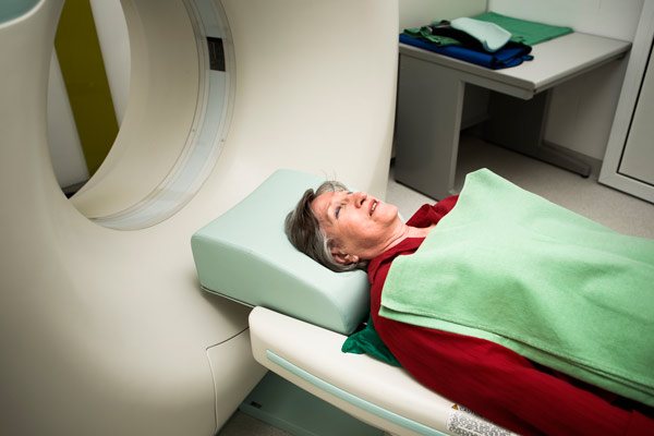
MRI
Magnetic resonance imaging is the main method for identifying and determining tumors in the brain. Using a special apparatus and electromagnetic waves, a three-layer image of the tumor . MRI provides high-quality images of even very small tumors.
In addition, this method allows us to determine the presence of cancer at an early stage . MRI is used during surgery to specify the size of the tumor and establish important characteristics: structure, localization, prognosis of spread.
CT
To obtain an accurate image of healthy structures using computed tomography, fairly specific x-ray equipment is used, as well as a computer setup . It is worth noting that this diagnostic method is not very sensitive compared to magnetic resonance imaging.
CT is not effective enough to detect tumors at the first stage of disease development.
But still, in some specific cases, such an examination is considered quite useful. This is ensured by the introduction into the patient’s body of a substance with contrast properties, which greatly facilitates the detection of tumors.
Computed tomography allows you to determine the location of the tumor, as well as its type . In addition, CT can detect swelling of organ structures, internal hemorrhages and other symptomatic manifestations, and is often used to assess the usefulness of treatment measures and identify the possibility of relapse.
Pneumography
The essence of pneumography is based on the use of air as contrast, which is introduced into the space of the ventricles of the brain. volumetric neoplasms in the area of study changes in the main characteristics of the ventricles and subarachnoid spaces.
Such indicators are determined in accordance with the location, size and structure of the existing tumor.
Angiography
Angiography is performed using X-ray equipment and a contrast agent that is injected into the vessels. After this, a picture is taken, on the basis of which further examination is carried out. In particular, a significant indicator is the change in the position of the vessels and the displacement of their branches - this is a sign of the presence of a neoplasm.
Another important characteristic is the presence of newly formed vessels that form in the body of the tumor. This diagnostic method allows you to accurately and very quickly determine not only the location of the tumor in the brain, but also its type.
Neurosonography
The technique is based on ultrasonic waves. Used to determine cancer formations in children under 12 months .
In fact, waves of this type do not have the ability to penetrate the bones of the skull. But a small child has some physiological characteristics - fontanelles are natural expansions between parts of the skull.
It is through such expansions that ultrasonic waves penetrate. Neurosonography allows you to determine the presence of formations in the brain, as well as changes in the position of organ structures.
Average price for laser lipoma removal in Russian clinics. This article contains information about poorly differentiated squamous cell carcinoma of the esophagus.
The link https://stoprak.info/vidy/zhenskix-polovyx-organov/matka/opisanie-operacii-po-udaleniyu-miomy-laparoskopicheskim-metodom.html describes the stages of the operation to remove uterine fibroids using the laparoscopic method.
Ultrasound of the brain
It was previously noted that ultrasound has low penetrating ability - it cannot pass into the brain through the bones of the skull. Obviously, in a different situation, many other methods would be irrelevant due to the undeniable advantages of ultrasound.
But still, there is a special technique for determining formations in the brain using ultrasound - “M-echo” . Based on the results of its implementation, it is possible to establish the degree of displacement of the midline structures of the organ in question. Such changes in most cases indicate the presence of a tumor.
SPECT
Single photon emission computed tomography is performed using a standard gamma camera. It is not sufficiently effective in identifying tumor cells and scar structure after treatment. In most cases, it is used after a CT or MRI to determine the degree of malignancy of the existing tumor.
Electroencephalography
The essence of this technique is based on the temporary specification of the electrical potentials of the brain. Electroencephalography uses special plates that are attached to the patient's head. In some situations, it is possible to use needles - and inject them directly into the brain. Using the results obtained, it is possible to identify foci of impulse pathology for a specific part of the organ.
Magnetoencephalography
The characteristics of the magnetic field formed by nerve cells in a state of electrical activity are measured. Magnetoencephalography is used to determine a specific assessment of the activity of the parts of the brain of interest.
Lumbar puncture
Lumbar spinal puncture is a specific procedure, the principle of which is based on the removal of cerebrospinal fluid from the human body in order to determine the presence of pathological diseases or disorders in the system through further research.
The needle is inserted exactly between the 3rd and 4th lumbar vertebrae. Once the correct positioning of the instrument in the subarachnoid space is achieved, it becomes possible to measure the pressure of the cerebrospinal fluid and, accordingly, the fluid intake itself.
Subsequently, the resulting sample is sent to the laboratory to be examined for the presence of tumor cells and markers - substances that indicate the disease. But it is worth noting that tumor markers have not yet been identified that indicate the initial stage of brain cancer.
In addition, for safety reasons, it is highly recommended that you undergo a CT or MRI scan before undergoing the procedure in question.
How does kidney cancer manifest in women? Symptoms, stages, how long they live after treatment. Let's discuss the types of intestinal lymphoma here.
How is massage useful for small uterine fibroids? Here https://stoprak.info/vidy/zhenskix-polovyx-organov/matka/mioma-nebolshix-razmerov.html expert opinion.
Biopsy
The technique is surgical in nature. Its essence consists in taking a small fragment of tissue for further examination under a microscope for indicators that will determine the degree of malignancy. The final results may also provide information about the type of tumor cells. A biopsy can be performed either during surgery or separately.
But it is worth considering some contraindications for this procedure. For example, in the presence of gliomas, a standard biopsy is quite dangerous. It can cause damage to healthy structures, which can lead to disruption of important functions.
In such cases, the doctor uses other types of biopsy: fine-needle or stereotactic. The latter takes place using computer technology, which carries out control.
Possible complications
The consequences of cerebral edema are very diverse, but the most common of them are:
- Disturbances in sleep and wakefulness
- Depression
- Headache
- Meteor dependence
Swelling of the left hemisphere of the brain. CT, axial slice. On the left, the sulci are worse visualized compared to the right hemisphere, and the differentiation of gray and white matter is less clear.
Hypoxia. CT, axial slice. The boundary between gray and white matter is not differentiated. The grooves are no longer visualized, indicating irreversible damage to the brain parenchyma.
Kinds
Among all types of headaches, migraine and standard tension were the most common. Both types can be accompanied by mild stiffness, or maybe serious and severe pain.
The pain is felt in the temple area or radiates to the eyes, to the back of the head, or to the area of the cranial vault.
Quite often, with various pains in the head, there may be a vegetative disorder, that is, problems during the respiratory process, rapid fatigue, and so on.
Migraine has a paroxysmal form, which is also accompanied by severe pain in the head. The pain comes through pulsation. Such attacks can be caused by bright light or other types of irritants, as well as constant fatigue. As a rule, such headaches last up to several hours; at best, they end in half an hour.
Experts also identify a third common type of headache, which is called cluster pain or, in other words, beam pain . The main distinguishing feature of such pain is its immediate appearance. In addition, a person feels a cluster headache exclusively on one side of the head. These types of attacks can be repeated over several months, or can end in a week.