Brief description of the procedure Time: from 10 minutes, with contrast up to 35 minutes Necessity of using a contrast agent: as prescribed by a doctor Necessity of preparation for the study: no Presence of contraindications: yes Restrictions: available Time to prepare a report: 60-120 minutes Children: over 14 years old
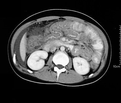
CT of internal organs is a gentle and highly informative diagnostic method based on layer-by-layer scanning of an area using a fan-shaped release of X-rays. The result of the study is the visualization of the anatomical area in high-quality photographs or three-dimensional images.
Due to the fact that the scanning step during a CT examination of internal organs ranges from 0.1 to 10 mm, the method makes it possible to study in detail the disorders and pathological processes of internal organs, to determine the exact localization of neoplasms and foci of inflammation. Despite the fact that CT diagnostics is based on the use of ionizing radiation, the technique is much safer in comparison with traditional X-ray examinations.
How to carry out the procedure
Spiral K appeared in the early 90s, and after a short time the developers created computed tomography, which was no longer just multi-layered and multi-slice, but multi-slice, with greater accuracy and reliability. The procedure was carried out with contrast and was called MSCT angiography. To understand what it is, you need to understand the essence of the procedure. This is an examination of a specific part of the human body, which is carried out using a special device with radiation with the simultaneous introduction of a magnetic substance that penetrates even the smallest vessels. With MSCT of the brain, the images display information on every millimeter of the vascular system, which makes it possible to identify tiny anomalies, diseases, tumors and neoplasms.
All information is displayed in the pictures. What does this photo show? All data about the scanned area. The radiation records the movement of the contrast and makes a layer-by-layer scan of the vessels, displaying information on the monitor. With contrast, not a single vessel or its change will go unnoticed.
Equipment for MSCT can be equipped with a different number of rows of detectors: from 2 to 64. Equipment with 4 and 16 rows is most often used, but 64-row ones provide more accurate information. With MSCT of the brain, the speed of image fixation is high, which makes it possible to obtain a large number of images. Thanks to this, a three-dimensional image of the organ’s vascular system is formed. At the same time, the procedure is fast, doing its job in one human breath, because it is this breath that needs to be held. And when you exhale, everything will be over.
In terms of technology, the procedure is similar to taking an X-ray of the lungs, except that the study is carried out on the brain, which means that the person’s head is placed in a special device. A light dose of radiation is safe for the head and overall health.
How is MSCT with contrast agent performed? In the process, the patient is injected with an iodine-based substance directly into a vein. In this way, even the smallest neoplasms are found, namely:
- their presence and location;
- how much the tumor has metastasized and the designation of its boundaries;
- whether parenchymal organs and lymph nodes are affected.
The procedure also allows you to:
- identification and diagnosis of the spread of tumor foci not only in the brain, but also in the kidneys, spleen, liver and pancreas;
- identification of injuries to the peritoneum, chest and pelvic area.
Studying the brain as the most important structure of the body using MSCT has a number of advantages:
- the session takes place in a short period of time;
- the number and speed of images allow you to accurately determine the problem even at an early stage;
- it is possible to carry out any scaling of the problem area, which is impossible with conventional CT;
- the first procedures lasted about half an hour, now the session does not take even 5 minutes, despite the fact that the irradiation period lasts no more than 30 seconds;
- MSCT of the peritoneum and sternum is done in 20 seconds, while a person can calmly hold his breath, which is important for restless children, people suffering from claustrophobia, with severe painful syndrome, disruption of the respiratory process and even heart failure;
- enormous opportunities for diagnosing patients in severe stages of the disease, when contact with them is limited, for example, during forced ventilation or continuous monitoring of heart function;
- Thanks to multi-layer research, images make it possible to build a visual chain of data processing in two- or three-dimensional space, not only for vessels, but also for joints, bones and internal organs.
The advent of computed tomography scanners
The first mathematical algorithms for CT were developed in 1917 by the Austrian mathematician I. Radon (see Radon transform). The physical basis of the method is the exponential law of radiation attenuation, which is valid for purely absorbing media. In the X-ray radiation range, the exponential law is satisfied with a high degree of accuracy, therefore the developed mathematical algorithms were first applied specifically to X-ray computed tomography.
In 1963, the American physicist A. Cormack again (but in a different way from Radon) solved the problem of tomographic reconstruction, and in 1969, the English engineer-physicist G. Hounsfield designed the “EMR scanner” - the first computer X-ray tomograph, clinical trials of which were carried out in 1971 - designed for head scanning only. Funds for the development of CT were allocated by EMI, in particular, thanks to the high income received from the contract with The Beatles[1].
In 1979, Cormack and Hounsfield were awarded the Nobel Prize in Physiology or Medicine for their development of computed tomography.
Do I need to prepare for the procedure?
The preparatory process is simple, but important, making it possible to obtain only truthful data about the patient’s condition. He must adhere to the following rules:
- Do not eat 4 hours before the examination, if it is carried out with a contrast agent; this procedure is done only on an empty stomach.
- Avoid coffee, tea, alcoholic and caffeinated drinks.
- No smoking.
- Get at least 7 hours of sleep per night so that your body reflects its condition in the pictures as accurately as possible.
When using a contrast agent, the patient will first be tested for compatibility with it. And if necessary, other related tests will be prescribed to make sure there are no contraindications to the procedure. Usually this is blood from a finger for a general analysis, checking the functions of the thyroid gland, a reaction to the iodine component of a contrast agent. When all the results are in hand, the doctor will analyze them and allow the patient to undergo the procedure.
Possible complications after
The most common consequence of CTA is the penetration of the contrast agent into the tissue from the vascular bed. If the volume of such intake does not exceed 10 ml, then it can gradually resolve on its own, but with large quantities an inflammatory reaction and even necrotic changes in tissues develop. Such complications occur in weakened or unconscious patients who are unable to report pain during contrast infusion. For treatment, you need to elevate the limb and apply cold. If blisters or severe redness appear on the skin, or numbness or tingling is felt 3 hours after the procedure, it is recommended that you be examined by a surgeon to determine the indications for surgical treatment. The onset of an allergic reaction to contrast may be indicated by itching and swelling of the skin, local redness, and breathing problems. The long-term consequences of CTA can be a negative impact on kidney function, so urine tests are monitored after 5 and 14 days.
Who is it shown to?
It’s clear how MSCT is done, but who should undergo it? The procedure is prescribed by the attending physician, who can be either a therapist or a neurologist. It is indicated for:
- identifying the cause of pain in the middle ear area;
- if there are signs of subatrophy;
- to find an aneurysm or vascular malformation;
- to understand why the patient’s consciousness is confused or their limbs are numb, they are struck by paralysis, they suffer from migraines, visual system failures or vertigo, which may indicate a tumor, hemorrhage in the brain or be a consequence of injury or rupture of an aneurysm;
- determining the size of the brain cavities, which indicate the presence or absence of hydrocephalus or intracranial hypertension;
- in the preoperative period;
- monitoring the effectiveness of therapy after identifying a problem;
- visual determination of damage to bone and soft tissues of the organ;
- determining the causes of fainting;
- brain biopsies to monitor the process at each stage;
- for diagnosing skull fractures or soft tissue injuries after injuries and accidents;
- to find tumors;
- in case of stroke to detect bleeding or blood clot;
- to check the integrity of the temporal bone, such an injury can cause hearing loss, both partial and complete;
- diagnostic measures to identify meningitis and its various complications.
When the procedure is impossible
Doctors have not identified any absolute reasons why a head examination cannot be performed using MSCT. But each attending physician, at his own discretion, can determine whether the procedure will be safe. Most often, it is not performed on pregnant women and young children, replacing it with MRI. The doctor must accurately determine the line between the necessary harm and the patient’s health. And this is provided that the load of radiation on the body is minimal. The list of relative contraindications includes patients:
- pregnant women;
- asthmatics;
- with kidney disease and heart failure;
- with any form of diabetes;
- having multiple lesions of the body with myeloma spots;
- with a negative reaction to the contrast agent gadolinium.
Research results
The radiologist's report and the resulting images usually take no more than 2 hours to prepare. But in some cases, the interpretation of images can take longer, sometimes up to a day.
CT can determine:
- atherosclerotic plaques, their size and location;
- the condition of the walls of all vessels (veins, arteries, peripheral system);
- stenosis and occlusion (blockage) of the vessels of the head and neck (risk of developing acute cerebrovascular accident);
- ischemic lesions, heart failure, aneurysms;
- hemorrhages, blood clots, neoplasms, metastases;
- traumatic damage to the walls;
- dissection of the aorta and other large vessels.
Risks of examination
Despite the relative safety of MSCT, even here there are risks associated with its implementation, which later manifest themselves in the form of side effects, causing failure in various systems of the human body. Even if we consider that the risk of side effects after the procedure is minimal, you need to know about them in advance:
- The most difficult thing for young people and children to endure is even the small amount of radiation exposure given by modern devices. It is they who experience relapses of oncological processes, which has been confirmed by repeated studies that have shown the presence of a small percentage of such cases.
- Implanted electronic devices are a particular contraindication for the safer MRI procedure. But even with MSCT, although in rare cases, insulin pumps and neurostimulators can malfunction, causing a deterioration in the patient’s health.
Due to the fact that the capabilities of MSCT are so wide, many hospitals and health centers prefer not to use conventional CT and ultrasound, preferring multilayer angiography. It makes it easy to examine the entire large circulatory system, consisting of the carotid artery, pulmonary vessels, liver, as well as the iliac and caval veins of the lower and upper types. The results are incredibly accurate and informative, helping not only to make an accurate diagnosis, but also to select effective treatment.
Indications for CT angiography
The patient will be referred for this examination with the following symptoms and diseases:
- detection of various types of tumors, determination of the presence of metastases;
- in cases of persistent headaches of unknown origin, sudden decrease in vision, the appearance of dizziness, tinnitus and other symptoms that give reason to suspect a circulatory disorder in the vessels of the brain and neck;
- intracranial hemorrhages;
- anomalies in the structure and development of the vascular system;
- atherosclerosis, vasculitis, varicose veins;
- heart disease, ischemic heart disease, angiopathy;
- aneurysms and dissection (dissection) of large blood vessels;
- stenosis, occlusion;
- thrombophlebitis, thrombosis, embolism;
- pathology of the renal arteries;
- injuries;
- pulmonary embolism.
CT is performed for postoperative monitoring, in cases where it is impossible to make a diagnosis using other diagnostic methods.
Differences between MSCT and MRI
In the first case, diagnostics are carried out using X-rays, which generate electronic pulses that are recorded by the device. They allow you to find out the condition of a particular organ. MRI can diagnose any problem with internal soft structures without radiation exposure. Here the device generates a magnetic field, which resonates with the cells and shows their condition. But MSCT is better at checking bone structures.
Both procedures do not require special training and are currently considered to be as safe as possible. MRI is strictly prohibited for people with implanted devices, but for MSCT this is not a problem. Both studies provide accurate information about the layer-by-layer state of blood vessels or organs. Doctors often prescribe them in tandem in order to obtain the maximum possible information about the patient’s condition. Most often this happens after an accident or serious traumatic brain injury, so that not only soft but also bone tissues are analyzed. Both procedures can be performed one after the other, but it is preferable to start with an MRI and then do an MSCT the next day. The latter is possible only if the annual level of radiation exposure does not exceed the permissible norm.