What is proctalgia?
Anorectal pain (proctalgia) is a common disorder and affects 6.6% of the population.
Proctalgia can develop as a result of a number of organic processes or, more often, due to an underlying functional disorder. In almost all cases, the patient’s well-being suffers greatly, and the quality of life decreases. A person experiences serious psychological stress without receiving significant relief, since many doctors look at his complaints as insignificant, and often simply cannot cope with the diagnosis and treatment of a very unpleasant symptom complex. This chapter discusses the most common pathological processes in the anorectal region with the main manifestation being proctalgia. Diagnostic criteria
Chronic proctalgia
Diagnostic criteria include all of the following.
- Chronic or recurrent, acute or dull aching pain in the rectum.
- Seizures lasting minutes or longer.
Further, based on the results of digital rectal examination, chronic proctalgia can be more accurately characterized as levator ani syndrome or nonspecific anorectal pain syndrome
Levator ani syndrome
Criteria for the symptom complex of chronic proctalgia + pain on palpation of a tense puborectalis muscle.
All criteria must be recorded within the last 3 months, and the first symptoms must appear no later than 6 months before diagnosis.
Symptoms of osteochondrosis: nausea, dizziness, radiating to the stomach, intercostal neuralgia
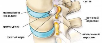
With the help of the spine, a person is able to stand and move. This is where the spinal cord is located, from which nerves extend to all parts of the body. Not a single internal organ would function without the spine.
Unfortunately, the ridge is often susceptible to various diseases. One of these ailments is considered to be osteochondrosis, a disease that causes degenerative changes in the vertebrae, as well as intervertebral discs.
The human spine is divided into the thoracic, cervical and lumbar zones, and pathology can affect any of these zones.
Gives to the stomach with osteochondrosis
Almost all organs of the peritoneum are connected to the nerve endings of the thoracic spine. Patients suffering from thoracic osteochondrosis most often note the formation of pain radiating to the abdomen.
Abdominal pain with osteochondrosis is distinguished by the following symptoms:
- non-diffuse pain;
- pain is localized in the area of innervation of the affected area;
- pain occurs and also intensifies after the slightest movements or when turning, as well as coughing;
- pain is felt at the level of muscle tissue;
- usually the pain is one-sided, constant, dull or aching.
Pain in the abdomen in people suffering from osteochondrosis limits and impedes their movements. Usually the pain radiates to the stomach with such lesions of the ridge:
- in case of damage to the mid-thoracic nerves, pain is observed in the stomach;
- if the pathology is localized in the 8th and 9th roots, then the pain forms in the area of the duodenum;
- if osteochondrosis affects the 7th, 8th, 9th thoracic roots on the right, then pain is felt in the hypochondrium on the right.
Read: Exercises for osteochondrosis of the thoracic region
Cervical and thoracic osteochondrosis, in addition to abdominal pain, can provoke problems with stool, impaired intestinal motility, and pathologies of gas formation.
Soreness in the stomach
When the spine is affected by osteochondrosis, patients often experience pain in the organs of the digestive system. Often such stomach pains are mistakenly diagnosed as gastroduodenitis.
Infringement of the spinal root in the vegetative part of the ridge causes spasm or irritation of the nerve. The result is heartburn and nagging pain. Over time, pain in the digestive organ becomes constant.
Stomach pain caused by osteochondrosis is easily differentiated from gastric disorders: pain due to pathologies of the spine increases with the slightest movements or turns.
Pain in the lower abdomen
Typically, pain in the lower abdomen is caused by pathology progressing in the lumbar region. If the disease affects the thoracic area of the spinal column, then pain is felt on the right in the abdominal area. The symptoms resemble those of appendicitis, but without an increase in body temperature. In addition, pain in the lower abdomen is similar to manifestations of gastritis and colitis.
Osteochondrosis in this case is accompanied by the following symptoms:
- heartburn and nausea;
- feeling of heaviness in the right hypochondrium;
- cramps and bloating;
- stabbing pain in the right lower abdomen;
- constipation occurring with nagging pain;
- pain in the epigastric zone.
Read: How to treat osteochondrosis at home
It is very difficult to independently distinguish pain syndrome due to osteochondrosis from pathologies of the digestive organs. It is recommended to consult a specialist in a timely manner to avoid the development of complications.
Anal fissure
An anal fissure is an ellipsoidal abrasion or fissure in the epithelium of the anal canal, located below (distal) the dentate line. Passing stool through the anal canal causes severe pain and is accompanied by bleeding in the form of a small amount of scarlet blood in the stool or on toilet paper. The pain begins during bowel movements and continues after it. Patients describe the pain as sharp, cutting. One study analyzed 15,000 outpatient visits to a proctologist; the prevalence of anal fissure was 10%; men and women were affected with equal frequency. Children and older people suffer from anal fissure less often than middle-aged adults.
Cracks can be divided into acute (lasting up to 6 weeks) and chronic. In many cases, acute anal fissures are small in size and heal without medical intervention.
In terms of pathophysiology, it has always been believed that fissures arise as a result of constipation, straining, passage of hard feces (“high pressure fissures”) or, as with IBD and diarrhea, due to too frequent work of the organ (“low pressure fissures”). In addition, recent studies have shown that fissures can develop as a result of decreased blood flow to the anal canal (mainly along the posterior midline) and increased pressure in the anal canal (especially under the action of the internal anal sphincter). All together causes local ischemia, which creates the ground for the formation of ulcers. The ulceration breaks at the slightest injury. As soon as a crack appears, spasm of the internal anal sphincter begins to stretch the edges of the wound in different directions, thereby preventing healing.
Physical examination of the perianal area in patients with a fissure can be difficult, since in this situation the anal sphincters are usually in a state of spasm, and in some patients, rectal examination is completely impossible due to very severe pain. This pain can be reduced by using topical anesthetic gels. A more complete examination of the rectum, including anoscopy, should be performed under sedation. Fissures in most cases (90%) are located along the posterior midline, less often - along the anterior (mainly in women). A soft fibropapilloma (“sentinel tubercle”) is often formed along the lower (distal) edge, while a hypertrophied papilla can be seen along the upper edge. In a typical case, an acute crack has clean edges, while in a chronic case, the edges are inductively changed and raised. Cracks on the side surfaces are rare. This localization can be found in Crohn's disease or other less common diseases, such as acquired immunodeficiency syndrome and tuberculosis. Young people (under 40 years of age) without warning signs (anemia, colorectal cancer, family history of IBD) can be treated without additional diagnostic testing. Patients over 50 years of age should undergo a screening colonoscopy (if not previously performed) before starting treatment.
We recommend treating chronic anal fissures step by step, ensuring, first of all, relaxation of the internal sphincter, atraumatic passage of feces and elimination of pain. To begin with, patients should consider lifestyle changes that include adequate dietary fiber intake (25-30 g per day). Dietary fiber is needed to avoid constipation and straining during bowel movements. Fibers have been shown to promote healing of anal fissures in placebo-controlled studies. Another key point is sitz baths. Baths ensure cleanliness of the perianal area and help relax the anal sphincter. They have also been shown to increase local blood circulation and thereby stimulate the healing process. Drug treatment is the second line of therapy, and there are several options available. Local anesthetics (eg, lidocaine) reduce pain but do not affect closure of the defect. Drugs are contraindicated. The “gold standard” of therapy is considered to be “chemical sphincterotomy.” A recently published Cochrane meta-analysis showed that topical nitroglycerin promotes healing in 48.6% of patients (0.2-0.3% nitroglycerin ointment applied twice daily for 4-6 weeks). Other studies report recovery in 88% of cases. From a physiological point of view, topical application of nitrates can be considered an excellent therapeutic approach, as they relax smooth muscle, allow the edges of the defect to approach each other, and improve blood flow in the anoderm. Unfortunately, the use of nitroglycerin is limited by frequent side effects: headaches (up to 70% of patients), hypotension and nausea. CCBs (nifedipine, diltiazem, etc.; forms for local and oral use) relax the anal canal and help heal fissures in 2/3 of cases. Injections of botulinum toxin (a complex of botulinum toxin type A with hemagglutinin, the drug Botox) facilitate the closure of the defect in approximately 65% of patients. It appears that changing the dose of botulinum toxin in the range of 60 to 100 units does not affect the rate of complete healing. Among the side effects, transient NK appears in 3% of patients. A recent meta-analysis noted that for chronic fissures, botulinum toxin (see below) and topical nitroglycerin were only slightly better than CCBs for healing; this difference turned out to be insignificant. Another meta-analysis of three randomized controlled trials with 180 participants showed that botulinum toxin, with fewer side effects, is as effective as nitroglycerin preparations. CCBs and botulinum toxin have fewer side effects than nitroglycerin.
If drug therapy does not lead to healing, then the next step is surgery. The “gold standard” is lateral internal sphincterotomy, which gives a recovery rate of 90-95%. Incision of the internal anal sphincter allows the edges of the epithelial defect to come into contact and heal. The most significant complication in this case is NK, which occurs in approximately 10% of patients, although the problem is usually limited only to the passage of gases, and this symptom is expressed to a small extent. A recent meta-analysis of four studies involving 279 patients found that lateral internal sphincterotomy was more effective than botulinum toxin for the treatment of chronic anal fissures (hazard ratio, 1.31; p < 0.0001).
Anal itching
Anal itching is another benign condition that can cause pain in the anorectal area. Anal itching affects 1-5% of the US population; In terms of frequency, men predominate over women in a ratio of approximately 4:1. Symptoms of intense itching in the perianal area and burning do not go away after defecation, and the itching is most often not accompanied by bleeding, but if excoriation occurs, pain and blood may appear after defecation. In some patients, the disorder goes away on its own (acute anal itching), and the culprits in this category of patients may be side effects of drug therapy or nutritional factors.
In terms of pathophysiology, anal itching appears secondary to local irritation. Later, an inflammatory response develops, which is either limited to the superficial layers of the perianal skin or extends deeper. Thus, contamination with feces can lead to tissue maceration, then this area becomes infected with Candida, which provokes chronic manifestations in the form of itching and burning. Skin papillomas and cracks can interfere with good hygiene and thus lead to skin irritation. At the same time, too frequent washing of the perianal area can provoke irritation of the inflamed tissues. Regardless of what was the trigger, irritation forces a person to scratch the affected area, resulting in trauma, excoriation, and even the formation of ulcers, further aggravating local inflammation and symptoms.
In order to determine the true cause of anal itching, it is important to carefully collect anamnesis. In acute cases, examination of the perianal area reveals redness and irritation of the skin, areas of excoriation, sometimes with linear superficial or deep tissue defects. The patient should be carefully examined at the moment of straining - this is how rectal prolapse and hemorrhoids are detected. In cases where itching persists for a long time, the skin may thicken and become whitish in color due to lichenification. Contrary to popular belief in the United States, pinworms (Enterobius vermicularis) are unlikely to cause itching. In the absence of warning signs (unexplained weight loss, anemia, rectal bleeding, colon cancer in close relatives), no additional tests are required at the patient’s initial visit, although anoscopy may be necessary.
Ideally, treatment should be aimed at eliminating the underlying cause. Unfortunately, in many cases the etiology remains unclear. As a first step, it is advisable to explain to the patient the essence of the problem and advise him to avoid constant scratching, which, by incessantly affecting this area, creates a vicious circle, again and again triggering inflammation, which is caused by the release of histamine. An antihistamine, especially taken at night, can reduce nighttime itching. Incontinence or chronic diarrhea should be treated with dietary changes, Kegel exercises, and the use of loperamide or diphenoxylate-atropine. The perianal area should be kept dry at all times. The patient should avoid excessive wiping and washing. The soap or other detergents used should be fragrance-free. Sitz baths help keep the perianal skin clean. Tampons and pads can improve hygiene and minimize irritation. Applying zinc ointment to the skin before bowel movements, using hygiene sprays, using a bidet, not wiping, and drying the skin with a hair dryer will all help break the vicious cycle that maintains inflammation.
In cases where simpler measures do not help, you can use hydrocortisone cream (1%). Topical steroids should not be used for more than 2 weeks, as there is a risk of causing pathological thinning of the perianal skin. Many patients benefit from TCAs: they improve sleep and minimize nighttime itching and scratching. Finally, one randomized placebo-controlled trial involving 44 patients found that a four-week course of topical capsaicin (0.006%) compared with placebo relieved symptoms in 31 patients (p < 0.0001). Patients who responded to such therapy required continued local applications to maintain remission. In refractory cases, patients are referred to proctological surgeons for intradermal infiltration of the skin with methylene blue. This method of treatment can have a positive effect in 80% of patients.
Causes of anal itching
- Tonic stimuli
- Soap
- Deodorants
- Perfume
- Dry cleaning products
- Allergy to textile dyes, fabric softeners
- Tight underwear (insufficient air permeability, pressure on the skin)
- Mechanical factors
- Cracks
- Fistulas
- Abscesses
- NK, smearing
- Haemorrhoids
- Prolapse and/or intussusception of the rectum
- Infections
- Candida albicans
- Herpes simplex virus
- Papillomavirus (genital warts)
- Staphylococcus aureus
- Group A beta-hemolytic streptococcus
- Corynebacterium (erythrasma)
- Pinworms
- Scabies
- Syphilis
- Gonorrhea
- AIDS virus
- Skin diseases
- Psoriasis
- Seborrhea
- Lichen planus
- Guttate scleroderma
- Atopic dermatitis
- General diseases
- Diabetes
- Lymphoma
- Leukemia
- Aplastic anemia
- Malignant tumors
- Bowson's disease
- Extramammillary Paget's carcinoma
- Squamous cell carcinoma
- Other
- Food allergies (to tomatoes, citrus fruits, beer, coffee, tea, Coca-Cola)
- Medicines (mineral oil, quinidine, colchicine, neomycin)
B vitamins

These vitamins play an important role in metabolism. Essentially, we are talking about thiamine (vitamin B1), riboflavin (B2), as well as vitamins B6 and B12. Standard requirements are covered by the usual diet, and therefore the consequences of deficiency are not very noticeable. Vitamin B deficiency can occur as a result of impaired absorption due to certain diseases of the gastrointestinal tract. Examples include celiac disease. The consequences of this disorder include sudden loss of vision or blindness. However, these problems can also be caused by inflammation of the optic nerve!
Transient proctalgia (proctalgia fugax)
The prevalence of transient proctalgia (proctalgia fugax) (from the Latin fugax - “transient”) in the general population ranges from approximately 3 to 14%. Among the patients, there appears to be a slight predominance of women. The disease-defining symptom, proctalgia, manifests itself as a sudden attack of severe pain deep in the anorectal area. The pain is described as sharp, stabbing, twisting or cutting. The attacks occur unpredictably at any time, but nighttime episodes are more memorable because they force the person to wake up. In a typical case, the pain is localized, although sometimes it can radiate to the buttocks or perineum. The attacks are usually short (lasting seconds or minutes) and go away on their own each time. Most patients (85%) experience fewer than 1–2 attacks per month.
The exact etiology of proctalgia is unknown. The pathophysiology is believed to be spasticity of the smooth muscle of the anal canal. Studies have shown that during a painful attack, an increase in pressure inside the anal canal and an increase in the frequency of slow waves of the canal are observed. Along with this, it was also possible to detect high-frequency myoelectric activity in the anal zone, coinciding in time with an attack of proctalgia.
The diagnosis of transient proctalgia is established based on the patient’s medical history without any additional expensive laboratory tests or instrumental studies. A physical examination, as a rule, does not reveal anything unusual: there are no cracks, prolapses, space-occupying formations, thrombosed hemorrhoids, etc. Nevertheless, in the current medical and legal realities, we recommend that sigmoidoscopy be performed every time with a flexible probe with careful retroflexion in the rectum. Anorectal manometry in resolving the issue of the cause of rectal pain for clinical practice does not help much, but if it is performed, as a rule, the results indicate one or another type of pelvic floor dyssynergia.
Treatment begins with explaining to the patient the benign nature of his illness. Drug therapy is difficult to implement, since attacks, as a rule, quickly pass on their own even before the patient tries to take anything, and long before the medicine has time to be absorbed and act. The authors’ recommendation is to begin conservative therapy at the time of the onset of an attack with a sitz bath or an enema with warm water. Prolonged attacks are usually treated with sublingual nitroglycerin (0.3 mg) or topical nitroglycerin (0.1%) applied to the perianal skin. You can try to treat persistent symptoms with short-acting anxiolytics, antispasmodics (hyoscyamine, dicyclomine, etc.) or topical forms of CCB (for example, diltiazem cream). A prospective, double-blind study showed that the duration of a painful attack was reduced by inhalation of a P2 agonist (salbutamol). Patients with frequent, severely life-threatening attacks are sometimes helped by an injection of botulinum toxin into the anal canal, but there are very limited case series studies and no well-controlled studies devoted to this method.
Neuralgia: causes
Neuralgia can affect any part of the body, causing mild to severe pain. At the same time, the nerves send impulses to the brain. This may be due to:
- nerve damage caused by infection, mechanical injury, or stress;
- oppression of the nerve by tumor changes or swollen tissues;
- inflammatory changes in the nerves (for example, herpes zoster);
- nerve damage through toxins (such as carbon dioxide);
- deficiency of B vitamins;
- diseases such as diabetes, Lyme disease, lupus erythematosus or rheumatoid arthritis;
- hypothyroidism;
- discopathy or scoliosis.
- Taking certain medications can also cause neuralgia. This is the case with tuberculosis.
Levator ani syndrome
These muscles surround the anus to form a suspension that supports the rectum. They are easily palpated during digital rectal examination. It is believed that the cause of the pain characteristic of levator ani syndrome is prolonged spasm and tension of the mentioned muscles. Levator ani syndrome is estimated to affect 6% of the population in the United States. There is a slight predominance of women and a decrease in prevalence with age after 45 years. The characteristic pain is described as dull. Sometimes we are talking only about discomfort in the form of pressure in the rectum, which lasts for several hours. Factors thought to be associated with symptoms include prolonged sitting and defecation. Some patients report difficulty defecating or a feeling of incomplete bowel movement. An important clinical finding during the examination is pain on palpation along the excessively tense levator ani muscle when running a finger in the direction from the coccyx to the pubis. Often the pain is asymmetrical, and it predominates on the left. This syndrome is also called puborectal syndrome, chronic proctalgia, piriformis syndrome, and pelvic tension myalgia.
Initially, the idea of a correct diagnosis appears already during the collection of anamnesis, physical examination, with the exclusion of other disorders in the anorectal area mentioned earlier. The international committee proposed the following diagnostic criteria: “chronic or recurrent course of the disease in the form of attacks of rectal pain lasting more than 20 minutes, observed for at least the last 3 months in the absence of ischemia, inflammatory diseases of the colon, cryptitis, abscess, fissure, hemorrhoids or coccydynia." The diagnosis is very likely if pressing with a finger in the posterior direction along the puborectalis muscle reveals tension in the components of the levator anus muscle and causes pain. To date, the value of anorectal manometry for diagnosing the syndrome in question has not been precisely established, but, as in other cases, the study reveals a peculiar form of pelvic floor dyssynergia.
All treatment options are aimed at reducing pressure in the anal canal or tension in the levator ani muscle. They can be implemented with greater or lesser success. Positive effects should be expected from finger massage (performed 3-4 times a week), sitz baths and exercises to train the pelvic floor muscles. Diazepam, methocarbamol, baclofen and cyclobenzaprine are widely used to relax muscles with the help of medications. The method of introducing botulinum toxin into the puborectalis muscle, which can cause NK and gas, has been poorly studied to date, but it can be kept in mind: when implemented in severe, persistent cases, a certain effect can be achieved. Blockade or ablation of the internal pudendal nerve may help some patients who are refractory to drug treatment. Surgery such as pudendal nerve decompression is not recommended due to the unpredictable risk of developing NK.
Read also
Chiari malformation
What is Chiari Malformation?
Chiari malformation (formerly Arnold-Chiari malformation) is a congenital defect of brain development that involves the dislocation of the cerebellar tonsils into the spinal canal through a large... Read more
Consequences of traumatic injuries to the skull, spine, brain and spinal cord
Traumatic brain injuries, or traumatic brain injuries, are damage to the brain that impair brain function. Brain injuries are divided into two large groups - closed...
More details
Carpal tunnel syndrome
Clinical manifestation of carpal tunnel syndrome This disorder occurs in the median nerve. The causes are tenosynovitis of the flexor tendon in the hands, acute articular rheumatism, pregnancy...
More details
Intracranial hypertension
Intracranial hypertension is a condition (syndrome) associated with increased cerebrospinal fluid (CSF) pressure inside the skull. The main symptoms of increased intracranial pressure are...
More details
Rehabilitation after stroke
It is very important to begin rehabilitation after a stroke as early as possible, since further recovery and the ability to avoid disability depend on this. For rehabilitation after a stroke in the clinic “First...
More details
Solitary rectal ulcer syndrome
Solitary rectal ulcer syndrome (SRUC) is a defecation disorder associated with pelvic floor dysfunction and internal prolapse. Young people usually get sick (third to fourth decade of life) with a frequency of 1-3 per 100,000 population per year. Among the cases, women are slightly more prevalent than men. As with many other functional gastrointestinal disorders, patients with SSPC are more likely than the general population to have comorbid psychiatric conditions, although this intestinal pathology is not a direct result of anxiety or depression. Manifestations of SSPC are nonspecific. The most commonly mentioned clinical signs are rectal bleeding and mucus discharge. The name of the disease is incorrect, since only 25-30% of patients have a solitary ulcer. The majority (30-40%) have multiple ulcers; in 15-20% of patients, only hyperemia of the mucous membrane is observed; Polypoid formations are also found.
Ulcerations are usually located on the anterior wall of the rectum, 4-10 cm from the anus. The diameter can range from 0.5 to 6 cm, although in most cases the size does not exceed 1 - 1.5 cm. Some ulcers with rounded edges, induration, bleed. Sometimes, when examining a patient, the impression of a voluminous process is created, that is, the changes can be confused with a malignant neoplasm.
The pathophysiology of SSPC is not well understood, although one recent study found that excess anorectal segment length and weakened fixation of the rectosacral region, as in complete rectal prolapse, may play a role. This causes rectal invagination (“internal prolapse”), which has a local traumatic effect and creates ischemia with subsequent formation of an ulcer. Excessive straining, especially in patients with pelvic floor dyssynergia, reduces blood flow and leads to even greater ischemia. This theory is supported by studies showing improvements in rectal blood flow and ulcer healing when biofeedback is used.
In all cases, when SSTP K is detected, a biopsy must be performed to exclude a malignant tumor. Pathognomonic histological findings in SSPC include hyperplasia of smooth muscle cells in the lamina propria with penetration between collagen fibers (also known as fibromuscular obliteration), stretching of the crypts, disorientation and thickening of the muscular lamina mucosa, increased number of goblet cells, dilation of the glands. Anorectal manometry and failure to expel the balloon indicate pelvic floor dyssynergia. In this case, patients are advised to take pelvic floor training courses. Traditional or magnetic resonance defecography can reveal intussusception and the formation of enteroceles and cystoceles in connection with it. Some clinical information can be obtained from barium-enhanced radiography. It makes sense to reserve endorectal ultrasound with puncture biopsy for those patients in whom it was not previously possible to completely exclude a malignant tumor.
Treatment begins with explanatory work. The patient is explained what the essence of his illness is; provide information on lifestyle changes, including advice to avoid straining too much and to consume adequate amounts of dietary fibre; consult a physiotherapist to organize pelvic floor training designed to establish proper coordination of the bowel movement mechanism. You should not count on a positive effect from the use of steroids and 5-aminosalicylic acid preparations. Retention enemas with sucralfate can reduce bleeding. Surgical treatment is indicated for the category of patients who are not amenable to therapy, despite everything, continue to lose blood. Surgery is also discussed in cases where the biopsy results do not exclude malignant growth. Previously, anterior resection with rectopexy was always performed, but such an operation does not in any way affect the functional genesis of the pathology. Recently, some coloproctologists have recommended endorectal circular proctoplasty according to Longo (stapled transanal rectal resection). There are no publications on studies that would directly compare different options for surgical tactics.
Diagnostics
Confusing symptoms with “female” and “male” diseases, patients mistakenly go to see a gynecologist or urologist, who do not see any abnormalities and cannot make any diagnosis.
You cannot wait until the pain goes away on its own, as the discomfort will only intensify.
Experienced gynecologists and urologists, seeing the picture of the disease, immediately refer their patients to a neurologist. Since the most important symptom is pain, the doctor must study all its characteristics: duration, nature, frequency, reasons causing it. After this, the neurologist prescribes additional therapy to determine the inflamed nerve.
The main types of diagnostics are:
- radiography,
- MRI,
- electroneuromyography,
- Ultrasound with dopleography.
Frequent pathology with pain in the anorectal area
| Types of pathology | Symptoms | Diagnostics | Treatment |
| Anal fissure | Rectal pain, bleeding | Examination of the perianal area may require anesthesia; when spreading the buttocks, a tissue rupture is detected in the anal canal; soft fibropapilloma of the skin (“sentinel tubercle”) | More than 90% of cases are treated with a high-fiber diet, sitz baths, and emollient laxatives; chronic anal fissures - fissures that do not heal for more than 6 weeks: topical nitroglycerin, topical or oral CCBs, botulinum toxin injections, lateral internal sphincterotomy |
| Transient proctalgia (proctalgia fugax) | Sudden short-term acute pain (lasting seconds or minutes), unpredictable, absence of anal pain in the interictal period | Anamnesis; when examining the perianal area - normal | Explaining to the patient the essence of the pathology; conservative treatment: sitz baths, warm water enemas; for prolonged attacks - nitroglycerin under the tongue, topical nitroglycerin preparations; for persistent symptoms - anxiolytics, antispasmodics, topical CCBs, botulinum toxin injections |
| Anal itching | Intense itching in the perianal area, burning sensation that does not go away after defecation | Anamnesis; hyperemia, perianal skin irritation, scratches, chronic lichenification | Treatment of the underlying pathology, hygiene talk, antihistamines, topical glucocorticoids |
| SSYAPK | Nonspecific symptoms, rectal bleeding, mucus discharge | Multiple ulcers (1-3 cm in size) localized on the anterior wall; histology: hyperplasia of smooth muscle cells in the lamina propria with their penetration between collagen fibers, expansion of crypts, disorientation, thickening of the muscular lamina of the mucous membrane, increased number of goblet cells | Consuming enough dietary fiber; recommendation to avoid straining; a biofeedback training program to establish proper coordination of the bowel movement mechanism; surgical treatment methods for those who have not responded to conservative therapy |
| Haemorrhoids | Bleeding, pain, itching | Rectoscopy/anoscopy: internal hemorrhoids are located above the dentate line, external hemorrhoids are located below the dentate line; mixed version | Increasing the amount of dietary fiber in the diet, topical glucocorticoids, sclerotherapy, latex ring ligation, cauterization, direct current electrotherapy, cryotherapy, infrared photocoagulation, surgical treatment |
| Levator ani syndrome | Dull pain or pressure that gets worse when sitting | Anamnesis; pain on palpation along the tense muscles that lift the anus (more pronounced on the left than on the right) | Explanatory work, finger massage, sitz baths, striated muscle relaxants, anxiolytics, botulinum toxin injections |
General information
Proctalgia is a pain syndrome in the anus and rectum caused by muscle spasm. It is characterized by attacks of sharp pain, radiating to the abdomen, perineum, and tailbone. The pain may go away on its own after defecation or a warm sitz bath. The disease can be long-term, debilitating, with the development of cancerophobia. Identification and treatment of other diseases of the intestines and genitourinary system, and psychoprophylaxis are important.
Many diseases of the rectum are manifested by pain. Pain can be of a different nature, associated with defecation (occur during or after it) and of varying intensity. If organic causes of pain in the rectum cannot be detected, the proctologist diagnoses the presence of proctalgia syndrome (pain in the rectum of unknown etiology). Proctalgia can be caused by psycho-emotional experiences, spasms of the rectum of a neurological nature.
Proctalgia is more typical for middle-aged men. It usually manifests itself as attacks of sudden pain in the rectum of varying intensity and duration (from several minutes to half an hour). Painful attacks can occur several times a night and contribute to the development of sleep disorders. Pain in the rectum requires a visit to a specialist for a thorough examination and identification of possible proctological pathology.
Proctalgia