Aneurysm clipping is a surgical intervention during which the sac-like formation of brain vessels is removed microsurgically. During this operation, special devices are used - clips, which are self-clamping microdevices made of a special titanium alloy. This device is used to press the neck of the aneurysm at a certain pressure. Thanks to a specially designed clamping surface, the clip does not slip off the surface of the vessel, thereby ensuring a reliable clamp.
Important! Clipping is a rather complex type of surgical intervention, since it is often very difficult to reach the affected vessel. That is why all examinations must be carried out before prescribing such a procedure.
Clipping a cerebral aneurysm can certainly be called the most effective method of treating this type of pathology. But since the procedure involves an open operation with craniotomy, it may not be prescribed to every patient, since this method of therapy has certain contraindications. Moreover, it is important to understand that this treatment method requires long-term rehabilitation, and in the postoperative period the patient may experience very serious side effects.
What is an aneurysm
Any artery, from the aorta to the smallest, is a three-layer tube. The inner layer, or intima, is the endothelium (epithelial cells), the middle layer consists of elastic and muscle fibers. The outer one is a mixture of collagen and connective fibers. If one of them becomes thinner or damaged, the vascular wall bulges outward under blood pressure. A bubble forms - an aneurysm - which poses a serious health threat.
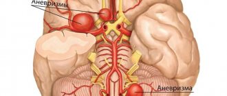
Most often, defects in the arterial wall appear at the site of their branches, where the blood pressure is highest. A cerebral aneurysm consists of three structural elements: neck, body, and dome. In the cervical area, the protrusion has a three-layer structure, and its dome consists only of intima. This is where a rupture is most likely. According to statistics, it occurs mainly in patients aged 30 to 50 years, accounting for more than 80% of all cases of cerebral hemorrhages.
Classification
Aneurysms are divided according to their structure into two groups. They can be fusiform or saccular. In the first case, they look like a uniform expansion of a section of the vessel. In the second, they protrude on one side, forming a kind of bubble. Saccular can consist of one or more chambers and are much more common than fusiform.
There is another classification - according to the location of the cerebral aneurysm. It can be localized in the vertebrobasilar system or on the following arteries: anterior or middle cerebral, as well as on the internal carotid. In more than a tenth of cases, multiple protrusions are diagnosed, on several vessels at the same time. By size, formations are distinguished as miliary, the diameter of which does not exceed 3 mm, small - no more than 1 cm, medium - from 1 to 1.5 cm, large - up to 2.5 cm. If the size exceeds 25 mm, we are talking about the so-called giant aneurysm.
Terms and rules of rehabilitation
The duration of rehabilitation is determined by the patient’s condition. In planned patients, recovery takes from 2 to 6 weeks, in patients operated on for complications - up to 6 months. The rehabilitation period after complicated cerebral aneurysms can extend up to 1 year and include recovery after a stroke.
The early period takes place in the intensive care unit, where patients remain until vital signs normalize (from 1 day to 2 weeks).
The late period of rehabilitation is carried out sequentially:
- In the hospital - until clinical improvement;
- In the clinic - until complete clinical recovery;
- At home - for several years, sometimes for life.
General rules of rehabilitation:
- Security mode;
- Monitoring vital functions (respiration, blood pressure);
- Raising the head end of the bed;
- Rational pain relief during all manipulations;
- Maintaining normal body temperature;
- Nutrition through a tube (according to indications);
- Control of urination and defecation;
- Maintaining hemodynamics (systolic blood pressure at 120-140 mm Hg).
After clipping on the cerebral vessels, an additional warning is given:
- Cerebral ischemia (calcium channel blockers);
- Dehydration (parenteral solutions);
- Cerebral edema (diuretics).
How quickly does the condition return to normal after clipping an aneurysm?

In planned patients, improvement occurs within 2-3 days. Such patients are transferred from intensive care to the cardiology department for a period of 14-28 days. Residual effects (for example, headaches) disappear by 2-3 months.
In patients who have undergone clipping due to complications, improvement occurs by the end of 1-2 weeks of the resuscitation period. Such patients are transferred to the department for up to 4 weeks. Residual effects disappear within 3-6 months, some of them can be determined for life.
Causes
The pathology can be congenital or acquired. Vascular wall defects due to intrauterine developmental anomalies are often accompanied by other diseases. These include congenital coarctation of the aorta, multiple renal cysts, arteriovenous malformations, and underdevelopment of connective tissue.
Acquired changes are the result of various traumatic brain injuries or previous diseases. Aneurysm of cerebral vessels often develops against the background of hypertension, atherosclerosis, and hyalinosis. If the protrusion is formed after the introduction of an infectious embolus into the artery, it is called mycotic. In addition, aneurysms form due to uneven blood flow or arterial hypertension.
Symptoms
In most cases, a slight expansion of the artery section does not manifest itself in any way. Such an aneurysm is most often discovered by chance or during a routine examination. When clinical signs appear, they are divided into tumor-like or apoplectic. In the first case, the formation quickly increases in size, compressing nearby brain structures as it grows.
All symptoms add up to the standard clinical picture of the tumor and directly depend on the location of the protrusion. Typically, a tumor-like aneurysm is localized in the cavernous sinus or in the area of the chiasm, the intersection of the optic nerves.
A defect in the arterial wall located in the chiasmal region negatively affects visual function: visual acuity decreases and its fields are disrupted. A long-term and fairly large cerebral aneurysm often leads to nerve atrophy followed by complete blindness. If it is located in the cavernous sinus, which is located at the base of the skull and regulates venous outflow from the eye sockets and brain, cranial nerve palsy occurs. When the 3rd, 4th and 6th pairs are affected, oculomotor disorders such as strabismus and inability to focus on an object occur.
Damage to the branches of the trigeminal nerve is manifested by the characteristic symptoms of its neuralgia. In advanced cases, even the bone tissue of the skull is deformed, which is revealed during an X-ray examination.
When the disease proceeds according to the apoplexy type, any clinical signs are most often absent. An apoplectic aneurysm is discovered only after it has ruptured and hemorrhaged into the brain. Among the symptoms that may precede this, painful sensations in the area of the eye sockets and forehead are occasionally mentioned.
Surgery
The operation is performed if:
- the size of the formation exceeds 7 mm;
- educational growth progresses;
- there is a hereditary predisposition to hemorrhagic stroke.
Before the operation, a comprehensive examination of the patient is carried out:
- Collection of tests (general blood and urine tests, biochemical, to identify infectious diseases).
- Fluorographic examination of the chest, ECG.
- Consultations with a neurologist, therapist, cardiologist.
- MRI allows you to obtain data about an aneurysm larger than 3 mm in size.
- CT scan provides data on a pathological formation exceeding 5 mm in size. The study makes it possible to view the internal structure of the aneurysm.
- Digital subtraction angiography makes it possible to detect formations smaller than 3 mm.
- Additional measures: measuring blood pressure, body temperature, in case of chronic diseases - bringing the body to normal levels.
Actions before surgery:
- After all the tests have been carried out, the person is hospitalized.
- The surgeon talks about the progress of the operation and possible complications. The anesthesiologist clarifies whether there are any allergic reactions to the anesthetic substance. The patient's consent is taken to undergo surgery.
- Before the operation, a person must refrain from eating and drinking water, take a shower, and wash his hair.
Aneurysm rupture
When the bulge ruptures, the first thing that occurs is a very severe headache. At first, it can be felt locally, where the damage is located. Then gradually the painful sensations become diffuse, covering the entire head. These are accompanied by the following signs of hemorrhage:
- Nausea with repeated vomiting.
- Increased sensitivity and tone of the occipital muscles.
- Kernig's sign is the inability to straighten an elevated leg bent at the knee (in a supine position).
- Brudzinski's symptom complex, when when pressure is applied to different points of the body, the limbs move involuntarily.
- Loss of consciousness for varying periods of time.
- Mental disorders (from minor to severe), seizures of the epileptic type.
Hemorrhage between the meninges - subarachnoid - causes a prolonged spasm of nearby arteries. In most cases, it leads to damage to the brain matter, as in an ischemic stroke.
If, when a cerebral aneurysm ruptures, blood enters its ventricles or substance, focal symptoms are added to the general symptoms. Specific signs depend directly on the location of the damage. When it is located in the area of the bifurcation of the carotid artery, visual function suffers. Damage to the anterior cerebral artery leads to mental disorders with simultaneous paresis of the legs, and damage to the middle cerebral artery leads to speech disorders and paralysis of the opposite side of the body. An aneurysm located in the vertebrobasilar system, as a result of rupture, causes incoordination, dysphagia, frequent eye fluctuations, paresis of the trigeminal and facial nerves.
The incidence of subarachnoid hemorrhages is about 80% of the total. Intracerebral hematomas are less common, but have more pronounced negative consequences. The most dangerous thing is bleeding into the ventricles, since the consequence is most often death. When hemorrhaging into the cavernous sinus, located outside the dura mater, the medulla is not damaged.
Side effects after surgery
Although this procedure prevents the development of a fatal anomaly, clipping an aneurysm has certain consequences that you need to be aware of. They can manifest themselves either singly or complexly, to some extent it depends on the presence/absence of provoking factors.
Possible complications after surgery include:
- general state of weakness;
- loss of sensitivity, tingling;
- impaired ability to speak, see and remember;
- seizures;
- infectious lesions;
- side effects from general anesthesia (impaired body orientation, decreased blood pressure, rapid breathing);
- the formation of blood clots, which threatens stroke;
- There is also a risk of aneurysm rupture during surgery.
Factors that can provoke the above phenomena include smoking tobacco products, excess body weight and high blood pressure. Therefore, if after surgery you notice a deterioration in your body’s capabilities or signs of infection, you should urgently consult a doctor. This also includes persistent headache, redness with swelling, bleeding and discharge from the incision site, fainting, inability to control the activity of the bladder and intestines.
Note! Particularly dangerous consequences of the operation are convulsions, chest discomfort, fainting and difficulty breathing. In such a situation, you need to call an ambulance as quickly as possible.
Diagnostics
Detecting a cerebral aneurysm, if it is small in size and does not manifest itself clinically, can only be done by chance. An asymptomatic disease in most cases is detected during special examinations, which are carried out for another reason. If pathological symptoms appear, you need to contact a neurologist.
Diagnosis begins with collecting anamnesis based on the patient’s complaints. A neurological examination is then performed, during which the location of the aneurysm is determined based on specific tests.
The next diagnostic stage is an instrumental examination. It includes the following manipulations:
- X-rays can be used to detect characteristic deformations of the skull bones at its base. Petrified protrusions in which calcium salts are deposited are also visible on x-ray.
- Magnetic resonance imaging and computed tomography are more informative methods of examining the brain, but they may not be enough to confirm the diagnosis.
- Angiography provides the most complete picture of the condition of the vessels without introducing contrast agents into them. With its help, a three-dimensional image of the damaged artery is obtained, which allows one to judge the type, size and shape of the protrusion.
If it is not possible to use high-precision equipment, a lumbar puncture (cerebrospinal fluid sampling) is performed. Based on the presence of blood in the cerebrospinal fluid, a conclusion is made about intracerebral or subarachnoid hemorrhage.
Particular attention is paid to differential diagnosis, since with a cerebral aneurysm the symptoms may be similar to other diseases. These include tumors, abscesses or cysts if the pathology is of a tumor-like type. In the case of apoplectic symptoms, differentiation is made from ischemic stroke, epilepsy attack, meningitis, transient ischemic attacks (spasm).
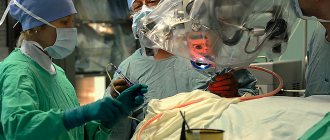
How is endovascular intervention performed for vascular aneurysm of the brain?
The operation is performed under general anesthesia, as full control of blood pressure and the patient’s position on the operating table is necessary.
All manipulations on blood vessels are carried out under x-ray control in the cath lab. The intervention is carried out mainly through a puncture in the area of the femoral fold, from where a catheter is passed through the femoral artery towards the aneurysm, the aneurysm is completely filled with platinum microspirals and is disconnected from the blood flow.
Currently, for endovascular correction of aneurysm with a wide neck, methods of protecting the aneurysm neck are used to prevent microcoils from falling out into the supporting vessel:
endovascular treatment of aneurysm
Temporary protection of the neck of the aneurysm with a balloon (balloon-assistance method), when a catheter with a balloon is inserted into the area of the supporting vessel, which is inflated and then microspirals are introduced into the aneurysm, after which the balloon is removed;
- Permanent protection of the neck of the aneurysm with a stent that is inserted into the vessel and remains in the vessel permanently. The stent has cells through which microspirals are introduced into the cavity of the aneurysm and the aneurysm is disconnected from the blood flow;
- Introduction into the vessel of a flow redirecting stent , which has a high density and directs blood through the vessel in such a way that blood does not enter the aneurysm and the aneurysm is thrombosed, that is, the possibility of its rupture is excluded. Complete thrombosis of the aneurysm occurs within 4 to 6 months after the intervention.
After installation of stents of any type, medications are required for three months to prevent stent thrombosis, which must be taken into account when choosing this intervention technique.
Treatment
An aneurysm is treated primarily surgically, since such a formation requires removal. However, if the protrusion is small in size and does not manifest itself clinically, then the neurologist is limited to systematic observation. A patient with this diagnosis is registered and examined regularly. Additionally, measures are being taken to eliminate the causes that provoked the pathology. If there is an increase in the brain aneurysm with a simultaneously increasing risk of its rupture, surgery is performed using one of the modern techniques.
General recommendations
The leading causes of this disease are atherosclerosis, hypertension, and diabetes. If there are chronic pathologies, it is necessary to treat them, as well as adjust your lifestyle. First of all, you should stop smoking and reduce your alcohol consumption. These bad habits have an extremely negative impact on the condition of blood vessels for all of the listed diagnoses.
The transition to a healthy diet is one of the most important conditions for stabilizing the condition and the effectiveness of drug therapy for major diseases. It is necessary to exclude unhealthy fatty foods from the diet, minimize sugar consumption, eat more fresh vegetables and fruits, lean meat, and dairy products. Heat treatment is also important: it is advisable to boil, stew or bake dishes, and avoid fried foods.
Moderate physical activity is required. If sports are contraindicated, you need to move more, walk down the street, climb stairs without an elevator. Special gymnastics are effective in normalizing blood circulation and restoring muscle and vascular tone. In addition, with a cerebral aneurysm, it is very important to maintain a stable emotional background, be less nervous, and avoid stress.
Medicines
It is impossible to cure the pathology itself with tablets or injections, but in most cases drug therapy is necessary. Medicines are prescribed according to the established cause of the aneurysm:
- If the vascular wall becomes thinner due to atherosclerosis, special drugs are used to lower cholesterol levels. These include nicotinic acid, statins, and bile acid sequestrants. By normalizing lipid metabolism, the process of deposition of cholesterol plaques and artery deformation slows down.
- If you have hypertension, you must take medications that lower your blood pressure. They reduce the risk of rupture of an existing brain aneurysm and the formation of new ones.
- Antibiotics are used if the cause of the pathology is an infectious embolus. Antibiotic therapy is intended to treat an infectious-inflammatory process that has caused pathological changes in blood vessels.
In case of rupture with hemorrhage, drug therapy is carried out as for a hemorrhagic stroke. Drugs are used intravenously to stabilize intracranial and arterial pressure, reduce vascular permeability, as well as hemostatic agents and barbiturates. At the same time, the question is raised about the advisability of surgical treatment. Doctors, based on information about the location and volume of hemorrhage, prescribe surgery or continue treatment with medications.
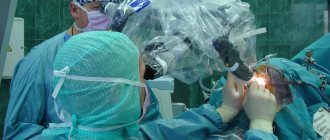
Surgical intervention
The most effective method of treating a cerebral aneurysm is a microsurgical operation to remove it. The following methods of varying degrees of invasiveness are used. Cervical clipping is performed microsurgically after craniotomy. Through a hole with a diameter of no more than 4 cm, the meninges are opened, the aneurysm is isolated, and a special clip is applied to its neck. Thus, the lumen of the protrusion is blocked.
Endovascular occlusion is a less traumatic operation indicated for elderly patients, as well as in the presence of somatic diseases or a deep location of the aneurysm in the brain tissue, its fusiform shape?
What are the advantages of endovascular surgery?
Endovascular operations began to be practiced recently, but quickly gained great popularity among doctors and patients due to many advantages:
- The main advantage of endovascular interventions is the low risk of complications, due to the fact that during such an operation no incisions are made and general anesthesia is not used. One small puncture under local anesthesia is enough.
- After the procedure, patients recover very quickly. You can return to your normal life after a maximum of 4 weeks (often within a few days), while after open surgery on large vessels - after 3 months.
- Hospitalization time is reduced. Typically, it is enough for the patient to spend only 1–3 nights in the hospital. During and after the procedure, the patient experiences much less pain and discomfort than after classic open interventions through an incision.
- Due to the very low risk of complications, endovascular surgery is often the best choice for elderly patients and people with severe concomitant diseases.
- Excellent cosmetic result. There are practically no traces left at the puncture site.
Endovascular surgery helps to safely cope with various pathologies, even in severe cases.
Take care of yourself, make an appointment with a doctor now