Compression of the nerves between the toes leads to compression (tunnel) neuropathy of the common plantar nerves.
The common plantar nerves are involved in the innervation of the toes. Nerve fibers pass below the deep transverse metatarsal ligament, which connects the heads of similar bones, and this in turn creates conditions for their compression when the foot is deformed.
Tunnel neuropathy has another name - neuroma or Morton's neuralgia. Therefore, this article will focus specifically on the pathological manifestation under this name
Anatomy and location
The anatomy of this nerve has no peculiarities. The fiber consists of sensory (afferent) and motor (efferent) components. Individual bundles of nerves are covered with a connective membrane (perineurium). Inside it there are septa that cover individual fibers (endoneurium). To form a single sciatic nerve, the epineurium surrounds the entire ligament. Inside the connective tissue there are vessels that supply the nerves. In addition, the shell softens and absorbs vibration.
The human sciatic nerve begins in the nerve plexuses (connections) of the sacrum, emanating from two lumbar and three sacral segments of the spinal cord. Its location explains the return of pain in case of damage to the lower back (hernia, injury, osteochondrosis).
The human sciatic nerve pathway is quite simple. Having originated in the sacral nerve connection, which is located in the small pelvis, it passes through the exit there into the gaps of the buttock muscles. Next, the fiber descends to the back of the thigh from the gluteus maximus muscle and branches. The branches diverge to the thigh and gluteal muscles and joints. The nerve then travels to the back of the knee. The processes spread in two directions, forming the tibial and fibular branches. They fit the skin, muscles and joints of the lower leg and foot. The picture shows how the nerve is located.
Human foot, its structure and diseases and points + Schemes of bones and muscles
Human evolution has made the foot a unique and complex mechanism that performs spring and balancing functions, softening shocks when moving.
Thanks to the limbs, a person gained the opportunity to move, maintain balance, and resist movements.
There are 26 bones in the foot and they are all connected into one mechanism by ligaments and joints.
In addition, there is a huge amount of muscle tissue and tendons.
Bones
The foot and hands are similar in structure. Anatomy divides the foot into the following bone sections:
Tartarsal
Includes 7 dice. The most bulky ones are the talus and heel. The talus is located between the lower leg and relates more to the ankle. This includes:
- - club-shaped;
- - scaphoid;
- - sphenoid bone.
Metatarsus
This is a collection of five tube-shaped bones. This section is middle and is responsible for the functioning of the fingers and the correct position of the arch. The bones ending at the joints lead to the beginning of the fingers.
Distal section
There are 14 bones in it. Each finger has 3 bones, except the thumb, which has only two. Between the bone formations there are joints to ensure mobility.
Thanks to this area of the foot, the human body maintains balance and can move. Interestingly, in case of loss of arms, the toes perform a replacement function.
Joints are located between the bones. In addition, the foot contains muscles, ligaments, nerves, and blood vessels.
How are the bones arranged?
The bones require a more detailed consideration, since they are the main component of the foot.
The heel bone is the most powerful
It is located in the rear and carries a huge load. Despite the fact that this part has nothing to do with the ankle, it plays a large role in the distribution of pressure. The shape of the calcaneus resembles a triangle in three dimensions with a long axis.
The role of the connector between the calcaneus and the talus is performed by the joints. A strong connection of these two bones is necessary to give the foot a normal shape. The back of the bone holds the Achilles tendon. This place can be found by a small ledge. And the lower part is a support when walking on the surface of the earth.
On the front part you can find the tubercle where the scaphoid bone and the joint meet. On the surface you can notice many protrusions and, conversely, depressions. These are places where blood vessels, muscles, nerves, and ligaments are attached.
The talus is several times smaller than the calcaneus
But it is massive and forms part of the ankle. It faces the heel. It mainly consists of cartilage and, surprisingly, does not hold anything together except ligaments. Its surfaces, consisting of 5 pieces, are covered with a thin layer of hyaline cartilage.
This bone consists of the following parts:
- - a body that belongs to the ankle and performs a connecting function with the foot thanks to ligaments and joints;
- - the head, which is the front part of the bone with the articular surface. This part is needed to ensure a reliable connection with the boat;
- - neck - the thinnest part located between the head and the body.
Despite the strength of the bone, it is often injured or diseased.
Cuboid
You can find it on the outside of the foot at the outer edge. Located behind the 4th and 5th metatarsal bones. It is shaped like a cube, hence its name. From the back it comes into contact with the calcaneus, and that is why it has a saddle shape and a calcaneal process.
Scaphoid
Located directly on the foot at the inner edge.
Its ends are flattened, the upper part can bend, and the lower part is sunken.
Thanks to the joints, it interacts with the talus and serves as the former of the foot.
Wedge-shaped
Consists of three bones:
- - medial, which is also the largest;
- - intermediate, smallest;
- - lateral - middle.
They are all small and located quite close to each other. In front of them are the metatarsal bones, and behind them is the navicular bone. The entire system is strong and rigid, forming a solid base for the foot.
Metatarsals
They are tubes bent at an angle. They have the same structure and perform similar functions in both young and adult years. The bends of the bones give the arch the desired position. If you look at the surface, it is lumpy due to the connection of ligaments, joints and muscles.
Phalanx
The same as on the fingers. The only difference is the size. The thumb is assembled from 2 phalanges, and is much thicker in shape due to the load that occurs when walking. The rest consist of three phalanges and are much thinner and shorter.
Joints
What are joints made of?
The feet are distinguished by the presence of a large number of joints that play a connecting role between the bones.
If we compare them by size, the largest is the ankle joint, which connects three large bones together. This allows a person to raise and lower the foot and make rotational movements.
The remaining joints are much smaller, but essentially their function is similar. They provide the necessary flexibility.
Let's talk a little about the ankle joint. It includes the greater talus and two smaller tibias, which include the ankles. The edges of the joint are attached by strong ligaments, and it itself is securely connected to cartilage.
The transverse or subtalar joint plays a huge role. It is inactive, but connects three whole bones - the navicular, talus and calcaneus. For more reliable fixation, participation in the connection of ligaments is provided.
The subtalar joint is helped to form the arch by the cuboid and calcaneal joints. Sometimes this joint is called the Grecian cavity, and in medicine it is called the talonavicular joint.
One of the most significant joints is the metatarsophalangeal joint. They take part in every movement of the human body.
The least important joints are the scaphoid and sphenoid bones.
Ligaments
In first place in importance is the plantar ligament. It originates from the heel bone and ends at the origins of the metatarsal bones.
The ligament is distinguished by a large number of branches that carry the fixing function of the longitudinal and transverse arches.
This connection is responsible for the correct condition of the arch throughout a person’s life.
To strengthen the skeletal system and joints, smaller ligaments are needed. Thanks to them, the human body is able to maintain balance and loads during movements.
Muscles
The foot can only move with the help of muscles. They are everywhere - in the area of the foot, lower leg and ankle. The muscular structure of the lower leg provides movement of the feet during walking and in an upright position.
The anterior part consists of the extensor longus muscle group and the tibialis muscle. Thanks to them, the phalanges on the legs can be bent and unbent.
The long and short fibulae provide lateral flexion of the foot and pronation.
A very bulky muscle group is located in the back. These muscles consist of several layers. This includes the following muscles:
- triceps, including gastrocnemius and soleus;
- flexor digitorum;
- plantar;
- tibial (partially).
When this muscle group works, the sole bends with the help of the Achilles tendon. Muscle tissue also helps with bending and straightening the fingers.
For the movement of four fingers, not taking into account the thumb, the short-type extensor muscle, which belongs to the dorsal muscle group, is responsible. Small muscles on the foot allow it to perform the functions of abduction and flexion.
Blood
To allow blood to flow to the feet, there are tibial arteries in front and behind. They stretch along the very foot on the sole. Small connections and circles extend from these large arteries.
When the foot is injured, the functioning of one of the circles is disrupted, but the others continue to provide the necessary blood flow to the limbs.
The veins on the back side are responsible for the outflow. They appear intertwined and supply blood to the greater and lesser saphenous veins in the lower leg.
Nerves
They form an integral part of the normal functioning of the human foot. They are responsible for sensations:
- - pain;
- — vibrations;
- - touch;
- - cold or heat.
Nerve signals leaving the central nervous system along the sural, peroneal, superficial and tibial nerves reach the spinal cord and are processed there.
Nerves transmit signals to muscles, being essentially reflexes - voluntary or involuntary (independent of human will). Involuntary ones include the work of the glands (sebaceous and sweat), vascular tone.
As for the skin, there are several zones on the foot that differ in density, structure, and elasticity. For example, the sole leather is high density and the heel leather is thick. Initially, the skin of the palms and feet is the same, but over time and with increasing loads, additional layers appear. The dorsum of the foot is smooth and elastic, with nerve endings.
Drawing a conclusion, we can say that nature has done everything to ensure that the foot can withstand enormous pressure.
Foot diseases
The foot is regularly exposed to loads, either static or shock. Injuries are a common occurrence for her. They are almost always accompanied by pain, enlargement of some epiphyses, swelling, and curvature. Pathology can be detected by x-ray.
Arthrosis
This is a disease during which cartilage loses its elasticity. This often disrupts metabolic processes. Pain, crunching, swelling appears.
Causes of arthrosis:
- - infectious diseases;
- - allergies;
- - systemic diseases - lupus erythematosus, scleroderma;
- - tuberculosis;
- - syphilis;
- - dislocation or bruise.
You can often find arthrosis of the first toe.
The disease develops in 3 stages:
- Pain occurs at first, but goes away after rest. Sometimes deviation of the thumb becomes noticeable. There is a crunching sound when moving.
- Painkillers and anti-inflammatory drugs are taken to dull the pain. The toe becomes severely bent and it becomes impossible to pick up shoes.
- The pain does not go away even when taking analgesics. The deformity spreads to the foot, causing problems with walking.
Arthrosis also greatly affects the ankle, deforming the joint and affecting the cartilage.
This disease can be treated conservatively only at an early stage. Then you will need surgical intervention - endoprosthetics, resection, arthroplasty.
Flat feet
There are congenital or acquired flat feet. Reasons for appearance:
- - excess weight;
- — heavy loads;
- - diseases of nerve endings;
- - injuries;
- - wrong shoes;
- - history of rickets or osteoporosis.
Flat feet exist in two types:
- Transverse - with a decrease in the height of the arch, when the heads of the metatarsal bones contact the ground.
- Longitudinal - that is, the entire foot has contact with the ground. Increased leg fatigue and pain.
Arthritis
A joint disease that affects the entire human body. There are primary and secondary arthritis. The reasons for its appearance are the same as for arthrosis. Symptoms include:
- - pain;
- - leg deformity;
- - swelling, redness;
- - fever, rash, fatigue.
Treatment methods depend on the root cause of the disease and can be physiotherapeutic, medicinal, manual, etc.
Clubfoot
As a rule, it occurs from birth. The cause is a subluxation of the ankle joint. Acquired clubfoot becomes a consequence of trauma to the lower extremities, paralysis, and paresis.
Disease Prevention
Preventing the development of diseases is much easier than treating them. Prevention includes:
- performing special strengthening exercises;
- practicing gentle sports - cycling, skiing, swimming;
- wearing comfortable shoes made from natural materials;
- walking on pebbles, sand, grass;
- use of special orthopedic insoles;
- providing rest for the legs.
Source: https://artritsystavov.ru/artroz/kistej-i-stop/stopa-cheloveka-stroenie-shema.html
Pain
The pain that occurs when the largest human nerve fiber is damaged is peculiar:
appear and pass abruptly, unexpectedly. After relief has occurred, sometimes it hurts at a certain point in the lumbar spine, the center of the buttock, or behind the knee. In other cases, the pain is constant, aching, and periodically intensifies; the nature is burning, piercing, cutting, shooting, pulling; felt in the area where the sciatic nerve is located; worsen after stress (physical or emotional overload, hypothermia); accompanied by redness, swelling, impaired (weakened or increased) skin sensitivity, increased sweating; intense pain forces you to put less strain on the affected side, limp, disturb sleep and can cause loss of consciousness.
Do not make a diagnosis yourself, even if you know the location of the sciatic nerve and the nature of the pain when it is affected. Various diseases may be hidden behind the symptoms.
Causes of defeat
The sources of damage to the sciatic nerve are:
infectious diseases; poisoning; long stay in the cold; pinching; injuries (fractures, dislocations, sprains, operations in areas adjacent to the nerve); tumors; diseases of the pelvic organs, especially of an inflammatory nature; metabolic or blood supply disorders in the spine; hereditary diseases or abnormal intrauterine development leading to nerve vulnerability.
To treat the sciatic nerve, a blockade is used; the nerve is blocked for a while, then it is unblocked.
Very often, many people experience a crunch in the neck, there can be many reasons for this, including salt deposits and problems with the spine.
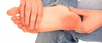
conclusions
Numbness of the big toe is a sign that can have the most unexpected consequences. Sometimes the nature of such a symptom is temporary and most harmless, in other cases it signals the beginning of a severe pathological process. To get rid of doubts and, most importantly, to detect the disease in time, it is necessary to undergo a preventive examination - it will help identify and eliminate the causes of possible pathology, and will also allow the use of conservative treatment methods instead of surgical intervention.
Diseases
The main diseases affecting this nerve are:
sciatica. The fiber becomes inflamed or compressed, causing sharp cutting pains that are felt at the back of the thigh. The condition worsens with movement when the nerve becomes stretched. Sensitivity in the leg changes. The rate at which the disease develops depends on its source; sciatica. This is pain in the lower back, caused by injuries to the spine of various natures, involving the sciatic nerve. Sensitivity is not impaired, pain is of moderate intensity.
The sciatic nerve is an important component of the human body. Its anatomy is simple: motor and sensory fibers are surrounded by connective tissue. The nerve is mostly located in the leg, starting in the pelvis from the nerve plexus of the sacrum and ending in the foot. If the nerve area hurts, this is a serious reason to see a doctor. Unpleasant sensations are caused by sciatica and lumboischialgia. Damage to the nerve fiber can lead to loss of mobility and sensation in the legs, control of urination and defecation.
The symptoms of sciatic nerve pathologies directly depend on its anatomy and physiology. Let's take a closer look at where the sciatic nerve is located, how it is located in relation to the muscular structures of the lower limb, and what pathologies are associated with nerve fibers n. ischiadicus.
Video
We offer you to watch a video on the topic of the article.
Education: Rostov State Medical University, specialty “General Medicine”.
Found an error in the text? Select it and press Ctrl + Enter.
Dentists appeared relatively recently. Back in the 19th century, pulling out diseased teeth was the responsibility of an ordinary hairdresser.
Four pieces of dark chocolate contain about two hundred calories. So if you don’t want to gain weight, it’s better not to eat more than two slices a day.
Regular use of a solarium increases your chance of developing skin cancer by 60%.
Many drugs were initially marketed as medicines. Heroin, for example, was originally brought to market as a cure for children's coughs. And cocaine was recommended by doctors as an anesthesia and as a means of increasing endurance.
74-year-old Australian resident James Harrison has donated blood about 1,000 times. He has a rare blood type whose antibodies help newborns with severe anemia survive. Thus, the Australian saved about two million children.
A person taking antidepressants will, in most cases, become depressed again. If a person has coped with depression on his own, he has every chance to forget about this condition forever.
According to WHO research, talking on a mobile phone for half an hour every day increases the likelihood of developing a brain tumor by 40%.
Over the course of a lifetime, the average person produces no less than two large pools of saliva.
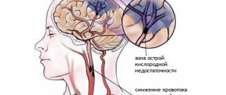
The human brain weighs about 2% of the total body weight, but it consumes about 20% of the oxygen entering the blood. This fact makes the human brain extremely susceptible to damage caused by a lack of oxygen.
Each person has not only unique fingerprints, but also tongue prints.
Anatomy of nervus ischiadicus
Nervus ischiadicus (sciatic nerve) is a collection of long nerve fibers of the sacral plexus, its main extension, providing innervation to the muscles of the lower limb. Plexus sacralis (sacral plexus) is a thick nerve formation in the shape of a triangle, the apex of which is directed downward. It is formed by the fusion of the anterior branches of the fourth and fifth lumbar spinal nerves, the first and fourth sacral spinal nerves (according to Prives). Plexus sacralis is located in the pelvic region between the fascia of the piriformis muscle and the superior pelvic fascia. This plexus produces both long and short branches.
The sciatic nerve passes through the greater sciatic foramen below the musculus piriformis (piriformis muscle), where it is covered by the gluteus maximus muscle. It is in this place that damage and compression most often occur. Next, the nerve emerges from under the lower edge of the gluteus maximus muscle and passes between the muscles of the posterior thigh to the popliteal fossa.
In the upper half of the thigh it goes under one of the ends of the biceps femoris (lat. m. biceps femoris), then between the m. biceps femoris and m. semimembranosus. In the popliteal fossa (fossa poplitea) it passes into the nervi tibialis and fibularis. In some cases, the division into these two nerves occurs in the pelvic area. This structure is considered a normal variant; the innervation zones remain the same. And both nerve formations are located in a common membrane, dividing only in the fossa poplitea.
The tibial nerve (nervus tibialis) is the thickest branch of the nervus ischiadicus. It continues its course, passing between the two ends of the gastrocnemius muscle, under the popliteus muscle, reaching the ankle on the medial side of the leg. Here the nervus tibialis divides into terminal branches and supplies innervation to the skin and muscles of the foot.
The peroneal nerve (nervus peroneus communis) is a relatively thin branch of the nervus ischiadicus. After leaving the fossa poplitea goes in the middle between m. biceps femoris and m. gastrocnemius. The most vulnerable area of its location is the head of the fibula. The nervus peroneus communis wraps around the head and gives articular branches to the knee. But in this place it remains covered only with skin. Below it passes into the peroneus muscle, where it divides into a superficial and deep branch.
N. ischiadicus goes together with the inferior gluteal artery, which gives off arterial branches along the entire nerve, providing its trophism. The outflow of blood from it occurs through the inferior gluteal vein, which flows into the internal iliac vein.
It is important to note that the diameter n. ischiadicus is the largest compared to other nerve formations, about one centimeter.
Innervation of the foot: definition, possible disorders and their consequences
The foot is the most distal part of the human lower limb. This means that it is furthest from the center of the body. It is the feet that bear the entire load of the body's weight. Therefore, such a seemingly small part of the body has a very thoughtful structure. Read more about the anatomy, blood supply and innervation of the foot later in the article.
Topographic anatomy
The structure of any structure of the human body should be considered gradually. Therefore, before moving on to the anatomy of the innervation of the foot, it is necessary to disassemble its other parts. The foot, like any other musculoskeletal formation in the human body, consists of the following parts:
- bone frame;
- joints;
- striated muscles;
- vascular formations: veins, arteries, capillaries;
- nerves.
Bone frame
To fully understand the innervation and blood supply of the foot, one must understand what major bony structures it consists of. After all, large nerves and blood vessels are predominantly located along the bones and have similar names.
There are three areas on the foot:
- tarsus;
- metatarsus;
- phalanges of fingers.
The tarsal region is located most proximally, that is, directly under the ankle joint. The line that demarcates these two formations is also the upper edge of the human foot. This line runs along the posterior edge of the heel bone.
The tarsus contains two rows of small bones. The first row, which is located closer to the edge of the foot, consists of the talus and calcaneus. They are larger.
In the second row, which is closer to the metatarsus, there are five bones at once, placed in two more rows. The first is represented by four bones: three wedge-shaped and one scaphoid.
The second row contains only one cuboid bone.
The metatarsal part of the foot is located in the middle between the other two sections. It consists of five bones of approximately the same shape and size. Each of them includes three parts: head, body and base.
The phalanges of the fingers consist of the smallest bones. Each phalanx includes three bones. The only exception is the big toe, which consists of only two bones. This finger is also called the first and is designated by the Roman numeral I. The little finger, accordingly, is designated by the number V.
Core muscles
The main task of the nerves involved in the innervation of the foot is aimed precisely at transmitting impulses to the muscle frame. After all, it is precisely due to the receipt of nerve impulses that muscle contraction is possible, and therefore, a person can walk.
There are five muscle groups in the foot:
- lateral;
- back;
- front;
- surface layer;
- deep layer.
The lateral group includes the long and short peroneus muscles. Their contraction ensures abduction, outward rotation (pronation) and flexion of the foot.
The anterior group consists of the following muscles:
- extensor hallucis longus, due to which it is possible to extend both the first toe and the foot as a whole by lifting its upper edge;
- anterior tibial, which provides extension of the foot;
- extensor digitorum longus, due to which it is possible to extend the second to fourth toes, as well as raise the outer edge and abduct it to the side.
The muscles of the superficial layer are involved in the formation of the Achilles tendon, which ensures movement in the ankle joint.
The deep layer of muscles consists of the flexor digitorum longus (provides external rotation of the foot and its flexion), the long flexor of the first toe (performs the function in accordance with the name), the posterior tibial muscle (flexion of the foot and adduction inwards).
Features of blood supply
The innervation of the foot and the course of the arteries in it are inextricably linked, since in most cases the artery, vein and nerve go in the same direction. Therefore, you should know the main vessels of the distal limbs. These include:
- posterior tibial artery;
- anterior tibial artery;
- lateral plantar artery;
- medial plantar artery;
- dorsal artery of the foot.
The posterior and anterior tibial arteries are continuations of the popliteal artery.
The lateral and medial plantar arteries, true to their name, carry blood to the plantar part of the foot. The medial vessel has two branches: deep and superficial. The deep one carries blood to the muscle that abducts the big toe and the flexor digitorum brevis muscle. The superficial branch supplies blood only to the abductor pollicis muscle.
The lateral plantar artery supplies blood to most of the sole. At the level of the base of the metatarsus, it forms a plantar arch, from which many small branches extend to various structures of the foot. From this arch branch the plantar metatarsal arteries, which, in turn, give off branches called “perforating”.
From the plantar metatarsal artery at the level of the phalanges of the fingers, the plantar digital artery is formed, each of which is then divided into two own arteries.
The dorsal artery of the foot carries blood to the dorsum. It eventually divides into two branches: the first dorsal metatarsal artery and the deep plantar branch. The tarsal vessels also depart from it: lateral and medial. They carry blood to the lateral and medial surfaces of the foot, respectively.
Another branch of the dorsal vessel of the foot is the arcuate artery. From it, by analogy with the plantar vessels, the dorsal metatarsal arteries depart, which are divided into digital arteries.
Nerves of the dorsum of the foot
Let's start looking at the nerves of the most distal part of the limb with the innervation of the dorsum of the foot. But first you need to figure out what the external landmarks of this site are. The inner edge is limited by the tuberosity of the navicular foot, which is easy to palpate, especially in thin people. The tuberosity of the fifth metatarsal is easily visible at the outer border.
The innervation of the skin of the foot, namely its dorsal parts, is carried out by the following nerves:
- saphenous nerve;
- medial cutaneous dorsal nerve;
- intermediate cutaneous dorsal nerve;
- lateral dorsal cutaneous nerve.
The first three are branches of the superficial peroneal nerve, the last one arises from the tibial nerve. From the saphenous nerve, impulses go to the middle part of the ankle and the medial part of the tarsus. In some people, this nerve is longer and ends right at the base of the first finger.
The medial cutaneous dorsal nerve passes along the middle area of the foot and is divided along its length into branches that go to the skin of the dorsum of the big toe and partially to the second and third toes.
The intermediate cutaneous dorsal nerve is divided into digital branches, which extend to the areas of the third and fourth, as well as the fourth and fifth toes facing each other.
The lateral dorsal cutaneous nerve carries impulses to the lateral surface of the fifth digit.
A feature of the innervation of the human foot, namely its rear, is its significant variability. For example, some people lack the dorsal cutaneous nerve.
Nerves of the sole of the foot
Innervation of the foot muscles of the plantar part is provided by the plantar nerves: medial and lateral. Both of these nerve trunks arise from the tibial nerve.
The medial nerve runs along the median plantar canal and forms a small arch. The beginning of this arch corresponds to the base of the first metatarsal bone, and its end corresponds to the middle of the fourth metatarsal bone. Along the median nerve, the medial calcaneal branches depart from it. They ensure the transmission of nerve impulses to the midplantar part of the heel.
The medial nerve carries impulses to the muscle that abducts the pollicis and also to the flexor digitorum brevis muscle. It is interesting that in young children several branches go to the superficial flexor muscle at once.
Then branches emerge from the medial plantar nerve, which innervate the facing surfaces of the first to fourth fingers. These branches are called the first, second and third common digital plantar nerves.
The innervation of the toes of the sole of the foot is carried out to a greater extent by these branches.
The lateral nerve is located between the quadratus muscle and the flexor digitorum brevis muscle. It also has two branches: superficial and deep. They arise from the nerve at the base of the metatarsal bone. The superficial nerve gives off several branches: the digital nerve of the lateral edge of the fifth finger, the common digital nerve. They innervate the skin on the surfaces of the fourth and fifth fingers facing each other.
What is neuropathy?
Neuropathy of the lower extremities is not a diagnosis, but a collective concept for diseases in which the peripheral nervous system is damaged. First of all, the distal parts of the extremities are affected - the innervation of the lower leg and foot.
There are indeed many reasons for this problem, and clinical symptoms are also variable. Neuropathies are manifested by disorders of movement, sensitive areas, trophism of the skin and muscles.
It is possible to develop mononeuropathy (damage to one nerve) or polyneuropathy (multiple damage to several nerve fibers at once).
Causes of neuropathy
There can be many reasons that lead to disruption of the innervation of the foot. The main ones are listed below:
- alcohol abuse;
- drug use;
- prolonged exposure to toxic substances, especially salts of heavy metals: lead, mercury, arsenic;
- endocrinological diseases: diabetes mellitus, thyroid pathologies;
- severe liver disease;
- prolonged deficiency of vitamins and nutrients;
- side effects of certain medications: Amiodarone, Isoniazid, cytostatics;
- severe infectious diseases: diphtheria, HIV infection, mumps;
- autoimmune diseases in which antibodies are produced against the body’s own cells: systemic lupus erythematosus, dermatomyositis, rheumatoid arthritis;
- genetic predisposition.
Symptoms of neuropathy
Clinical manifestations of neuropathy depend on which nerve function is impaired: sensory, motor or trophic (nutritive). It is noteworthy that the most distal sections are the first to suffer. Therefore, the innervation of the toes will suffer first. As the disease progresses further, the symptoms will spread higher.
https://www.youtube.com/watch?v=yABmxYzrp_8
Sensitive disorders manifest themselves as follows:
- Painful sensations of a pulling or aching nature that correspond to the zone of innervation of the affected nerve.
- So-called paresthesia - a sensation of goosebumps crawling on the skin, tightening, twisting of the foot. Sometimes these sensations are so unpleasant that patients would prefer leg pain to them.
- Impaired sensitivity. Moreover, there is a simultaneous loss of all types of sensitivity in the zone of innervation of the affected nerve: pain, temperature, tactile.
- Sometimes sensory ataxia develops. This is a condition in which a person experiences unsteadiness when walking due to the fact that they cannot feel the position of their feet. This occurs due to a violation of the deep sense of orientation of body parts in space.
The following manifestations are characteristic of movement disorders:
- tremor and spasms in muscles whose innervation is impaired;
- with a long-term process, muscle weakness develops;
- flaccid paralysis – the patient loses the ability to move the foot;
- decreased reflexes, which are detected during a neurological examination.
Due to impaired innervation of the muscles, foot deformation develops due to muscle atrophy. Atrophy occurs both due to inactivity of the muscle during paralysis, and due to damage to the trophic function of the corresponding nerve.
Consequences of innervation disturbance
Long-term disruption of the innervation of the toes and other parts of the lower extremities can lead to irreversible consequences. Restoring nerve function is a rather complex and not always feasible process, especially if treatment is untimely and incorrect.
Atrophic changes in the foot area first lead to dry skin. Then ulcers and cracks appear, which are very difficult to heal. If you do not follow the rules of personal hygiene, an infection can get there.
With prolonged inactivity of the foot, restoration of its function is difficult. Thus, paralysis of the lower extremities may remain for the rest of life. Therefore, when treating neuropathy, attention is paid not only to drug treatment methods, but also to physical therapy.
Pain and unpleasant paresthesia can lead to psychological problems in the patient. Therefore, sometimes there is a need to take antidepressants.
Conclusion
The feet are a truly important part of the human body. Therefore, not only a medical professional, but also an ordinary person should know the general principles of the anatomy of the foot, the features of its blood supply and innervation. It is also necessary to have an understanding of what neuropathy is and how it manifests itself in order to seek medical help in time.
Source: https://autogear.ru/article/422/572/innervatsiya-stopyi-ponyatie-topografiya-funktsii-krovosnabjenie-vozmojnyie-narusheniya-i-ih-posledstviya/
Innervation zones
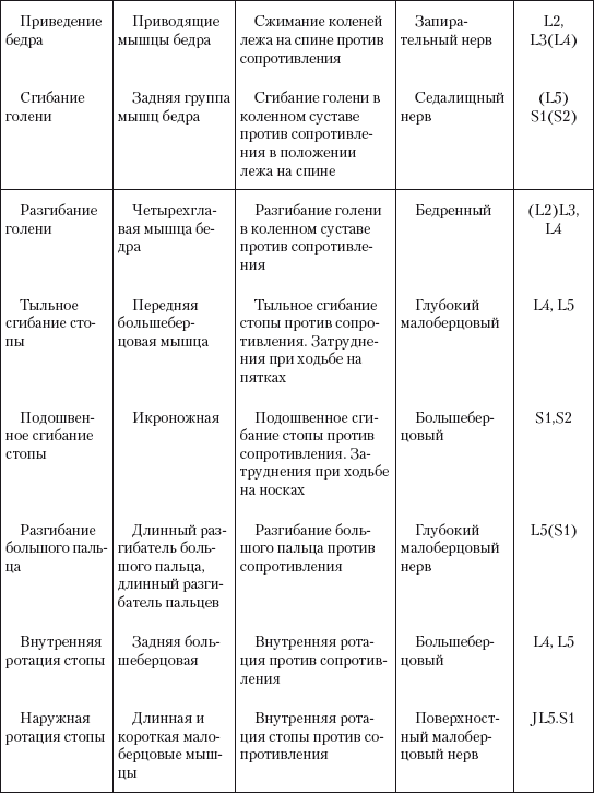
The innervation of the muscles of the lower limb by the nervus ischiadicus and its branches is as follows:
Muscle branches n. ischiadicus - semimembranosus, semitendinosus muscles and long head of the biceps femoris; Nervus tibialis - posterior tibialis muscle, long flexors of the fingers and thumb, mm. gastrocnemius, plantaris, soleus, popliteus. Nervus peroneus communis - tibialis anterior muscle, extensor digitorum longus and brevis, extensor pollicis longus.
The innervation of the muscles of the lower limb is presented in more detail in the table below.
A diagram of the cutaneous innervation of the lower limb is presented below.
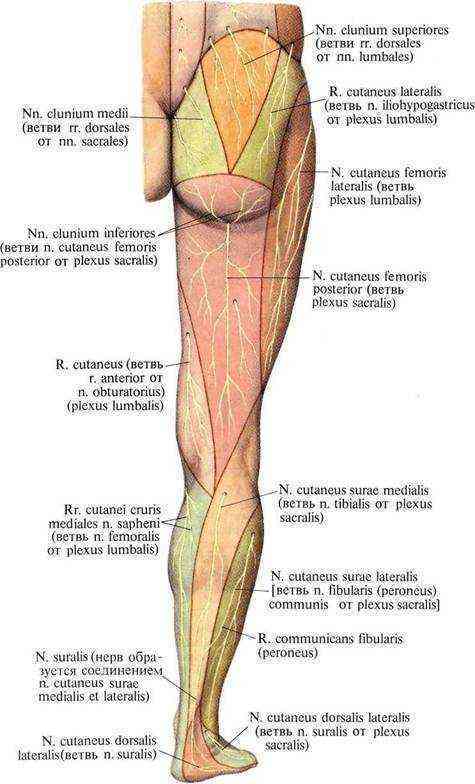
Pathology of Nervus ischiadicus
The presence of pain in the lower spine, which spreads across the gluteal region, the back of the thigh to the foot, is a signal for a neurologist to evaluate the condition of the sciatic nerve. The pain associated with its pathology is called sciatica.
The main diseases of the sciatic nerve include:
direct damage to the structure of nerve fibers due to injury, unsuccessful injection; radiculitis of the lumbosacral region, causing aseptic inflammation and swelling of the nerve; tunnel neuropathy, such as piriformis syndrome; intoxication with certain types of chemicals, infection; diabetes; tumors of the nervus ischiadicus or surrounding tissues.
Injuries to the girdle of the lower extremities, the extremities themselves, as well as the pelvic bones can lead to direct damage to the nerve, that is, rupture of its tissue. Or to its compression by surrounding tissues.
Lumbosacral radiculitis is an aseptic inflammation of the nerve roots against the background of degenerative diseases of the spinal column, intervertebral joints, and its ligaments. With radiculitis of the last two roots of the lumbar region and four roots of the sacral region, vertebrogenic sciatica occurs (another name for sciatica). The reason for this is most often intervertebral hernia, osteochondrosis, spinal canal stenosis, spondylolisthesis, and osteoarthritis of the facet joints.

Tunnel neuropathy occurs due to nerve compression in narrow anatomical spaces. For example, compression in the infrapiriformis space has a special name - piriformis syndrome. With this syndrome, compression of the nerve occurs due to inflammation and spasm of the muscle itself. There is severe pain in one leg, which increases with load on the piriformis muscle, walking, and standing. The patient feels a decrease in symptoms.
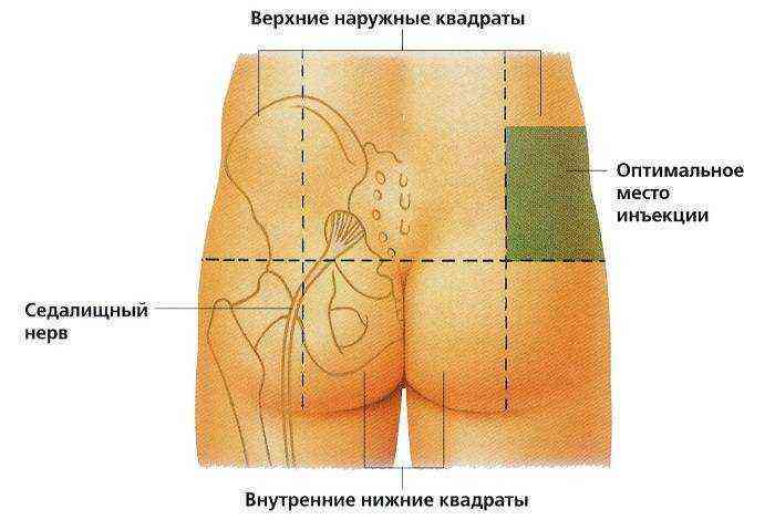
Injection neuritis occurs as a result of incorrectly placed injections. The reason may be the toxic effect of the drug on nerve fibers after an injection into nearby tissues or mechanical damage to the nervus ischiadicus itself. Also, sciatica can occur due to post-injection pathologies of surrounding tissues. These include swelling, infiltration or abscess.
Tumor formations directly in the nervus ischiadicus are quite rare. But a tumor in nearby structures can cause compression of nerve fibers and disrupt their structure.
It can be concluded that pathologies associated with the nervus ischiadicus are also associated with its topographic anatomy. Knowing the course of the nerve from the sacral plexus to the foot helps to correctly diagnose the cause of the pathology and avoid it in the future.
Human anatomy. Gain M.G. and others. Moscow, Medicine, 1985; Sciatic nerve neuropathy. Piriformis syndrome. M.V. Putilin. Journal of the Attending Physician, 02/06; Visual neuroscience. R. Barker et al. GEOTAR-Media, 2006.
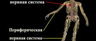
The anatomy of the human body is designed in such a way that the nervous system plays a major role in the normal functioning of the body, for example, the vascular nerve bundle of the neck, the sciatic nerve, etc. Many of them originate in plexuses emanating from the spinal cord. They are called spinal. These include the sciatic nerve - the thickest and longest in the body.
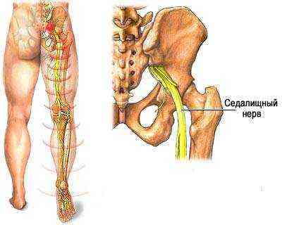
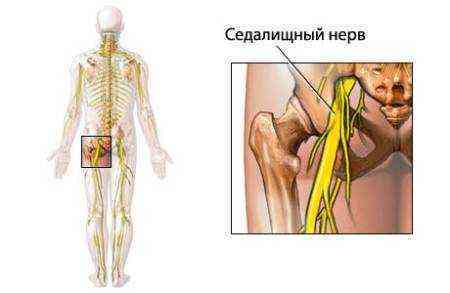
Diagram of the location of the sciatic nerve
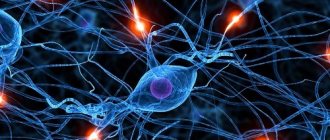
What is neuritis
It is necessary to understand what is inflamed and where it hurts. Nerves - anatomical cords - form the peripheral nervous system and connect the central nervous system with the organs of the human body. In the peripheral nervous system, a distinction is made between the somatic nervous system, which innervates (connects) muscles and skin, and the autonomic nervous system, which innervates blood vessels, internal organs, etc.
There are three types of nerve fibers:
- vegetative;
- sensitive;
- motor.
Inflammation of each of them has its own symptom. But a common manifestation of any neuritis is paroxysmal pain, which intensifies along the innervation of the diseased nerve. Muscles may lose sensitivity and go numb. The skin on the leg where the neuritis occurred may turn blue. Swelling and sweating appear. Polyneuritis is characterized by larger localizations of pain in the lower extremities.
The symptoms of mononeuritis are the same in any part of the leg - pain and numbness. In severe forms, tendon reflexes are lost. Muscle atrophy occurs at the site of nerve damage, but paralysis is practically excluded with local neuritis. An endemic form of neuritis is common among pregnant women, caused by a lack of B vitamins.
Neuritis is inflammatory damage to a peripheral nerve. If one nerve is inflamed, then they speak of mononeuritis, or local neuritis. When several nerves are affected, we are talking about polyneuritis.
What is the sciatic nerve
The science of anatomy defines the sciatic nerve as the largest in the human body. It significantly exceeds the size of the nerve fibers of other organs and systems that provide innervation. The thickness of this nerve can reach more than one centimeter. Its length is from the waist to the toes.
The sciatic nerve is a bundle of myelinated nerve fibers wrapped in auxiliary sheaths. The inner layer, the endoneurium, contains a network of capillary blood vessels. The middle enveloping layer of the nerve is called the perineurium. It contains large vessels covered with loose connective tissue, which serves as a cushion. The outer sheath of the nerve is called the epineurium. It consists of dense connective tissue.
This nerve fiber is one of the most important in the human body. Its main physiological functions include providing muscular sensitivity, movement of all flexor and extensor muscles of the torso, thigh, lower leg, and foot. Thanks to the presence of the sciatic nerve, we have the ability to walk, run, jump, and perceive various sensations.
Location of the sciatic nerve
The nerve begins in the pelvic cavity. It arises from the lumbosacral nerve plexus. From the pelvis it passes through the sciatic foramen, in which it has a lateral location. The posterior cutaneous nerve of the thigh and the gluteal neurovascular bundle lie medially. After emerging from the gluteus maximus muscle, it lies near the fascia lata, which lies across the nerve, and passes between the biceps and membranosus muscles. Next, the fiber descends to the popliteal fossa, where it divides into two branches: the common peroneal and tibial nerves. In addition, the sciatic nerve has muscle and articular branches.
The peroneal and tibial nerve endings provide sensitivity to the muscles, joints, and skin of the lower leg and foot. The tibial branch promotes movement and sensation in the back of the lower leg, the muscles, the skin of the sole, the plantar side of all five toes, and the knee and ankle joints. The peroneal nerve is divided into two nerves: deep and superficial. The first activates and causes sensitivity in the anterior muscles of the lower leg and the back of the foot. The second one forces you to move and perceive sensations in the muscles of the outer surface of the lower leg and foot.
Diagnosis
The diagnosis is made based on the patient's existing symptoms.
Sometimes an X-ray examination, MRI or computed tomography is performed in order to differentiate concomitant pathologies with similar symptoms and identify path processes that contribute to damage to the interdigital nerve bundle.
In some cases, it is possible to diagnose during a trial treatment in the form of local administration of an analgesic drug, which leads to a decrease or disappearance of pain.
Pinched sciatic nerve
Currently, many people suffer from neurological diseases, accompanied by nagging pain in the spine and gluteal region, with unpleasant pain radiating to the leg. All these symptoms indicate sciatica of the sciatic nerve. In medicine, it is also called radiculitis of the lumbosacral region, neuropathy, neuralgia or neuritis. Pain occurs when the sciatic nerve is inflamed or pinched.
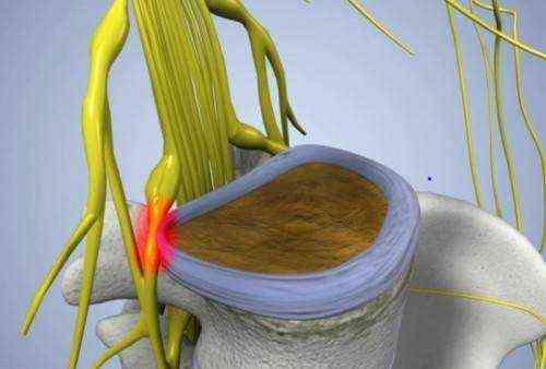
The inflammatory process can be caused by various injuries to the spinal column, injuries, infections, and inflammation of the enveloping tissues. It can also be triggered by heavy physical activity, hypothermia, colds, viral diseases, diabetes, tumors, their metastases, late pregnancy or high fetal weight. The causes of a pinched nerve are structural changes in the spine - osteophyte, muscle spasm, herniated disc, narrowing of the spinal canal. All these phenomena increase pressure on the nerve endings.
Diagnosis and treatment
A pinched sciatic nerve can be diagnosed based on the symptoms of the disease, laboratory tests, and x-rays. In case of acute inflammation, a study is performed of the spinal cord fluid, which contains the maximum number of cellular elements. These examinations indicate the presence of the disease. An X-ray examination will help identify the cause of pinching in the spine. To diagnose soft tissue disorders, magnetic resonance imaging is prescribed.

Sciatica is treated with medication in combination with various therapy methods. For nerve inflammation, painkillers, anti-inflammatory, and sedatives (tablets, injections, ointments) are used. To quickly relieve pain, novocaine blockades are performed. Along with basic drug treatment, doctors recommend physiotherapy, massage, exercise therapy, and vitamin therapy. In some cases of injury or to eliminate the cause of inflammation, surgical intervention is necessary. For colds of the sciatic nerve, various folk remedies are also used: rubbing with alcohol solutions, applying compresses.
Treatment of numbness of big toes
There is no need to try to cope with this problem on your own; you need to contact a specialist who will carry out all diagnostic measures and find out the cause of this condition.
- NSAIDs and painkillers are necessary for severe pain.
- Insulin therapy is mandatory for diabetes mellitus and to some extent stops degenerative processes in the lower extremities.
- Drug therapy - to relieve discomfort in the foot area (paresthesia), the following groups of drugs are used:

statins - stop atherosclerotic vascular damage;- antispasmodics - promote vasodilation;
- antiplatelet agents - improve the rheological properties of blood;
- vitamins - improve the general condition of the body;
- chondroprotectors - promote the restoration and regeneration of connective tissue;
Manual therapy and therapeutic exercises are indicated for any diseases of the lower extremities.
In some cases, physical therapy and massage help completely restore movement in damaged joints.