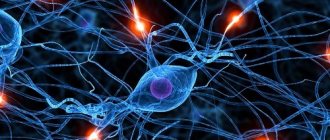
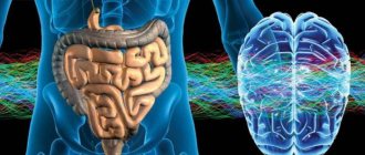
The human body works as a whole. The coherence and interaction of all organs is ensured by the central nervous system. It is found in all living beings and consists of nerve cells and their processes.
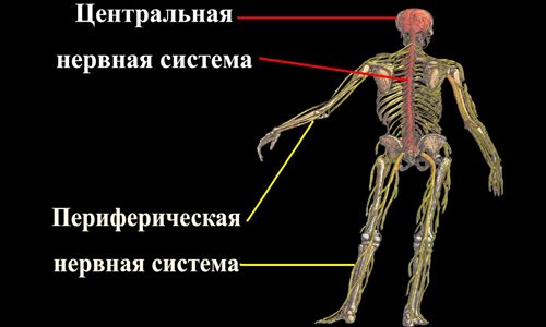
The central nervous system in vertebrates is represented by the brain and spinal cord, in invertebrates – by a system of unified nerve ganglia. The central nervous system is protected by the bone formations of the skeleton: the skull and spine.
Structure of the central nervous system
Anatomy of the central nervous system studies the structure of the brain and spinal cord, which are connected to each organ through the peripheral nervous system.
The central nervous system is responsible for feelings such as:
- hearing;
- vision;
- smell;
- touch;
- emotions;
- memory;
- thinking.
The brain structure of the central nervous system mainly contains white and gray substances.
Gray are nerve cells with small processes. Located in the spinal cord, it occupies the central part, encircling the spinal canal. As for the brain of the head, in this organ the gray matter makes up its cortex and has separate formations in the white matter. The white substance is located under the sulfur. Its structure contains nerve fibers that form nerve bundles. A number of these “ligaments” make up a nerve.
The brain and spinal cord are surrounded by three membranes:
- Solid. This is the outer shell. It is located in the internal cavity of the skull and spinal canal.
- Arachnoid. This cover is located under the hard part. In its structure it has nerves and blood vessels.
- Vascular. This membrane is directly connected to the brain. She goes into his furrows. Formed from many blood arteries. The arachnoid is separated from the choroid by a cavity that is filled with medulla.
Lips and tongue
The huge number of nerve endings in the area of the tongue and lips is explained by the abundant innervation of the entire oral cavity:
- The lingual, hypoglossal and mylohyoid nerves provide sensitivity and motor activity of the floor of the mouth (muscles, mucous membrane, root of the tongue).
- The trigeminal nerve innervates the skin, mucous membrane and muscles necessary for chewing food.
- The glossopharyngeal nerve leaves thousands of endings in the tongue, parotid gland and muscles of the pharynx.
- The palate is controlled by the vagus nerve.
Thus, the endings from many cranial nerves end in various parts of the oral cavity, which is why it is so richly saturated with receptors. The lips and tongue are able to sense taste, temperature, pain, pressure, stretching, and touch.
Spinal cord as part of the central nervous system
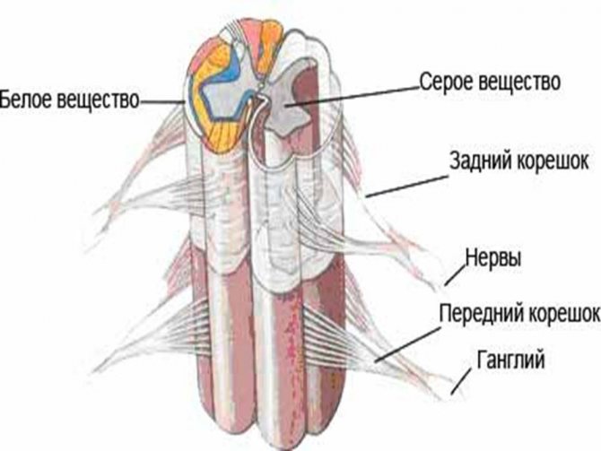
This component of the central nervous system is located in the spinal canal. It stretches from the back of the head to the lumbar region. The brain has longitudinal grooves on both sides, and the spinal canal in the center. On the outer side of the brain of the back there is a white substance.
The gray element mainly consists of the lateral, posterior and anterior horny areas. The anterior horns contain motor nerve cells, the posterior ones have intercalary ones that produce contact between sensory (lying in the nodal sections) and motor cells. Attached to the anterior horny areas of the motor particles are processes that make up the fibers. Those neurons that create the dorsal roots join the posterior horny zones.
These roots are intermediaries between the peripheral nervous system and the spinal brain. The excitation coming to the brain enters the interneuron, and then through the axon it goes to the desired organ. Reaching the opening between the vertebrae, the sensory cells connect with their motor counterparts. After this, they are divided into posterior and anterior branches, which also consist of motor and sensory fibers. 62 mixed nerves extend from each vertebra in two directions.
Spinal nerves.
The spinal nerves extend from the spinal cord in both directions in the correct order, so that sections similar to each other can be identified - segments containing a section of the spinal cord and one pair of spinal nerves. A person has 8 pairs of cervical, 12 pairs of thoracic, 5 pairs of lumbar, 5 pairs of sacral and 1 pair of coccygeal spinal nerves. The posterior branches of the spinal nerves contain sensory and motor fibers and are directed to the skin and muscles of the back and neck. The anterior branches of the spinal nerves are the most powerful. They contain sensory and motor fibers intended for the muscles and skin of the neck, the anterior and lateral surfaces of the torso, the upper and lower extremities. The anterior branches of adjacent nerves are connected to each other in the form of loops, exchanging fibers and forming plexuses. The exception is the anterior branches of the thoracic nerves, which run segmentally in the intercostal spaces. The anterior branches of the remaining nerves form plexuses: cervical, brachial, lumbar, sacral and coccygeal.
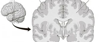
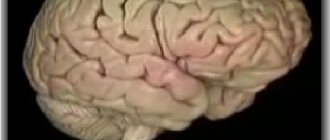
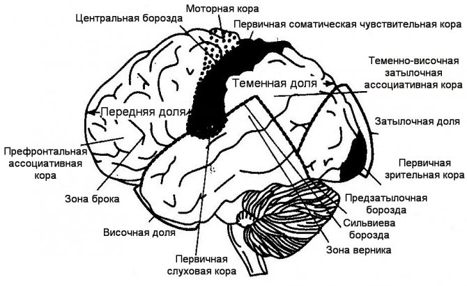
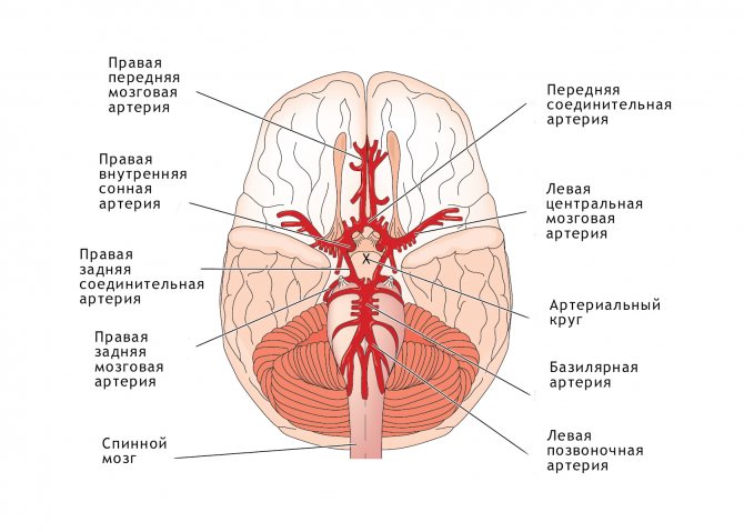
Human head brain
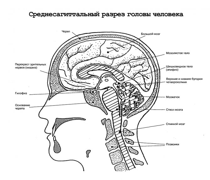
This organ is located in the brain section of the skull. Conventionally, it has five sections; inside it there are four cavities that are filled with cerebrospinal fluid. The majority of the organ consists of the hemispheres (80%). The second largest share is occupied by the trunk.
It has the following structural sections:
- average;
- cerebral;
- oblong;
- intermediate.
Fingertips
Slightly fewer nerve endings are contained in the thickness of the skin on the tips of the fingers. It is worth noting that tactile analyzers in the fingertips are the most ancient structures of living organisms, so in the process of evolution their number has increased. With our fingers we perceive touch, temperature, pain, pressure, shape, and surface features of objects. This is called touch.
On one square centimeter of the surface of the skin of the fingertips there are about 1.5 thousand tactile receptors (touch), 200 pain receptors, 15-20 baroreceptors and 15 temperature ones.
Localization of the tumor in the central nervous system
A primary brain tumor (that is, one that was originally born in a given location and is not a metastasis of a tumor that arose elsewhere in the person's body) can be either benign or malignant. A benign tumor does not grow into neighboring organs and tissues, but grows, as if pushing them aside, displacing them.
The division of CNS tumors into benign and malignant is conditional, from the point of view of both the patient and the doctor.
The fact is that a benign tumor developing in another part of the body can grow for years without causing dysfunction or posing a threat to the life and health of the patient. The growth of a benign tumor in the cranial cavity or spinal canal, where there is little space, quickly causes displacement of brain structures and the appearance of life-threatening symptoms.
Primary tumors are divided into low- and high-grade tumors. The former, as well as benign ones, are characterized by slow growth and, in general, a favorable prognosis. But sometimes they can develop into aggressive (high-grade) cancer. Read more about the types of brain tumors in the article.
Why don't we feel pain?
Throughout the human body (skin, mucous membranes, internal organs, blood vessels) various types of nerve endings are scattered that respond to pain, touch, stretching, temperature, and so on. Perceiving receptors, when irritated, send signals to the brain along nerve processes, so a person immediately experiences certain sensations.
Each body is individual and perceives stimuli, such as pain, differently. There is such a thing as a pain sensitivity threshold. The higher it is, the less pain the body experiences. At a low threshold, even a minor stimulus can cause a strong impulse and cause pain (this is how a person perceives it).
Main functions
The main sections of the central nervous system are formed by brain structures consisting of a large number of interconnected and continuously interacting nerve cells. The peripheral section, formed by nerve fibers that innervate skeletal muscles (muscles of the limbs and trunk), receives impulses coming from the central parts of the brain. Thanks to this structure, the functions of the human central nervous system are diverse and ensure the interaction of all parts of the human body.
The structure of the central nervous system maintains the integrity and unity of the body’s vital functions, and the main function of the central system is reduced to the formation of nervous reactions (reflexes) to external stimuli. Interaction with the outside world is predominantly coordinated by the cortical and subcortical zones of the cerebral hemispheres. Functions of the central nervous system include:
- Sensitivity to environmental factors (temperature, humidity, tactile contacts, hearing, vision).
- Motor activity. The spinal and cerebral structures are involved in the organization of voluntary movements.
- Regulation of mental activity and behavior.
- Ensuring the functioning of the pelvic organs (processes of urination and defecation).
- Management of the respiratory and cardiovascular systems.
The central system includes such elements of nervous tissue as afferent and efferent neurons. The former perceive impulses coming from the periphery, the latter transmit them to effector (whose activity is determined by reflexes), executive organs. Effectors are compounds that cause physiological responses by binding to proteins and subsequent regulation of their biological activity.
Diagnosis and treatment of pathologies
Instrumental studies related to informative methods of differential diagnosis include MRI and CT, angiography, polysomnography, echoencephaloscopy, electroencephalography. The doctor selects pharmaceuticals and treatment regimens individually, taking into account the type of disease, age, and general health of the patient. Some diseases of the central nervous system (tumors, cysts, subarachnoid hemorrhages, hydrocephalus) require surgical intervention.
The central nervous system is the most important system that regulates the functioning of the entire organism. The correct functioning of the nervous system supports the harmonious interaction of all parts of the body. The central (main) nervous system is responsible for reflexes, shapes the body's reactions to external stimuli, coordinates cognitive functions and determines human behavior.
Common diseases
Cerebrovascular accidents occupy a leading position in the total number of diseases of the central nervous system. Acute disturbance of cerebral blood flow, transistor (transient) ischemic attacks, ischemic and hemorrhagic strokes are pathological processes that correlate with atherosclerotic damage to the vessels supplying the brain tissue and a sustained increase in blood pressure values. Pathologies that affect the central nervous system:
- Diseases of the blood vessels that supply blood to the brain tissue.
- Demyelinating diseases (associated with the destruction of the myelin sheath covering axons).
- Neurodegenerative processes (characterized by the progressive death of nerve cells - Alzheimer's and Parkinson's diseases, amyotrophic lateral sclerosis).
- Injuries in the head area (mechanical damage to the skull and brain tissue).
- Infectious diseases (encephalitis and meningitis, parasitic infestations, abscess, leukoencephalopathy, empyema - accumulation of pus).
- Vertebrogenic diseases (associated with osteochondrosis and other diseases of the spine, for example, pinching of the spinal nerve roots).
- Autonomic and neurotic disorders.
- Epilepsy and other paroxysmal disorders of consciousness.
- Oncological diseases.
- Hereditary diseases (associated with gene mutations, for example, Down syndrome).
Common diseases of the central nervous system include migraine, cluster and tension headaches, and other types of cephalgia. Chronic and acute intoxications (poisoning with ethyl alcohol or chemicals) are also often encountered in neurological practice.
Marsili syndrome
A rare hereditary disease in which the gene responsible for pain perception is missing. Patients with this pathology absolutely do not feel it under any stimuli. Pain receptors simply transmit signals to the brain incorrectly. Since pain is a protective reaction, people with Marsili syndrome are deprived of such protection and can easily break the bones of their limbs, constantly hit, burn themselves and receive other dangerous injuries. Ultimately, such situations can lead to disability or death.
Interesting Facts
- Our saliva contains an anesthetic substance that is more powerful than morphine - opiorphin. It is synthesized in small quantities only to reduce possible pain in the oral mucosa.
- On the surface of human skin there are more than 250,000 nerve endings that perceive low temperature (cold), and 30 thousand that perceive heat. There are about one million pain-sensing receptors.
- The tongue has 9 thousand endings responsible for determining taste.
- A huge number of receptors are concentrated at the boundary of the enamel and dentin of the tooth (75 thousand per tooth).
Structure, properties and age-related changes in nerve fibers
A nerve fiber is a process of a nerve cell covered with membranes. The central part of any process of a nerve cell (axon or dendrite) is called the axial cylinder. The axial cylinder is located in the axoplasm and consists of the finest fibers - neurofibrils and is covered with a membrane - the axolemma.
When examined under an electron microscope, it was found that each neurofibril consists of even thinner fibers of different diameters, having a tubular structure. Tubes with a diameter of up to 0.03 µm are called neurotubules, and those with a diameter of up to 0.01 µm are called neurofilaments. Neurotubules and neurofilaments carry substances that are formed in the cell body and serve to transmit nerve impulses to the nerve endings.
The axoplasm contains mitochondria, the number of which is especially high at the ends of the fibers, which is associated with the transfer of excitation from the axon to other cellular structures. There are few ribosomes and RNA in the axoplasm, which explains the low level of metabolism in the nerve fiber.
The axon is covered with a myelin sheath up to the point of its branching at the innervated organ, which is located along the axial cylinder not in a continuous line, but in segments 0.5-2 mm long. The space between the segments (1-2 µm) is called the node of Ranvier. The myelin sheath is formed by Schwann cells by repeatedly wrapping them around an axial cylinder. Each segment is formed by one Schwann cell, twisted into a continuous spiral.
In the region of the nodes of Ranvier, the myelin sheath is absent, and the ends of the Schwann cells are tightly adjacent to the axolemma. The outer membrane of Schwann cells covering myelin forms the outermost sheath of the nerve fiber, which is called the Schwann sheath or neurilemma. Schwann cells are given special importance; they are considered satellite cells, which additionally ensure metabolism in the nerve fiber. They take part in the process of regeneration of nerve fibers.
There are pulpy, or myelinated, and non-myelinated, or non-myelinated, nerve fibers. Myelin fibers include fibers of the somatic nervous system and some fibers of the autonomic nervous system. Non-pulp fibers are distinguished by the fact that they do not develop a myelin sheath and their axial cylinders are covered only with Schwann cells (Schwann sheath). These include most fibers of the autonomic nervous system.
Properties of nerve fibers. In the body, excitation is carried out through nerves, which include a large number of nerve fibers of different structure and function.
The main properties of nerve fibers are as follows: connection with the cell body, high excitability and lability, low level of metabolism, relative fatigue, high speed of excitation (up to 120 m/s). Myelination of nerve fibers occurs in a centrifugal direction, retreating a few microns from the cell body to the periphery of the nerve fiber. The absence of the myelin sheath limits the functionality of the nerve fiber. Reactions are possible, but they are diffuse and poorly coordinated.
As the myelin sheath develops, the excitability of the nerve fiber gradually increases. The peripheral nerves begin to myelinate earlier than others, then the fibers of the spinal cord, the brainstem, the cerebellum, and later the cerebral hemispheres. Myelination of the spinal and cranial nerves begins in the fourth month of intrauterine development. Motor fibers are covered with myelin at birth. Most mixed and centripetal nerves are myelinated by three months after birth, some by three years.
The spinal cord pathways are well developed at birth and almost all are myelinated. Myelination of only the pyramidal tracts does not end. The rate of myelination of cranial nerves varies; most of them myelinate by 1.5-2 years. Myelination of nerve fibers in the brain begins in utero and ends after birth. Despite the fact that by the age of three the myelination of nerve fibers basically ends, the growth in length of the myelin sheath and the axial cylinder continues even after the age of three.
Memory
Today it is believed that the basis of memory is neurons. Experiments on animals have shown that new skills acquired through learning are accompanied by certain chemical changes in DNA, RNA and proteins. Of course, their structure does not change, but some additional properties change - for example, DNA methylation and protein phosphorylation. According to some data, when memorizing information, the antigenic composition of brain tissue also changes.

Memory is a complex system based on numerous processes in the brain. This is the ability to remember and retain information, as well as reproduce it if necessary. Therefore, we can say that memory is an important part of our life.
If we lost our memory, we would have to learn everything again, from scratch.
Depending on the time of memorization and the amount of stored information, three types of memory are distinguished.
Sensory memory is direct, instantaneous, storing sensations. It lasts a few fractions of a second and stores all the stimuli that our senses have registered.
Short-term memory lasts up to 30 seconds. It stores a limited amount of information. For example, a person remembers a number from the phone book in a few seconds, but forgets it immediately after dialing it.
Long-term memory constantly stored a vast amount of information. For example, a person remembers his school days for many years.