The human peripheral nervous system is a conventional part of the general system of the body, which is usually classified from a medical point of view as nervous. In order not to get confused and bring some clarity, you should decide what is included in the list of the central nervous system, and what is in the list of the peripheral, i.e. what is it formed from? The central nervous system includes the brain and spinal cord, and the peripheral system includes nerve endings, nodes and nerves in general.
The peripheral nervous system is characterized by the absence of a general protective program that is inherent in the central nervous system, which is why it is exposed to external harmful toxic substances and various mechanical damage. Factors affecting the system can be the presence of various infections, poisoning, intoxication of the body, lack of vitamin supply to tissues and organs, disruption of the speed of the circulatory system, insufficient oxygen supply to tissues, damage caused by traumatic interference, etc.
Composition of the peripheral nervous system
The peripheral nervous system includes components: ganglia, nerve endings and sensory organs, nerves.
Ganglia are neurons localized in nodules in certain areas distributed throughout the body, which tend to differ in size; they are of two types:
- vegetative type;
- cerebrospinal type.
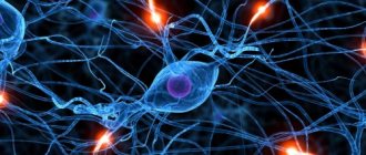
The shape of the neuron bodies is round, and the sizes themselves tend to vary from 15 to 150 microns. The structure of the neuron is due to the fact that in the central cellular part it has a nucleus containing a round nucleolus. Each individual neuron is separated from the connective contents by a layer of amphocytes, which are capsular cells belonging to the glial system. The terminal root of the proximal process of the cell includes two branches. The first is infused into the spinal cerebral nerve and goes to the end of the receptor. The second is located in the direct entry of the dorsal root, reaching the column of gray matter. At the locations of the orbits there are also autonomic ganglia, which are called multipolar, which is why they differ from the cerebrospinal ganglia. They are precisely the organ that provides sensitive, sympathetic and motor innervation to the eyeballs.
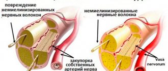
Peripheral nerves appear to be clearly predetermined solid formations of an anatomical nature. A sheath woven from connective tissue envelops the entire length of the nerve trunk and, from a medical point of view, is called epineurium. Group bundled nerve fibers are surrounded by a sheath of concentric layers of connective tissue called perineurium. The endoneurium consists of a layer of thin connective tissue formation separated from the perineurium, which covers the myelin sheaths of the nerve fibers of the spinal brain.
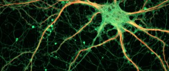
The peripheral part of the nervous system is abundantly supplied with blood vessels. The nerve fibers that form nerves are either straight or in the form of zigzags, thus protecting themselves from the process of stretching during movements of the limbs and body. There are myelinated and non-myelinated nerve fibers. The quantitative composition of both is different in each tissue and nerve region of localization. Myelin fibers are divided into thin, medium, and thick. The passage of arterial branches to the nerves is directed from the vessels that are in close proximity to them. The presence of blood capillaries, most of them directed along, in the endoneurium is due to their relationship to nerve fibers.
Main characteristics
The branches that extend from a specific nerve reproduce the functions of innervation of the nerve sheaths directly. The fibers that make up the systemic nerves are divided into two main groups: centripetal fibers and centrifugal fibers.
The former have the ability to transmit nerve impulses emanating from receptors to the spinal cord and brain. Centrifugal fibers are endowed with the ability to deliver impulses to innervated organs and tissues from the brain. Innervation of skeletal muscles occurs due to motor fibers. There is also the presence of such fibers as trophic ones, which ensure the usefulness of tissue metabolic processes.
The nuclei of the anterior horns of the spinal brain and cranial nerves are formed by the axons of neurons, taking into account the presence of motor nerves from the processes of nerve cells located in the sensory nodes of the cranial nerves and spinal cord. Mixed nerves contain motor and sensory fibers. Autonomic autonomic fibers emerge from both the brain and the spinal cord, formed by the neuronal processes of the lateral horns of the spinal cord and the autonomic nuclei of the cranial nerves.
The axons of neuron cells are ultimately directed to the peripheral part to the vegetative nodal formations of plexuses of nerves, where these fibers end. The processes of cells located on the periphery of the vegetative nodes are directed directly to the organs. The approach of autonomic innervation from the brain to the working organs occurs through two neurons. The first neuron in medical terminology is called the prenodal preganglionic neuron. And the second is a postganglionic neuron. The presence of autonomic nerve fibers is due to the inclusion of a general list of cranial nerves, branches and spinal nerves.
Some natural processes take place regarding the topography and characteristics of nerve branching. As they move towards the organs, the nerves have some similarities with the circulatory system and blood vessels. Both blood vessels and nerves have a segmental structure. Large nerves are most often located at the joint bends. The neurovascular bundles unite veins, nerves and arteries, while having one connective membrane, called the fibrous sheath, which is formed to provide thorough protection for the nerves. Superficial cutaneous, deep muscle and articular nerves are considered different.
Anterior branches of the spinal nerves: their zone of innervation and difference from the posterior ones
Anterior branches:
- Lost segmental structure.
- The truncopital and truncofugal muscles innervate - muscle fibers move, intersect, therefore => the anterior branches form plexuses.
But:
- The anterior branches of the thoracic SMN (Th2 - Th11 segments) - intercostal nerves - segmental structure + no plexuses, since they innervate parts of the body that have retained segmentation.
- Th12 – subcostal nerves.
Some features
As for the order of muscle branches departing from the nerves, it is usually in full accordance with the order of the arteries entering the muscles.
The place where the nerve enters the muscle is the middle third part of the belly of a particular muscle, and the nerve itself enters not from the outside, but from the inside. The possibilities of innervation peripheral processes depend on the distribution of nerves and branches, which belong to different spinal segments. Of no small importance are the connected neighboring nerves, which simultaneously form plexuses of them.
There are several types of these connections. In this case, the usual transition of fiber from nerve to nerve is not excluded. There are connections of a mutual nature where the exchange process occurs. There are cases when fibers separated from a nerve move on to the next one and for some time discover their presence in it, and then at the first possible transformation they return to their original form. In the connecting areas, the nerve has the ability to accept fibers that are different in their functional purpose.
A whole list of cases is caused by the fibers themselves leaving the trunk and passing through the tissue near the vessels, followed by a return to the original trunk. The presence of connections takes place in the spaces of the cranial nerves and elements of the spinal cord, stomatological and visceral elements and adjacent nerves of the spinal cord. These compounds can be found both inside organs and in individual tissues of the human body.
What plexuses do you know? Their innervation zone
The plexus is the place where nerves exchange fibers with each other. In the area of the plexuses, the nerves change the composition of the fibers (the number of nerves changes).
4 nerve plexuses:
- Cervical plexus - formed by the anterior branches of the 4 upper cervical SMNs. Located in front of the transverse processes. Innervates most of the muscles of the neck, the skin of the neck, the sides of the head, and the diaphragm.
- Brachial plexus - formed by the anterior branches of the C3-C8 SMN. It has supraclavicular and subclavian parts. Innervates the upper limb, including the upper limb girdle, as well as the superficial muscles of the chest and back.
- Lumbar plexus - formed by the anterior branches of the lumbar SMN. Innervates the skin, muscles of the lower abdomen, including the lumbar region, and the anterior medial part of the thigh.
- The sacrococcygeal plexus is formed by the branches of the sacral SMN. Innervates the muscles, skin and joints of the lower limb.
Functional features of the peripheral nervous system
The formation of the peripheral nervous system is due to the presence of nodes, nerves and their endings. As for nodes, their list includes nodes of the spinal cord, cranial, cerebral, and autonomic nodes. The nerves of the peripheral system include 31 pairs of spinal cord nerves, plus 12 pairs of cranial and cerebral elements.
The functions of the peripheral nervous system involve a predominance of mixed nerves. The endings themselves are receptors that have the ability to perceive and respond to stimuli coming from outside or inside the body. In addition, they are simultaneously effectors that transmit nerve impulses to the necessary organs responsible for certain functions.
The composition of each nerve includes unmyelinated and myelinated fibers. The outer part of the nerve is surrounded by connective tissue, which is a kind of sheath containing vessels designed to nourish the nerve. Based on the nature of the functions they perform, nerves are divided into sensory, motor, and mixed. Nerves can have significant external differences in their shape, thickness, and length. If they have a large diameter, such nerves are called the nerve trunk. The branches of nerves are called ramus. How thick a given organ can be depends on the presence of nerve fibers. The number of such fibers can reach several tens of thousands.
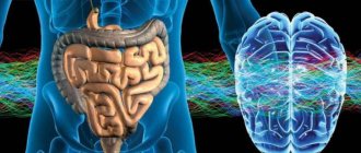
Brain
The adult human brain weighs approximately 1.3-1.5 kg. Scientists have found that until the age of 22 his weight gradually increases, and after 75 years it begins to decrease.
There are more than 100 trillion electrical connections in the average person's brain, which is several times more than all the connections in all the electrical devices in the world.
Researchers spend decades and tens of millions of dollars studying and trying to improve brain function.
What are the main functions of the system
The functional direction of the somatic system underlies the voluntary control of the activity of the skeletal muscles. The system plays a collecting role and receives information signals from the sensory organs, directing them to the central nervous system and transmitting them further to each individual muscle group. Thus, with its help, a relationship is established with the external environment and the motor processes of the human body. The facial muscles, tongue muscles, laryngeal muscles, as well as the muscles of the body and limbs of the body are subject to innervation. The peculiarity is that a person is able to control the situation and be aware of the processes taking place, regulating muscle activity.
The autonomic system is primarily responsible for regulating the activity of each individual organ and for homeostasis. As for this system, its peculiarity lies precisely in the fact that activity is determined by an independent regulatory flow and, in this case, is absolutely independent of human will. It has been defined as the autonomic nervous system, which includes the peripheral part of the nervous system. The autonomic system supplies nerves to internal organs, glandular tissues, muscles, skin, heart muscle and blood vessels. At the same time, the task of regulating metabolic processes in tissues, organs and their constituents is fulfilled.
The autonomic nervous system provides self-regulation through coordinated reactions while maintaining a constant state within the body. In addition, it directs, corrects and independently regulates the functioning of organs and metabolic processes between them. Its functioning is subordinated to the higher parts of the brain.