Good health to you, friends! If some kind of contraction begins in our body, then more often we treat it positively. Especially when it comes to significantly reducing the waist and other unnecessary fat volumes.
But doctors recognize a number of such conditions as pathological. And like all pathologies, they can bring us a lot of problems. One of the most unpleasant narrowings is called absolute spinal stenosis. We'll talk about it today.
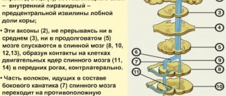
In medical practice, people often write about spinal stenosis, or more precisely, about stenosis of the intervertebral canal. Our spinal cord is located in this canal and our health will depend on its well-being. Such a natural tube protects one of the most delicate substances in the body, and even the smallest impacts can have a fatal effect on its functioning.
Symptoms of absolute stenosis
The signs of this serious disease will vary depending on where the critical narrowing of the spinal canal is located.
- Cervical region
Severe weakness of the arms, a feeling of partial numbness and “pins and needles” in the upper body, difficulty breathing. In some cases, a person loses sensation throughout the entire body from the neck down. Cervical absolute stenosis is the most dangerous, since there is a possibility of not only disability of the patient, but also death.
- Thoracic region
A rare pathology that manifests itself as chest pain, disorders of the genitourinary system, intestines, numbness of the arms and shoulder girdle.
- Lumbar
This is where stenosis develops most often. It is the lower back that is subject to the greatest stress in a person’s everyday life. Absolute stenosis manifests itself as severe pain – the pain radiates to the leg and buttock. So-called lumbago may be felt, walking is impossible or very difficult, and paralysis of the lower extremities develops - complete or partial. Stenosis also contributes to sexual dysfunction, spontaneous urination and defecation.
Prerequisites for development
To answer the questions surrounding the concept of “secondary spinal stenosis”: what it is, what causes its development, how to determine its occurrence and how to deal with it, it is important to know the structure of the human spine, especially its lumbar region. The spinal canal is formed from vertebrae, consisting of vertebral foramina, connected to each other by dense ligaments. This canal is the container in which the spinal cord is located. As a person moves, vibrations of this space occur, and the channel periodically narrows.
To prevent compression of the spinal cord and protect against mechanical stress, special cerebrospinal fluid and fatty tissue are provided between it and the walls of the canal itself.
In some diseases, including stenosis, nerve endings are pinched or the spinal cord is directly compressed. The most common cases of such narrowing is stenosis in the lumbar region.
There are primary and secondary forms of stenosis, caused by genetic and acquired processes, respectively. The first form, idiopathic, is the result of abnormal intrauterine anatomical development or abnormalities in genetics. An example of such a deviation is the incomplete development of the vertebral foramen, as a result of which this place becomes narrowed and compresses the spinal cord. This type appears at any age, often in young people.
- Interesting read: absolute stenosis
The prerequisites for the development of secondary stenosis are injuries to the spinal region, complicated processes in the postoperative period, or other diseases that a person has (spondylosis, oncology, arthrosis, disc protrusion, etc.).
The most common pathology is intervertebral hernia, which leads to compression of blood vessels and nerve endings.
Most causes of secondary pathology lead to a slow, gradual narrowing of the spinal canal, which makes it possible to successfully eliminate the disease and its consequences. The most dangerous prerequisite is injury, often accompanied by sudden and severe compression of the spine. Such damage is accompanied by a rapid decrease in the spinal space and causes difficult-to-treat consequences.
Diagnostics
- Studying the patient's complaints - the doctor asks about the location of the pain, its manifestation, and the presence of accompanying symptoms.
- Physical examination - the patient’s posture is studied, the presence of scoliosis, palpation and percussion diagnostics are performed.
- X-ray – X-ray will show bone formations and can also evaluate narrowing of the intervertebral canal to confirm the diagnosis of relative stenosis. If a narrowing of the lumen of less than 10 mm is detected, the patient is diagnosed with absolute stenosis.
- Multislice CT scan – detects degenerative processes of bone and cartilage tissue.
- A myelogram is an x-ray with contrast that allows you to evaluate the degree of pressure on the nerve roots.
- Magnetic resonance imaging – visualizes the condition of soft tissues and the spinal cord.
Causes of spinal stenosis
The pathology can be congenital, but most often it is a consequence of an incorrect lifestyle, mainly sedentary, or traumatic back injuries. Diseases that can lead to stenosis:
- Bone tuberculosis;
- Osteochondrosis;
- Arthrosis of the joints;
- Rheumatic joint pathologies;
- Neoplasms of the spinal cord - they can also fill the space and put pressure on the nerve endings, similar to cartilage and bone tissue.
Treatment of absolute stenosis
If the diagnosis of absolute spinal stenosis is confirmed, the patient must undergo surgical treatment as soon as possible. Drug therapy or exercise therapy are powerless in this case, and the time lost on them does not benefit the patient.
The operation protocol may be different:
- Discectomy – the disc at the level of which the maximum narrowing of the canal has formed is completely or partially removed. If a hernia is to blame for the narrowing, only it can be removed. Next, the patient receives a disc implant.
- Laminectomy - the surgeon removes part of the vertebral arch, resulting in the release of the spinal canal. Most often, such intervention is performed if the stenosis is of traumatic origin. After eliminating the compression factor, the patient is given an implant that ensures normal motor function of the spine and protects it.
Treatment
It should be noted that in each individual case the treatment of secondary stenosis will be different. Much depends on the degree and severity of the disease. If the disease was identified at the initial stage, then conservative treatment methods will be sufficient to eliminate it. If the situation is advanced, surgical intervention will be required.
Physiotherapy
For secondary spinal canal stenosis, the following physiotherapeutic procedures are recommended:
- electrophoresis;
- amplipulse;
- magnetic therapy;
- hydrotherapy;
- mud baths.
In addition, it is recommended to regularly take a contrast shower. It is best to alternate this procedure with physical activity. This helps reduce inflammation, reduce pain, and normalize blood circulation.
Drug treatment
An indispensable component of effective treatment of secondary stenosis is taking appropriate medications, among which are:
- anti-inflammatory drugs (for example, Naproxen, Ibuprofen, etc.);
- medications to relieve pain (eg, acetaminophen);
- anti-edema drugs;
- anti-inflammatory ointments and patches (for example, Nanoplast forte, Voltaren);
- medications that help normalize neuromuscular conduction (Mivacurium, Pancuronium);
- vitamins.
It should be remembered that any medications should be taken only as prescribed by the attending physician.
Therapeutic exercise (physical therapy)
If the patient’s condition allows, then he is prescribed special physical therapy. Performing exercise therapy for secondary spinal stenosis will help improve posture, reduce pain, and increase the strength of the spine. A physical therapy program should be developed by a rehabilitation physician, since only he can choose the optimal load.
Massage
In addition to exercise, massages also have a great effect. However, you should take into account the fact that massage should only be performed by a qualified specialist.
. Otherwise, the patient's condition may worsen. In order to do massages at home, today special exercise machines are sold that allow you to regulate the load.
Operation
Only surgical intervention will help to completely get rid of secondary spinal stenosis. As a rule, surgery is performed if conservative treatment does not help. The following types of surgical interventions are distinguished:
- decompression laminectomy – this operation involves removing areas that compress the nerve roots;
- implantation of stabilization systems is carried out with the aim of strengthening the supporting function of the spine.
Possible complications if left untreated
Without timely and adequate treatment, absolute stenosis can have extremely serious consequences. And irreversible paralysis of the limbs is far from the only thing. Thus, due to impaired nutrition of spinal cord cells and deterioration of its oxygen supply, an ischemic stroke of the spinal cord may occur. And if the spinal canal is critically narrowed at the level of the thoracic or cervical region, death may occur due to respiratory distress or sudden cardiac arrest.
How to diagnose
Before you begin treatment for secondary spinal stenosis, it is necessary to correctly diagnose it. Diagnostics is aimed at identifying the cause of the disease, as well as determining the consequences to which it led. So, after an external examination of the patient, the attending neurologist
prescribes additional methods for diagnosing stenosis. As a rule, the following procedures are performed for this disease:
- X-ray
– makes it possible to detect various bone formations (tumors, spinal injuries, etc.). The disadvantage of this diagnostic method is that it does not allow you to see soft tissue. In this regard, MRI is often additionally prescribed. - MRI
is an absolutely harmless method of visualizing the internal structure of the human body. It is based on the use of radio magnetic waves. This procedure allows you to quickly identify secondary spinal canal stenosis in the cervical, lumbar and spinal pathologies. - – it is based on the principles of MRI and radiography. In some cases, CT is supplemented with a myelogram. This allows for improved visualization of soft tissue.
Make an appointment with doctors at the Central Clinical Hospital of the Russian Academy of Sciences
The best specialists of the department of vertebrology
They receive patients with spinal canal stenoses of varying degrees of severity and origin.
The patient is examined directly on site, and an individual treatment program is developed for him, including drug therapy and physical therapy. In difficult cases, when absolute stenosis is accurately diagnosed, surgeons at the Central Clinical Hospital in Moscow
perform spinal surgeries, returning patients to motor activity and life without pain.
make an appointment
with
a vertebrologist, rheumatologist
or other musculoskeletal specialist by phone or using the online form.
Prevention
To prevent the occurrence of this disease, it is necessary to perform the following series of preventive measures:
- it is necessary to do exercises every morning, combining aerobic movements with resistance exercises;
- you should maintain correct posture;
- master the technique of correct body mechanics, which is aimed at minimizing the load on the spine.
Thus, this article has brought some clarity to the question of what it is - secondary spinal stenosis, what are its causes and what symptoms are characteristic of this disease. However, it should be remembered that self-medication is unacceptable here. The entire treatment plan must be prescribed by the attending physician.
Treatment of spinal stenosis
Many factors go into determining the optimal way to perform surgery. We are talking about the location of the pathologically narrowed area on the spine, the cause of root compression, concomitant diseases of the patient, etc. Most often the problem is solved by:
- Endoscopic surgery.
- One-stage or two-stage operation with thoractomy.
- Operations involving the installation of implants – metal or from a person’s own bone tissue.