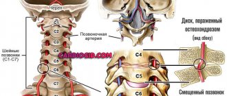
Over the past two decades, modern medical science has moved far forward, leaving behind the old methods of hardware diagnostics of all internal organs. Fundamentally new techniques for studying the functional activity of the human body have emerged, including methods such as ultrasound, as well as computed tomography and magnetic tomography. The latter examination method is of great value in diagnosing brain diseases, since with its help it is possible to quickly identify the formation of a pathological process and promptly carry out the necessary therapeutic measures.
However, when receiving a referral for a resonance imaging study, each patient is tormented by the question of whether MRI of the brain is harmful. In our article we want to provide accurate information about the principle of operation of a magnetic tomograph and its effect on human health. Talk about when it is worth doing an MRI of the brain, as well as about the features of this diagnostic procedure and the presence of contraindications for its use.
Principle of the MRI method
Diagnostic equipment works based on the influence of an electromagnetic field on hydrogen atoms contained in the cells of the human body. The tomograph has the shape of a space object and consists of the following elements:
- a retractable table on which the patient being examined is located;
- a complex system of scanners emitting electromagnetic waves of various lengths.
Oscillatory (or resonant) movements of tissues make it possible to obtain a reflection of the internal systems of the human body on the monitor of the device. As a result of its processing using computer programs, the scanned structures of the spinal cord, brain, internal and musculoskeletal organs receive a graphic or three-dimensional detailed image. The main applications of the MRI method are:
- identification of early stages of pathological processes;
- clarification and confirmation of the diagnosis;
- intermediate monitoring of the course of treatment.
If necessary, the resulting graphic image of the patient’s brain is enlarged in size and transferred to paper and digital media.
This study has many advantages over other diagnostic techniques, in particular:
- complete safety (it is prescribed even for children);
- obtaining more accurate final data (with the help of a magnetic resonance procedure, you can notice the slightest disturbances in the structure of the brain and changes in its cortex);
- The duration of the examination does not exceed 1 hour, and immediately after its completion the patient receives an image.
Whether there is harm from MRI or not depends on the patient’s compliance with all the instructions of medical specialists.
The effect of a magnetic field on the body
The operation of a tomograph is based on certain physical processes. A person is in a magnetic field with high intensity and is exposed to a radio frequency signal.
Tissues and organs respond to the signal with waves of different lengths, which are visible on the monitor in the form of a picture reflecting the state of the internal organs.
Is a strong magnetic field and electromagnetic radiation harmful or not, how do they affect human health?
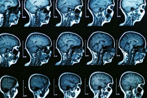
Photo:
Modern science does not have facts proving that such exposure has a harmful effect on living organisms.
MRI has been used for several decades, and during this time there has not been a single case recorded in which a person became ill during this procedure or his health condition worsened some time after being in electromagnetic fields.
However, as they say “just in case,” MRI is not recommended for pregnant women in the early stages of pregnancy, since the question of whether the effect of magnets on an embryo (an organism that is still developing) is harmful or not has not yet been sufficiently studied.
Skeptics doubt that MRI is not harmful, saying that the danger of magnetic fields to health can be traced not immediately after examination in a tomograph, but, say, several years later.
For example, some patients prescribe exposure to magnetic fields to cause headaches, irritability, vascular and even cancer diseases that arose years after the tomograph examination.
READ When is an MRI of the knee prescribed?
Particularly critical patients even put MRI and radiography on the same level.
In all these cases, we are not talking about specialists, but about ordinary people who do not have the necessary knowledge and factual data about the effect of the magnetic field on living organisms, about how harmful or not it is harmful to health.
Video:
The safety of MRI for health has been confirmed by numerous and long-term animal tests and decades of experience in clinical use.
Despite the safety of the method, we must not forget that MRI should only be prescribed by a doctor. The point here is not only that this procedure is expensive and takes a long time to do.
The need to obtain a referral from the attending physician is due to the fact that MRI is not the only way to make a diagnosis and for a correct diagnosis it must be supplemented with other methods.
In what cases do you not think about the dangers of diagnostics?
Unlike the computed tomography procedure, which uses radioactive radiation that negatively affects the patient’s body, MRI is absolutely not harmful to the child and pregnant women.
The study is necessary if a person has:
- head injuries;
- acute headaches;
- convulsive syndrome (involuntary muscle contractions);
- short-term loss of consciousness (fainting);
- increased intracranial pressure;
- speech dysfunction;
- loss of vision;
- inflammatory processes in the brain (for example, meningitis);
- thrombosis, atherosclerosis or vascular stenosis;
- concussions.
This procedure may be necessary if you suspect the formation of a tumor formation, determine the consequences of a stroke, or adjust drug therapy.
Features of diagnostics
A referral for an MRI of the brain is given by the attending physician, who must take into account some nuances. The patient is not allowed to have metal implants, special medical devices and devices:
- Elizarova;
- insulin pumps;
- pacemakers;
- artificial heart valves;
- prosthetic joints;
- titanium plates;
- braces;
- metal dentures.
A patient who has obvious signs of claustrophobia (fear of closed spaces) is advised to take sedative medications on the eve of the procedure.
Before performing the procedure, the patient must put on appropriate clothes, and the medical staff warns him about the possibility of using a “panic button” to stop the examination
Magnetic tomography is contraindicated in the following pathologies:
- insufficient renal function;
- various mental disorders;
- bronchial asthma.
Harmful effects on patient health from MRI of the brain
The use of strong electromagnetic field radiation, which is generated during the operation of powerful tomograph turbines, makes many patients think about abandoning the diagnostic procedure. However, science has proven the safety of MRI for the human body - the radiation when performing this technique does not exceed the portion of radiation that a person receives when using a mobile phone! From these results it follows that there is virtually no negative effect of electromagnetic waves on the human body.
MRI of the brain can be done several times a year if there are indications to determine an accurate diagnosis, monitor the severity of the disease and the effectiveness of treatment measures.
Can the use of contrast in MRI have harmful effects?
Brain scans using a contrast agent, which is injected into the patient intravenously, are used to detect tumor processes and visualize the vascular system. It is most often prescribed to diagnose cancer and evaluate treatment methods.
Such an examination may need to be carried out more than once. Many patients very often reported the occurrence of severe pain in the head and a deterioration in general well-being after an MRI. However, in fact, the diagnostic procedure itself does not have a negative impact. The appearance of such signs is associated with a side effect of the contrast and depends on the patient’s individual sensitivity to this drug, which is manifested by dizziness, headache, and fever.
The contrast used for MRI of the brain contains galidonium, a substance that penetrates the organ tissue during examination and improves the image on the device screen by affecting electromagnetic waves and hydrogen atoms
An allergic reaction of the body occurs quite rarely. The administration of a contrast agent is prohibited for patients with severe pathology of the urinary system. For minor disorders of excretory function, it is necessary to consult a urologist before performing contrast tomography.
How does a tomography device work: principle of operation
The operating principle of the tomograph will also help you understand how the diagnostic device works without radiation. The operation of the device is based on a physical phenomenon called nuclear magnetic resonance. Using this method, it is possible to obtain the dimensions of the electromagnetic responses from hydrogen nuclei. These properties are distinctive from more conservative diagnostic methods.
How MRI works
Nuclear Magnetic Resonance is based on the properties of protons. Further, created with the participation of radio frequency pulses, the electromagnetic field becomes a space for the release of energy, converted into a kind of signal, after which it is registered and processed in a computer system.
The magnetic nuclear resonance technique provides an opportunity to study the human body, essentially, due to the peculiarities of the magnetic properties of body tissues. Depending on the vector orientation of the protons (as a rule, two opposite phases are taken into account), it becomes possible to detect the projection in which one or another hydrogen atom is currently present. By influencing a specific area of the body with the help of electromagnetic waves, some of the protons periodically change their location. The task of the computer system is to collect and record information in parallel.
Comparison of MRI, radiography and CT in relation to harm to the body
It is hardly possible to compare the procedure of magnetic resonance imaging and conventional radiographs. First of all, the final data from fluoroscopy are inferior to organ scanning in terms of information content. In some cases, computed tomography provides sufficient results; its cost is much lower than electromagnetic diagnostics, but the harm to the patient’s health is much higher.
Of course, exposure to ionizing radiation, which is used to obtain an X-ray, is strictly regulated, and radiation dose standards are calculated for different age categories and depend on the state of human health. Medical specialists who refer patients for CT or X-rays calculate the doses of radiation and determine how many times the diagnostic procedure can be performed so as not to harm the patient.
Under certain circumstances (for example, if the patient has loose ferromagnetic implants or endoprostheses), an MRI may be unsafe, and a CT scan will solve this problem.
Is magnetic resonance imaging dangerous?
Despite the fact that the examination involves magnetic scanners that do not produce radiation, the procedure is absolutely safe. Based on the results of numerous experiments and studies, it became clear that magnetic waves and their radiation do not cause any harm to the human body, and there is no radiation at all during the operation of the tomograph. MRI also has no side effects.
There is a magnetic field in the tomograph, but the radiation dose in it is minimal. If we compare the radiation field on MRI and computed tomography or radiography, then in the first version its level is very low and is not harmful to humans. That is why diagnostics using a tomograph are used to examine any organ and the entire organism as a whole. MRI is allowed even for ischemic stroke.
Tomography does not expose the patient's body to radiation!
Important conditions for scanning
To correctly obtain the necessary information as a result of MRI, certain conditions must be met:
- it is important that the patient does not move during the diagnosis;
- the patient should not have any metal objects on his body;
- the patient should not bring gadgets and mobile devices into the tomography;
- the patient should not suffer from chronic renal or heart failure;
- It is important to listen to all the recommendations of the doctor and nurse, strictly following them.