Teshen's disease of pigs is a dangerous viral disease that affects young animals aged 1 to 3 months of life. During its development, the disease causes damage to the nervous system of animals, which causes inflammation of the brain and spinal cord. Lethal outcome with such invasion is observed in 30-50% of cases.
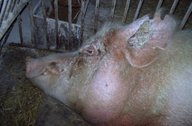
Sick pig
Pathogen
Scientists were able to identify the causative agent of Teschen's disease not so long ago, due to the low prevalence of the disease. A special type of picornavirus was identified in the second half of the 20th century. It consists of round microparticles with a diameter of no more than 30 nm. This family of enteroviruses, upon entering the body, immediately settles in the tissues of the nervous system, causing disruption of its functions and gradual destruction.
Thanks to its special structure and durable protective shell, the pathogen exhibits high resistance to external influences. In an acidic environment with a pH value from 3 to 9, the properties of the virion practically do not change. Only by increasing the pH value to 11.8 can the aggressiveness of the infection be reduced by 2 times.
The pathogen is also not affected by various lipid solvents. The virus survives under thermal influences:
- 1 year – in the temperature range from 0 to 4 degrees;
- 15-17 days – when exposed to a temperature of 37 degrees;
- 15 minutes – when increasing the value to 60 degrees;
- 10 minutes – when the temperature increases to 70 degrees;
- When completely frozen, the activity of the virion remains for several years.
Attention! The pathogen can be destroyed by rapid dehydration or by exposure to ether, to which the virus is less resistant.
Sources of the virus
In general, the process of infection of pigs with Teschen disease has not been fully studied. But it is known that this disease easily passes from infected individuals to healthy livestock. In the natural environment, this process is realized through the respiratory tract and gastrointestinal tract. Accordingly, the main source of spread of the pathogen is infected pigs.
The virus is released into the external environment from the animal's body along with feces in the acute form of the disease. Virions can also be contained in large quantities in discharge from the nose and mouth in the case of a latent form of the disease. The virus enters the body of healthy piglets:
The virus enters the body of healthy piglets by consuming infected water.
- when consuming infected food and water;
- in case of crowded keeping of animals;
- in some cases, when eating feces or body parts of dead pigs;
- through pig farm workers working with both sick and healthy animals;
- through care items and elements of the pigsty.
For a certain period of time, the pathogen actively develops in the intestines of infected livestock. It is also often found in the kidneys, liver, and lymph nodes. Then the infection gradually spreads to the nerve tissue. The highest concentration of virions can be traced in:
- trunks of large nerve endings;
- medulla oblongata;
- brain (less often spinal cord);
- cerebellum.
The virus can be detected in the blood at the initial stage of the incubation period of the disease. The infection is most likely not released into the external environment with urine.
Many researchers argue that the lethality of the disease directly depends on the piglet’s individual sensitivity to infection. If the virus is perceived acutely by the body, the animal dies. This is confirmed by the fact that even within the same age group and in the same litter, only a part of the animals die, while the rest successfully recover.
A contributing factor to the spread of the pathogen is non-compliance with basic sanitary and hygienic standards for keeping pigs. In farms with severe unsanitary conditions, the disease occurs much more often and is more widespread.
Teshen's disease
Teschen's disease
(Encephalomyelitis enzootica suum (Latin); Tesin disease, Talphan disease (English); Teschener Schweinelahmung, An-steckende Schweinelahme (German); Maladie de Teschen (French) - a viral disease of pigs, characterized by the development of non-suppurative encephalomyelitis and the appearance of paralysis The disease is manifested by clinical signs of damage to the central nervous system (hypersthesia, muscle tremors, nystagmus, opisthotonus, tonic-clonic convulsions, “stilted gait”, paralysis of the limbs of the muscles of the neck, pharynx). Very often, this disease of pigs was described under various independent names: equine polio, disease Talfan, piglet encephalomyelitis, Klobuk's disease, etc. Talfan's disease was described by Harding (1958) in England as a polyencephalitis affecting young piglets. Teschen's disease was first diagnosed by Trefni (1930, 1931) in Czechoslovakia, in the Teschen district. It was thoroughly studied by Klobuk (1931, 1933) The infection quickly spread in Czechoslovakia and entered Germany (1940), Australia (1939), Yugoslavia and Hungary (1945).
Currently it is found in Poland, Bulgaria, France, Italy. The detection of antibodies to Teschen-Talfan disease group viruses in pig blood sera indicates the widespread distribution of these viruses. In the CIS, the disease was first identified on farms in the Transcarpathian region. Information about the pathogen The Teschen disease virus was first isolated by Trefni (1930) in Czechoslovakia in the Teschen district. Morphology and chemical composition. Virions are spherical in shape, their diameter is 25-30 nm. The virions are non-enveloped, with a buoyant density in CsCI of 1.34 g/cm3, and contain a single-stranded RNA core surrounded by a cubic capsid. There are no lipoproteins and the virus is resistant to fat solvents. Sustainability. The virus is resistant to ether, chloroform and trypsin, does not change over a wide pH range (2.8-9.5). At pH 11.8, its infectivity quickly decreases by 50%. Trypsin at a concentration of 125-1250 μg/ml increases the virulence of the Teschen disease virus 10 times and promotes its reactivation after heat treatment (at 37 and 56°C). The virus can withstand heating at 60°C for 15 minutes, but is inactivated in 20 minutes. When heated to 70°C, it loses activity after 10 minutes. At 37°C, the virus can survive up to 17 days, but quickly dies with the development of putrefactive processes. Therefore, virus-containing materials should be immediately preserved with glycerin or Hanks’ solution with antibiotics and placed in a thermos with ice. The virus persists at 4°C and -79°C, as well as in 50% glycerin at 0°C for up to 20 months, in frozen form for years. Can withstand drying in the sun for up to 3 weeks. In salted and smoked products it remains active for more than 3 weeks. The virus is insensitive to the action of penicillin, streptomycin, nystatin and is quickly inactivated by 0.15% formaldehyde. Its reproduction and CPP are partially inhibited by 2-(?-hydroxy-benzyl)-benzimidozalone. Of the 10 disinfectants studied, only sodium hypochlorite and 70% ethanol completely inactivate the Talfan virus. The Teschen disease virus survives at 15°C for more than 168 days. Both viruses persist for a long period in liquid materials, in which they are more quickly inactivated with active aeration; Inactivation of viruses also occurs rapidly under the influence of ionizing radiation and during digestion. Antigenic activity. Specific VNA and CSA appear in the blood of sick and recovered animals. Antigenic variability and relatedness. In RN and RSC, the antigenic relationship between the Teschen disease and Talfan disease viruses is shown. Immunologically, Teschen disease viruses are a single type, within which two subtypes are distinguished: the first includes pcs. Bozen, Konratice, Repo-ryie, in the second - pcs. Tyrol, Sweden Ug, Talfan. GA properties have not been established. Localization of the virus, virus carriage and virus shedding. The virus is detected in the blood and organs for a short time and in small quantities. Viremia within 48 hours is observed between 4-6 days after infection, and virus titers in the blood are usually low. In the body of pigs, the virus is localized and multiplies in the tissues of the gastrointestinal tract, in which it appears 24-72 hours after infection and is detected within 5-7 weeks. The highest titers of the virus (106 TCD50/g) in fecal samples are detected 9 days after infection. In the tonsils, mesenteric and mesenteric lymph nodes, the virus appears 48 hours after infection and is detected within 6-8 days. In the brain and spinal cord it is detected after viremia. In maximum titers (105.5 - 10 6.6 TCD50/g) it is contained there even before signs of the disease appear and during the first days of the paralytic stage. The virus is found in the highest concentrations in the cervical and thoracic spinal cord and in the cerebellum, making these tissues the most suitable material for isolating the virus. By the 5th day after the onset of symptoms of the disease, the content of the virus in the tissues of the central nervous system decreases, and by the time the animal dies, so-called autosterilization can be observed. Less regularly, the virus is found in secretions and excreta (nasal discharge), sometimes in feces. The virus is released into the external environment, mainly in the prodromal stage and in the first 2 days of the disease. Experimental infection. Piglets up to 1-2 months of age are susceptible to experimental infection. Piglets of 2 weeks of age who do not receive colostrum are more severely ill than piglets of 3 weeks of age. Sometimes the disease can be reproduced only in suckling piglets of 3-5 days of age that do not receive colostrum. Experimental infection is easier to manage with intracerebral or subdural injection of virus-containing material. Intranasal or oral infection is less effective. Sometimes intracerebral and intranasal infection is practiced. Laboratory animals are insensitive to the Teschen-Talfan disease virus and the causative agents of other porcine polyencephalomyelitis. Cultivation. The virus multiplies in cell cultures of kidneys, lungs, hearts, testes, ovaries, etc. of piglets 3-8 weeks of age and pig embryos. Reproduction is accompanied by the formation of CPI plaques, which do not differ from the changes caused by other porcine enteroviruses. They are characterized by an accumulation of rounded cells with increased refractility, which become noticeable on the 3-5th day after infection. When using adapted “cultured” strains of the virus, the first signs of CPE appear much earlier (sometimes after 2-16 hours). The affected cells are separated from the surface of the glass; in addition to foci of degeneration, areas devoid of cells appear. When examining stained preparations of an infected culture, granularity and acidophilia of the protoplasm, chromatin condensation and eccentric position of the nucleus are revealed. Reproduction of the Teschen-Talfan disease virus is possible in continuous cell lines of the kidneys (IBRS-2) and other organs (SST). Some strains reproduce in HeLa and BHK-21 cells. Various strains cause 1 of 3 types of CPD in piglet kidney cell culture and, according to the characteristics of CPD in BHK-21, HeLa and Vero cells, the strains are classified into one of 4 groups. Epizootological characteristics.
The source of infection is sick animals.
More often, infection occurs through the consumption of feed, water, meat waste, slaughterhouse waste contaminated with the secretions of sick animals, as well as slops. Possible contact infection. the infection can be spread by convalescent pigs or animals that are asymptomatically ill. The gateway to infection is the olfactory area of the nasal mucosa. Spectrum of pathogenicity in natural conditions. Only pigs are susceptible. The disease is also found in Europe in wild pigs, in Madagascar in water pigs (Potamochoerus larvatus). Weaned piglets and gilts are most susceptible. Clinical signs and pathological changes
.
The incubation period lasts 1-4 weeks. In the prodromal period, which rarely exceeds 72 hours, low temperature, loss of appetite, sometimes vomiting, acute rhinitis, and loss of motor coordination are noted. Often these disorders go unnoticed. Then clinical signs of central nervous system damage appear. With encephalomyelitis, hypersthesia of the skin, convulsive contractions of various muscle groups, involuntary movements, rigidity of the muscles of the limbs, nystagmus, etc. are observed. Then symptoms of damage to the spinal cord develop - a staggering, tense gait, weakness of the limbs, incomplete support of them on the ground, progressive paralysis, gradually seizing limbs, neck and head. Due to paralysis of the pharynx, partial or complete loss of voice is noted. In severe cases, the disease usually ends in coma and death of animals. With a milder course, difficulty gait, development of semi-paralysis and paralysis without signs of excitement are noted. Animals are mainly infected through nutrition. Then the virus reproduces in the epithelial cells of the mucous membrane of the large intestine, and from there it penetrates the lymph nodes, the bloodstream and spreads throughout the body, including the central nervous system. Clinically, the disease occurs with symptoms of acute encephalomyelitis and meningitis, the most pronounced depression, increased skin sensitivity, spastic state of the labial, ocular and chewing muscles, as well as the muscles of the limbs, accompanied by swimming movements, vomiting, aphonia, hoarseness, strabismus, grinding of teeth, difficulty hard breathing. After the onset of the paralysis stage, body temperature drops to normal and reaches 32-34°C before death. Significant pathological changes are found only in the central nervous system. They are characterized by hyperemia of the meninges and serous infiltration. On sections of the brain, hemorrhages and perivascular round cell infiltration with the formation of foci of neuronophagia are noted in various places, and only the gray matter of the brain is affected. In the cerebellum, changes are found in Purkinje cells. The most characteristic changes are found in ganglion cells. The motor centers of the ventral horns are affected (“liquefaction” of cell nuclei, formation of vacuoles, the presence of karyorrhexis). Significant neuronophagy predominates in the ventral horns of the lumbar spinal cord. Histological changes are of diagnostic value. They are most pronounced in the middle and caudal parts of the brain (especially at its base - the cerebral peduncles and quadrigemina), as well as in the cervical and lumbar parts of the spinal cord. During a pathological examination, hyperemia of the soft membranes and swelling of the gray matter (mainly the rhomboid and midbrain) are observed. , cervical and lumbar thickenings of the spinal cord, catarrhal rhinitis, bronchitis and pulmonary edema, pinpoint and spotty hemorrhages under the epi- and endocardium. The stomach is usually filled with feed masses, the mucous membranes of it and the large intestine are folded, focally hyperemic and covered with thick transparent mucus. The bladder is always full. Microscopic examination of the central nervous system always reveals non-purulent, lymphocytic type polyencephalomyelitis and meningitis. When studying animals that died in the first days of the disease, focal and diffuse cellular infiltrates of glial elements, histiocytes and lymphocytes of varying sizes are observed in the gray matter of the brain and spinal cord. Small accumulations of red blood cells are sometimes found among the infiltrate cells. Polymorphonuclear and neutrophilic leukocytes are rare and only in animals that died (or were killed) in the first hours and days of the clinical manifestation of the disease. Many infiltrate cells are in a state of pyknosis and oexis. In lesions, disintegration of nervous tissue, dystrophic and necrobiotic changes in nerve elements, disintegration and disappearance of phospholipids are very often recorded. At the periphery of diffuse and large-focal lesions of the parenchyma, activation of microglia is noted, in places - small-focal accumulations of mononuclear cells. When the animals recovered, restoration processes were expressed in the lesions of the central nervous system, the composition of the infiltrate changed, the number of cells in it gradually decreased, macrophages and granular balls appeared. Many plasma cells accumulated around the vessels, and subsequently fibroblasts. In the soft meninges, changes were manifested by circulatory disorders, loosening and infiltration of the membranes with round cell elements (lymphocytes and histiocytes, subsequently plasma cells and fibroblasts). Pathogenesis. Primary reproduction of the virus is observed in the retropharyngeal lymph nodes and intestines. During the illness, leukocytosis is noted, the number of cells in the cerebrospinal fluid changes sharply, already on the 2nd day it increases to 44 (with a norm of 10-12), on the 3rd to 247-395 in 109/l (p <0.001), and polymorphic nuclear neutrophil leukocytes and lymphocytes predominate. The content of total protein in the cerebrospinal fluid increases 3-4 times, organic phosphorus increases 2-3 times, which indicates an intense inflammatory reaction in the medulla and membranes. Diagnostics.
To isolate the virus from the central nervous system, it is necessary to collect tissue samples from pigs with nervous syndrome at an early stage of its manifestation.
In animals with signs of paralysis, after a few days the virus is not detected in the central nervous system. The virus can be isolated in cell cultures from the spinal cord, cerebral cortex or cerebellum. Identification of the virus is carried out on the basis of physicochemical characteristics, IF or ELISA. ELISA is widely used to detect AT. In the differential diagnosis, Aujeszky's disease, rabies, CSF, and chloride and solanine poisoning should be excluded. The diagnosis of past infection, as well as latent virus carriage, is made based on the determination of AT titers in the blood sera of convalescents in RN in a culture of pig kidney cells using laboratory strains of the Teschen disease, Talfan and T-80 viruses. For accelerated diagnosis of the disease, as well as to detect the concentration of the virus in organs and tissues, IF is used. In the first 3 days of the disease, a specific glow is observed in the cytoplasm of neurons of all studied parts of the central nervous system, cells of the intervertebral nodes, individual cellular elements of the spleen, mesenteric lymph nodes, and the mucous membrane of the large intestine. But starting from the 4th day, the glow in the nerve elements and other tissues sharply decreases, and on the 5-7th day, enteroviral hypertension is detected only in the epithelium of the intestinal mucosa. That is why, to diagnose enzootic encephalomyelitis of pigs, in addition to the tissues (cerebellum, medulla oblongata and spinal cord) recommended by the instructions for IF, it is also necessary to select pieces of the colon mucosa. For intravital diagnosis and determination of virus carriage in animals, a direct method of fluorescent AT in smears - imprints from the mucous membrane of the rectum and feces - has been developed and introduced into production. Positive IF in smears is observed in epithelial cells and their fragments throughout the entire illness, and in those who have recovered (virus carriers) - up to 2 months. Immunity and specific prevention
.
For specific prevention of Teschen disease, GOA formol vaccine and live vaccine from the Tübingen mutant, representing pcs., are used. Konratice Teschen disease virus, attenuated by serial passage in porcine kidney cell culture. Veterinary and sanitary examination
.
Carcasses and slaughter products from sick and suspected diseases are prohibited from being released in raw form. In the absence of pathological changes, samples are sent for bacteriological testing for salmonellosis. If salmonella is detected, the internal organs are disposed of and the carcass is sent for boiling. In the absence of salmonella, it is allowed to process it into boiled, boiled-smoked sausages, canned food or parboiling. Cryodestruction - vaccination against cancer
Treatment of tumors without surgery and anesthesia
DOGSAUNA
A super warm thermos house for your dog!
Sections:
- Pig diseases
- Viral
Report an error, post material
Veterinarian consultation by phone
Symptoms and signs
The clinical picture of the disease in most cases is pronounced. After entering the body of a healthy pig, the virus goes through an incubation period. Its exact duration has not been determined, but it is generally accepted that it lasts 8-35 days.
After incubation for 1-2 days, the prodromal period lasts
After the incubation period, the prodromal period lasts for 1-2 days. It is accompanied by an increase in temperature to 40-41.5 degrees, a decrease in activity and appetite in animals. After it ends, the disease itself develops, the symptoms of which depend on the specific form of the disease.
Acute form
In the acute course of Teschen's disease, all signs are clearly expressed. The main symptoms of this form include:
- developing lameness of one of the hind limbs, which gradually turns into complete paralysis;
- the animal is often in a “sitting dog” position or lying on the ground, unable to stand up on its own;
- general agitation;
- excessive sensitivity of the skin, due to which the piglet reacts extremely sharply even to ordinary stroking;
- violation of general coordination of movements;
- appetite is maintained;
- signs of vomiting may be observed;
- convulsions resulting from the shock of the animal.
Gradually, paralysis from the hind limbs spreads to the body, neck and head of the pig. After damage to the pharynx and respiratory centers, the piglet completely refuses food and water. Breathing weakens and the animal falls into a coma. At this time, body temperature drops to 35 degrees.
In the acute course of the disease, death occurs within 4 days from the onset of the first symptoms. Mortality rate is about 90%.
Abortive (subacute) course
This form is much less common than the acute form. Clinical signs in this course are mild. Often they may be completely absent.
The most obvious symptoms in this case include:
A sick piglet is limping on its hind legs
- lameness in one of the hind limbs;
- lack of coordination;
- rare seizures.
The mortality rate for subacute manifestations is 30-50%.
Ultra-acute form
With this course of the disease, death occurs within 2 days from the moment the clinical picture appears. Signs can be bright or mild. As a rule, hyperacute Teschen disease is accompanied by general paralysis and signs of encephalitis.
Chronic course
The manifestations of this form are similar in nature to the subacute course. Symptoms are mild. Most often they are all represented by mild paralysis and lameness. A chronic course is observed, mainly in adult pigs. Mortality accounts for 20% of all cases of the disease.
Typically, against the background of developing Teschen disease, other complications may appear. In particular, these are diseases of the respiratory tract.
Symptoms
Symptoms of the disease are progressive paralysis and an increase in body temperature of 40.5-41.5 degrees.
The pathology is characterized by vivid symptoms. With Teschen's disease, pigs are often affected by progressive paralysis, eventually developing encephalitis. During the first two days, animals experience an increase in body temperature to 40.5-41.5 degrees. In the future, this indicator will normalize.
It is important to note that the body’s defense reactions, which caused a rise in temperature, cannot cope with the spread of the virus in nerve cells.
Pathology in animals occurs in four forms:
- Super sharp.
It is characterized by the fact that about 48 hours pass between the appearance of the first signs of pathology and the death that occurs as a result of the development of paralysis.
- Spicy.
The acute form is most often diagnosed in pigs. After infection, animals experience the following symptoms:
- impaired coordination of movements;
- decreased appetite;
- bouts of vomiting;
- rhinitis (in rare cases).
Paralysis develops from the hind limbs. The appearance of pathology is indicated by lameness that appears in pigs. Paralysis then affects the back of the torso, neck and head. At the last stage of the disease, the pharynx cannot cope with its basic functions.
Some animals experience increased skin sensitivity. Touching causes attacks of pain and convulsions in pigs. When complete paralysis occurs, the body temperature drops to 35 degrees. The acute form of the disease causes death in approximately 90% of infected pigs. Death is diagnosed 3-4 days after the virus enters the body.
- Subacute.
The subacute form is characterized by a mild clinical picture. Mortality reaches 50% of cases.
- Chronic.
The chronic form affects mainly adults. The pathology is asymptomatic, or its signs remain subtle. Infected animals exhibit impaired function of the hind limbs and partial paralysis. Mortality in the chronic form is relatively low and amounts to about 20% of the total number of virus carriers.
Treatment
Important! Effective treatment of the disease begins with correct diagnosis. To implement it, an analysis of clinical signs is carried out, supplemented by blood tests and histological examination.
When a positive diagnosis is made for this disease, a number of comprehensive treatment measures are carried out, which are aimed at preventing the spread of infection and strengthening the piglet’s body. There are no special treatments for the disease.
Of the proven and frequently used therapeutic measures, the following deserve special attention:
- the use of drugs containing an increased concentration of vitamin B;
- intramuscular injections of raw egg white, which is injected into the animal once a day in a dosage of 10 ml;
- intramuscular injections of vodka in a volume of 0.3 ml per kilogram of pig weight (twice a day);
- injections of Tetravit, which is prescribed twice a week, 7 ml.
Tetravit
The general course of treatment with these drugs is 3 weeks. In approximately 90% of cases, the implemented procedures lead to the recovery of animals. In case of secondary diseases, the process of their treatment is determined by the veterinarian.
“Noise pollution” of city residents’ streets and apartments
Now, in a competitive environment, large business owners are not interested in the opinions of city residents, and “noise pollution” in large cities has a steady tendency to constantly increase - on average, by 1 decibel per year. Unfortunately, the townspeople themselves are contributing to this increase in “noise pollution” by purchasing more and more cars for personal use, and standing for hours in endless traffic jams.
A bus, truck and even a motorcycle can produce noise of 80–100 decibels, while at the legislative level in many countries a standard value of 40 decibels has been adopted. In our cities, even at night this figure does not drop below 70 decibels.
A city dweller, getting into his apartment, naturally makes an effort to find silence. But, even if his apartment is located at a distance from factories and factories and there are no intense traffic flows nearby, then despite the walls and windows of the apartment, the city dweller is almost as defenseless from noise as on the street.
Even our ancestors, when they felt bad, turned to the Almighty for help, repented of speaking without due respect for such natural phenomena as the sound of the wind, the sound of raindrops, the howling of a snow blizzard, which often led to illness. All information on this topic can be found here.
On weekends and holidays, the city dweller wakes up and spends the whole day listening to the incessant accompaniment of an electric drill, the sound of a hammer, and the sound of dropped objects from neighbors. These noises are usually layered with sounds from household electrical appliances such as electric shavers, washing machines, and TV on, which many of us do not even notice.
This is how the noise that never subsides for a minute usually fills a city dweller’s day, and when the long-awaited night comes, which is destined by nature for a person to rest, the clang of the elevator, which delivered the neighbors who have been playing around, who are unceremoniously singing songs, or the noisy fun of other revelry companies can wake up even the dead.
When the first sleep is disturbed, the person is no longer allowed to sleep by television passions coming from behind the neighbor’s wall, the sound of water in the bath, the slamming of doors and the knocks made by the refrigerator when it is turned off. As soon as a city dweller loses himself in a short pre-dawn nap, the ringing of alarm clocks throughout the house will notify that a new working day has begun.
Prevention
Prevention of the disease involves three main directions:
- Compliance with veterinary and sanitary conditions for keeping livestock.
- Regular immunization of all livestock on the farm.
- Promptly limiting the focus of a developing epidemic.
To implement the second direction, several types of inactivated vaccines were developed. They are administered to animals twice with an interval of 2-3 weeks. After this, 80-95% of the livestock develop immunity, the duration of which ranges from 6 months to 1 year.
The third direction involves the urgent slaughter of all sick and suspected of having the disease pigs. At the same time, all healthy livestock are vaccinated. Also, all manure and bedding are removed from places where dead animals are kept, carefully isolating it from other animals. The pen itself and other parts of the pigsty are disinfected with bleach or caustic soda.