Rhythms of the brain EEG rhythm (English rhythm) is a regular (having a constant frequency) type of electrical activity that corresponds to a certain state of the brain and is associated with certain cerebral mechanisms. When describing a rhythm, its frequency is indicated, typical for a certain state and region of the brain, amplitude and some characteristic features of its changes over time with changes in the functional activity of the brain. The main EEG rhythms are associated with various human conditions.
Short description
Alpha rhythm is regular, sinusoidal in shape, with a frequency of 8-13 Hz (oscillations per 1 s) and an amplitude of 20-80 μV (microvolts). The alpha rhythm is recorded when biopotentials are withdrawn from all areas of the cerebral cortex, but more constantly from the occipital and parietal areas. The alpha rhythm is recorded in a person under conditions of physical and mental rest, always with eyes closed and in the absence of external irritations.
The beta rhythm has an oscillation frequency of 14-35 Hz. Amplitude 10-30 µV. It can be recorded in any areas of the brain, but is more pronounced in the frontal lobes. When opening the eyes, mental work and other stimuli, the alpha rhythm is quickly replaced by the beta rhythm. This phenomenon of changing a rare rhythm to a more frequent one is called the activation reaction (or desynchronization).
The theta rhythm has a frequency of 4-7 Hz and an amplitude of 100-150 μV. Observed in a state of shallow sleep, with oxygen deprivation of the brain, under anesthesia.
The delta rhythm is characterized by slow oscillations with a frequency of 0.5-3 Hz, high amplitude 250-300 μV, up to 1000 μV. It is found in all areas of the brain, during deep sleep, as well as during anesthesia. In children under 7 years of age, the delta rhythm can also be recorded while awake.
Comparison of EEG rhythms
| Rhythm | Frequency Hz) | Typical location | Common manifestations | For what pathologies |
| Delta δ | 0 — 4 | Frontally in adults, posteriorly in children; high amplitude waves | Deep sleep in adults In children Deep anesthesia and comatose states Present during attention tasks | Subcortical damage Diffuse damage Metabolic encephalopathy deep midline disorders Hydrocephalus |
| Theta θ | 4 — 7 | Hippocampus, cortex | In infants Drowsiness or awakening in adolescents or adults Simple (inaction) Associated with suppressed response to stimuli (has been found in situations where the subject is actively trying to inhibit some action or reaction) In hypoxia and shallow anesthesia | Focal subcortical lesions Metabolic encephalopathy deep midline disorders Some cases of hydrocephalus |
| Alpha α | 8 — 12 | Posterial areas of the head, on both sides, but with greater amplitude on the non-dominant side. Central locations (c3-c4) during rest | In a relaxed state With eyes closed Also involved in the control of inhibition, probably for the purpose of planning inhibitory activity in various parts of the brain Borderline state between sleep and awakening Meditation Immersion in dreams and fantasies | Coma |
| Beta β | 13 — 30 | Both sides, most frontally; small amplitude waves | Increased attention Active concentration, busyness or anxious thinking Mental activity Solving complex problems | Benzodiazepines Dup15q syndrome |
| Gamma γ | 30 — 100+ | Somatosensory cortex | Active learning Creative activity Cross-modal sensory processing (perception combines two different sensations, such as sound and a visual image) During short-term memory (recognition of objects, sounds, tactile sensations) During a conversation | Decreased cognitive abilities |
| Mu μ | 8 — 13 | Somatosensory and motor cortex | Motor neurons during inactivity | Autism |
EEG normal alpha rhythm
Rice.
1. Normal 10-Hz α-ri when the eyes open and is restored when the eyes are closed. Noteworthy is the increase in rhythm frequency immediately after closing the eyes (squeak phenomenon). The alpha rhythm remains the first step in EEG analysis. Normally, the EEG reveals a rhythm predominant in the posterior leads, with a bilateral distribution, with a frequency in the range of 8-13 Hz (α-frequency). If this rhythm weakens when the eyes open, it is designated as the α rhythm. During the normal development of a child, the α-rhythm with a frequency of 8 Hz is formed by the age of 3 years. The frequency of the α rhythm remains stable between 8 and 12 Hz even during the normal process of growing up and aging, even into old age. In approximately 10% of healthy adults, the alpha rhythm is poorly visualized, and in less than 10% its amplitude may be <15 μV. The alpha rhythm is maximally expressed in the occipital regions and shifts in the anterior direction in a state of drowsiness. Amplitude asymmetry >50% should be considered pathological, especially if the amplitude on the left is higher than on the right. The alpha rhythm is most pronounced in a state of relaxed wakefulness, and the differences in frequency on both sides do not exceed 1 Hz. Unilateral absence of a-rhythm indicates pathology on the ipsilateral side (Banquo phenomenon). Normally, the frequency of the α rhythm can transiently increase immediately after closing the eyes (squeak phenomenon). There are two variants of the α-rhythm: with a decrease in frequency by half (slow alpha variant) or with its doubling (fast α-variant), while maintaining the characteristic distribution and reactivity. According to the characteristics of the waves, the α-rhythm can have a sawtooth shape. The paradoxical α rhythm appears in a state of activation, and not in a state of relaxed wakefulness.
EEG norm of mu rhythm
Rice.
2. Pronounced mu rhythm in the left central leads when opening the eyes
The mu rhythm is localized in the central leads, which corresponds to the sensorimotor cortex, at rest, has an arched shape and a frequency characteristic of the alpha rhythm (usually 8-10 Hz) (Fig. 2). Although it resembles the α rhythm, it is not blocked when the eyes are opened; on the contrary, this rhythm is blocked during movements in the contralateral limb. It can be detected only on one side, and can be asymmetrical and asynchronous, despite the absence of an underlying structural lesion. The mu rhythm can slow down with age and usually has a lower amplitude than the α rhythm. A persistent, nonreactive mu-like rhythm associated with focal slowing is considered pathological.
EEG beta rhythm norm
Rice.
3. Breach rhythm in the right temporal region (with a maximum in T4) after craniotomy and temporal lobectomy
The beta rhythm exceeds 13 Hz in frequency. This is a common rhythm, normally its frequency varies between 18-25 Hz, and the amplitude does not exceed 20 μV. An amplitude above 25 µV is considered pathological. Benzodiazepine drugs, barbiturates and chloral hydrate provoke the appearance of pronounced generalized β-activity (fast activity) with an amplitude of >50 μV, which occupies >50% of the wakefulness recording and is in the frequency range of 14-16 Hz. Beta activity normally increases during drowsiness, light sleep, and mental activity. A persistent decrease in amplitude >50% suggests pathological changes in the gray matter of the cerebral cortex of the hemisphere in which the amplitude of the β rhythm is lower; however, less severe asymmetry may simply reflect normal cranial asymmetry. Defects of the skull can lead to the appearance of a breach rhythm (breach rhythm) of focal localization, asymmetric, high-amplitude (the amplitude can increase more than three times); β-activity in the area of skull defects can reach higher frequencies. This is considered normal in the absence of association with spikes (epileptiform activity) or focal slowing.
EEG norm of theta rhythm

Rice.
4. Normal θ-rhythm in the fronto-central leads in an 18-year-old patient in a state of wakefulness
The theta rhythm is characterized by a frequency of 4 to 7 Hz and varies in amplitude and morphology. In approximately one third of cases in healthy young adults, an intermittent theta rhythm is detected in a state of wakefulness, with a frequency of 6-7 Hz, an amplitude of <15 μV, maximally expressed in the frontal or fronto-central regions. Theta activity in the frontal areas can increase during emotions, concentration, and when solving intellectual problems. Theta activity normally increases during hyperventilation, in a state of drowsiness, and during sleep. Intermittent activity of 4–5 Hz, bitemporal, or even unilaterally predominant (usually left > right), can occur in approximately one third of cases in the elderly in the absence of clinical manifestations and is not considered pathological.
EEG norm of lambda rhythm
Rice.
5. Lambda waves with biocipital location in a 28-year-old patient with complaints of dizziness. Noteworthy is the frequent appearance of an artifact associated with pursuit eye movements in leads F7 and T8
Lambda waves were originally described as superficially positively directed peaked θ waves appearing in the bilateral occipital region. These potentials have a duration of 160 to 250 ms, and at times may be peaked, asymmetric, and have a higher amplitude than the predominant resting rhythm in the posterior leads. When λ waves appear asymmetrically, they can be confused with interictal epileptiform discharges, which can lead to erroneous interpretation of the EEG. λ waves are best expressed in young adults (in cases where they are detected), although they are more often found in children. Lambda waves are better expressed when the patient is viewing a textured or complex image with the appearance of rapid saccadic eye movements. If, in this case, a white sheet of paper is placed in front of the subject’s eyes, this leads to the cessation of the flow of visual information necessary for the formation of λ waves.
EEG norm of delta rhythm
Rice.
6. Intermittent δ-waves in the left medial temporal leads during the transition to drowsiness in a healthy 84-year-old patient undergoing evaluation for syncope.
The delta rhythm is activity of less than 4 Hz that accounts for <10% of the normal waking EEG recording in individuals aged 10 years or older. In the waking state, δ activity can be considered normal in very early and older ages. Normally, in older people, rare irregular δ-complexes can be detected in the temporal leads. This activity is similar in distribution to the temporal θ rhythm, often more pronounced in the left than in the right temporal areas, but in the Normal does not exceed 1% of the recording. In some cases, δ activity is considered normal - in people over 60 years of age, when a state of drowsiness occurs, in response to hyperventilation, and also during slow-wave sleep. Excessively expressed generalized δ activity is pathological and indicates encephalopathy of nonspecific etiology. Focal arrhythmic δ activity usually indicates a structural lesion involving the white matter of the ipsilateral hemisphere, especially if the activity is continuous and unreactive.
What are the rhythms?
The rhythms generated by the brain differ into five types depending on the amplitude and frequency of vibrations:
- The alpha rhythm is recorded in 95% of healthy patients at the moment of relaxation, when they close their eyes. Best expressed in the occipital regions. α-wave frequency 8-12 Hz. With visual stimuli, there is a deficiency of this radiation. At the same time, in people with congenital blindness or optic nerve atrophy, the α rhythm is absent.
- Beta rhythms are the fastest brain oscillations in the range of 12-40 Hz. They are associated with the processes of learning and concentration. Therefore, they are formed in a child during the development of logical thinking. Normally, this process ends by age five. Beta waves are generated naturally when you are awake. Under stress, their level increases, but a deficiency, on the contrary, is associated with absent-mindedness syndrome, depression and emotional disorders. Without β-activity, no meaningful human activity is possible.
- The gamma rhythm is clearly visible when solving complex problems. These are the highest frequency rhythms generated during an active thought process. Completely disappears in the deep sleep phase.
- The delta rhythm is characteristic of the recovery period and natural sleep. These are the slowest waves. They are formed in the prenatal period. Asynchronous delta waves appear during coma or drug intoxication.
- Theta activity manifests itself in the fetus already at 2-3 months and predominates in children under three years of age. They represent patterns of electrical activity in the range of 4-8 Hz. Normally, theta waves of an adult appear in the twilight state, during the transition from sleep to wakefulness. A significant number of them can be observed in a confused state, with mental disorders. A large amount of θ rhythm can signal a state of chronic stress.
The nature of rhythms and connections with brain functions
New discoveries in the field of electroencephalography (EEG) made over the past several decades have significantly changed the classical concepts on which the training of psychiatrists and neurologists in medical schools was based. According to classical concepts, information processing in the human brain is realized through the impulse (spike) activity of individual neurons. Oscillatory electrical activity (eg, EEG rhythms) was, at worst, rejected and ignored, and at best, considered background activity. An example is the book Principles of neuroscience, edited by E. Kandel, J. Schwartz and T. Jessel, which is considered the “bible” of scientists involved in the study of brain activity. The chapter on EEG electrical oscillations was missing from the fourth edition, and John Martin's chapter, “Collective Electrical Activity of Neurons: The Electroencephalogram and the Mechanisms of Epilepsy,” presented in the third edition, was discarded.
Over the past few years the situation has been slowly changing. We are now experiencing an EEG renaissance, driven by new methods for assessing human EEG and new experimental findings in animal studies that have allowed electrophysiologists to discover that changes in EEG oscillatory patterns play a critical role in maintaining brain function and can therefore be used to a powerful tool for diagnosing brain dysfunctions.
From a general point of view, oscillations are present in all physical and biological systems that strive to achieve equilibrium. In almost all cases, oscillations occur when a system is driven by two opposing processes: one that takes the system out of equilibrium and one that brings it back to equilibrium. In this respect, EEG fluctuations are no different from those in other biological systems. In the case of any observed EEG rhythm (such as alpha, beta or theta), we can always detect the factor that throws the neuron or neural network out of balance and the cause that brings it back.
However, oscillations can not only be a reflection of the action of two opposing forces in neural networks, but hypothetically can also serve as a source of a unifying principle in the organization of neural networks. For example, changes in the total local electrical potential created by neurons that generate a given rhythm can involve other neurons in this process that are not directly involved in rhythm generation. This entrainment synchronizes the activity of all neurons in the neural network with the rhythm generators. Despite attempts to prove this assumption, we still do not know whether it is correct or not.
Therapeutic effect
Brain wave therapy continues to be studied. According to already available data:
- Alpha rhythms help reduce anxiety, tension and restlessness, and improve memory. It accelerates blood supply to the brain structures, increasing the saturation of the body with oxygen and beneficial microelements. Facilitates the recovery period after illness. Reduces the negative impact of traumatic situations. Determines the release of the neurotransmitter serotonin, increasing its level in the body.
- Beta stimulation helps improve learning, communication skills and perseverance in patients. In addition, such exposure helps reduce fatigue.
- The effect of gamma rhythms has proven itself to be effective in enhancing the speed of information processing and stimulating short-term memory. These frequencies have also been found to have a beneficial effect on the brain in chronic migraine. In approximately half of the subjects, the frequency and intensity of attacks decreases.
- Stimulation of the δ rhythm helps reduce stress levels and relieve attacks of chronic pain, including cephalgia.
- Exposure to θ waves can enhance creativity, improve memory, control over the emotional sphere, and enhance intuition. It helps reduce anxiety and stress, strengthen the immune system, and enhance long-term memory, since the rhythm of theta waves is characteristic of the hippocampus, the part of the brain responsible for storing information and memories.
Activation reaction (test with opening and closing eyes)
One of the most important tests is to compare the EEG with eyes open and eyes closed. This procedure is as simple as possible. It is enough to ask the patient to open and close his eyes. You can also perform this procedure by placing cotton pads on the patient's eyelids.
In this case, EEG changes occur that make it possible to identify the degree of contact of the subject and the level of his consciousness. The eye opening test allows you to assess the reactivity of the recorded rhythms. The duration of the test is 15-20 s.
When the eyes are opened, the main occipital rhythm is usually suppressed. Eye opening does not have such a significant effect on other rhythms. When studying the reactivity of the α rhythm, attention is paid to the symmetry of changes.
The reactivity of α-oscillations when opening the eyes may indicate cortical or subcortical pathology.
➥ Main article: Functional EEG tests
In some patients, closing the eyes may trigger short-term epileptiform activity. At the same time, often at first the activity is in the nature of high-frequency oscillations, which gradually slow down and transform into a normal α-rhythm.
EEG artifacts that appear during tests with closing and opening the eyes reflect the movements of the eyeballs and are recorded mainly in the frontal regions.
Oculomotor potentials are recorded due to the fact that, from an electrical point of view, the eyeball is a dipole - the positivity of the cornea compared to the negative charge of the retina.
When the eyes are closed, the eyeball reflexively moves upward (Bell's phenomenon), which creates a positive potential in the frontopolar leads (Fp1 and Fp2). Accordingly, when the eyes are moved to the sides, the greatest fluctuations will be observed under the anterior temporal electrodes (F7 and F8).
The activation reaction is already well expressed in children over 3 years of age and manifests itself in the form of a decrease in the amplitude of the basic rhythm. Sometimes the activation reaction in children manifests itself in the form of increased background activity. This applies to children with delayed psychomotor development and reduced functional state of the brain as a result of brain disease or drug exposure.
When characterizing the functional state of the central nervous system, great importance should be attached to the criterion of stability. The need for an objective assessment of the degree of instability of the functional state is important for various diseases, both at rest and under standard load.
The manifestation of the activation reaction largely depends on the nature (pattern) of bioelectrical activity.
Reactions of background rhythms can manifest themselves in the form of depression of the main α-rhythm with the identification of more frequent potential fluctuations - the so-called activation reaction - and in the form of an increase in background rhythms or its detection.
The latter reaction is typical of a reduced initial functional state of the cortex. Normally, opening the eyes in people with a well-defined synchronized α-rhythm causes its depression and desynchronization (Fig. 1).
Some rhythms are masked by α activity and become visible when the α rhythm is suppressed by eye opening.
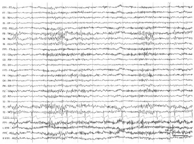
The activation reaction makes it possible to identify local changes (Fig. 2, 3). Epileptiform activity, which appears a short time after closing the eyes, usually concerns non-convulsive forms of seizures (Fig. 4, 5).
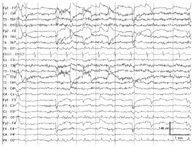
In some cases, people with post-traumatic syndrome (headaches, sleep disturbances) experience the appearance of unilateral generalized discharges of high-amplitude sharp waves and peaks (Fig. 6).
It is characteristic that the test with opening the eyes does not lead to a decrease in low-frequency ß-activity, and sometimes even increases its severity in the anterior parts of the brain. Against this background, flashes of high-frequency ß-rhythm often appear (Fig. 7).
In individuals with a low α-activity index, when closing their eyes, a rebound effect with increased α-activity is possible (Fig. 8). The activation reaction is weakly expressed in individuals with an initially high level of ß-activity, which may be associated with a low index of α-activity.
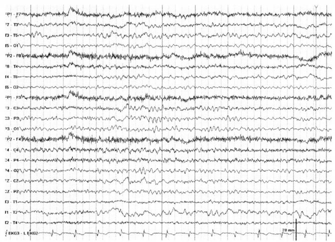
In healthy people, a perverted or paradoxical reaction manifests itself when irritation is applied against the background of drowsiness or shallow sleep and corresponds to the state of awakening. It is believed that the tendency to increase the α-rhythm in response to stimuli is more typical for people with increased basic anxiety (Fig. 9).
Rice. 1. Man, 72 years old. There are no neurological complaints.
When opening the eyes, there is a clearly expressed depression of α-activity from the level of background activity. After closing the eyes, the pattern of bioelectrical activity is restored
Rice. 2. Male, 69 years old. Secondary generalized convulsive attacks, acute cerebrovascular accidents in the left middle cerebral artery.
When opening the eyes, there is a pronounced depression of the α-rhythm. Against this background, a focus of ß-activity in the frontotemporal regions of the left hemisphere and δ-activity in the central-temporal regions of the right hemisphere is clearly visible
Rice. 3. Male, 20 years old. Traumatic brain injury 5 years ago.
Headaches, cerebrovascular accident in the area of the spinal artery on the left. When opening the eyes, depression of the α rhythm is clearly expressed. Against this background, discharges of groups of waves of ß-activity are detected in the fronto-central regions and local δ-activity in the temporal leads on the left
Rice. 4. Girl, 6.5 years old. Speech automatisms.
Against the background of open eyes, generalized discharges (acute slow wave) are clearly visible. After closing the eyes, an increase in paroxysmal activity is detected
The activation reaction can provoke various forms of paroxysmal and epileptic activity. Paroxysmal activity can appear only with the eyes open or closed, as well as at the moment of change - opening-closing of the eyes (Fig. 10).
Local (cortical) epileptic activity persists against the background of desynchronization caused by eye opening (Fig. 11). Epileptic activity, caused by a process in the deep structures of the brain, may disappear with open eyes (Fig. 12).
Rice. 5. Man, 58 years old. Traumatic brain injury.
Headaches, sleep disorder. An artifact is noted at the moment of opening and closing the eyes. Against the background of open eyes, there is a focus of δ-activity in the temporal region with an emphasis on the right and an increase in ß-activity. After closing the eyes, there is an increase in paroxysmal activity
Rice. 6. Male, 30 years old. Traumatic brain injury.
Headaches, sleep disorder. An artifact is noted at the moment the eyes open. Against the background of open eyes, a decrease in the amplitude of α-activity is observed. 2 s after closing the eyes, a generalized discharge of high-amplitude sharp waves and peaks appears in the left hemisphere
Rice. 7. Woman, 65 years old. Parkinson's disease.
When opening the eyes, there is a pronounced depression of the α-rhythm, but clearly expressed ß-activity remains in all leads of the right hemisphere
Rice. 8. Woman, 67 years old. Cyst of the right frontal region, symptomatic partial epilepsy.
When opening the eyes, there is a decrease in the amplitude of biopotentials; when closing the eyes, a short-term episode of activity appears in the occipital-parietal regions
Rice. 9. Man, 50 years old. Irritable bowel syndrome.
A slight decrease in the amplitude of bioelectrical activity with open eyes. Against this background, discharges of high-amplitude ß-activity are detected, and with the eyes closed, a clearly expressed α-rhythm appears
Rice. 10. Man, 50 years old. Cyst in the right fronto-parietal-temporal region, symptomatic partial epilepsy.
When opening the eyes, there is a generalized discharge of groups of high-amplitude θ-waves with an accent in the right hemisphere
Rice. 11. Male, 16 years old. Cyst in the right anterior region, symptomatic partial epilepsy.
Against the background of open eyes, a focus of δ-activity is clearly visible in the frontal regions of the right hemisphere and polyspike-δ-activity complexes in the occipital and temporal regions
Rice. 12. Male, 17 years old. Symptomatic locally caused epilepsy.
Against the background of open eyes, the suppression of generalized discharges of high-amplitude sharp and slow waves and their restoration with closed eyes is clearly manifested
Source: https://cmi.to/%D1%8D%D1%8D%D0%B3/%D1%84%D1%83%D0%BD%D0%BA%D1%86%D0%B8%D0% BE%D0%BD%D0%B0%D0%BB%D1%8C%D0%BD%D1%8B%D0%B5-%D0%BF%D1%80%D0%BE%D0%B1%D1%8B -%D1%8D%D1%8D%D0%B3/%D1%80%D0%B5%D0%B0%D0%BA%D1%86%D0%B8%D1%8F-%D0%B0%D0% BA%D1%82%D0%B8%D0%B2%D0%B0%D1%86%D0%B8%D0%B8-%D0%BF%D1%80%D0%BE%D0%B1%D0%B0 -%D1%81-%D0%BE%D1%82%D0%BA%D1%80%D1%8B%D0%B2%D0%B0%D0%BD%D0%B8%D0%B5/
Alpha wave stimulation
There are several ways to stimulate the body’s wave activity:
- Breathing exercises that allow you to maintain deep breathing saturate the brain cells with oxygen. Systematic breathing exercises promote the generation of alpha waves.
- Listening to stereo sounds with alpha waves is a technique accessible to everyone.
- Yoga and meditation are powerful activators of the α rhythm.
- Hot baths can help you relax and switch to alpha wave production mode.
Features of the EEG pattern in depression
In recent years, in connection with the development of electroencephalography, much attention has been paid to studies of bioelectrical activity in various functional states, including psychogenically caused ones. One of these pathologies is depression.
Depression is basically a disorder of emotions, the brain base of which is the limbic system and the old cortex. The main distinguishing signs of depression are mood disturbances (characterizing the internal emotional state) and affective (external) expressions. The physiological causes of this condition are a decrease in the regulatory function of the cerebral cortex and the promotion of pathological limbic-subcortical regulations to the first place.
The development of depressive states is accompanied by disturbances in the structure of all frequency ranges of the EEG. To a greater extent, these changes relate to changes in the basic rhythm of the EEG alpha rhythm (these are rhythmic oscillations mainly in the occipital and parietal cortex with a frequency of 8 to 13/sec and an amplitude of up to 30-70 μV; registration is carried out in a state of quiet wakefulness, with eyes closed and maximum muscle relaxation).
The alpha rhythm in depression can be significantly enhanced or, conversely, reduced, and the spatial distribution of the rhythm also changes. Changes in alpha activity in depression depend on the clinical picture of the disease.
Thus, an increase in the alpha rhythm index is characteristic of patients with “major depression”, and its decrease (a picture of a desynchronous EEG type with smoothed zonal differences and a predominance of faster potentials in the structure of polyrhythmic activity) is characteristic of dysthymic disorders.

During a functional test with opening and closing the eyes, a significantly smaller than normal decrease in the amplitude and power of the alpha rhythm is observed. that is, a decrease in the reactivity of the cerebral cortex. Against this background, there is an increase in the total power of the beta activity index in all areas of the brain. It is worth noting that the changes described above are not type-specific.
With the use of modern treatment methods (along with pharmacotherapy, biofeedback therapy, light therapy, and psychotherapeutic techniques are used), significant improvements are noted in both the psychophysiological and functional state of the patient.
(syn. alpha rhythm blockade) a change in the alpha rhythm of the electroencephalogram in the occipital regions of the human cerebral cortex by the beta rhythm, observed, for example, when opening the eyes.
The power of rhythm There is hardly a person who would not ask himself the question: “Why am I sleeping?” Active, impatient natures will continue their reasoning like this: “No, really, why? Why should I spend a whole third of my life on this strange and fruitless activity? Twenty years out
A MASTERPIECE OF HARMONY AND RHYTHM A time to cry, and a time to laugh; a time to mourn, and a time to dance. Ecclesiastes After another viewing of the play “Freilekhs,” the great Galina Ulanova said: “This performance is a masterpiece of harmony and rhythm.” Mikhoels was known as a kind of Don Juan. Really,
The concept of rhythm The law of associative montage is established anew each time or intuitively grasped by the director himself. Without solving this problem, the performance as a whole with a multi-episode composition created by an associative-montage method cannot arise. Measure
The duality of rhythm Trying to define the concept of “jazz,” Marshall Sterne calls jazz rhythm the only purely African element of this music[18]. Perhaps this is true only in relation to pre-avant-garde jazz. In the new jazz, the rhythm is already largely moving away from
Realization of rhythm A fundamental and profound change in the metro-rhythmic concept is the most obvious consequence of the jazz evolution of the 60s and 70s. This change was expressed primarily in the final transition of avant-garde jazz to an irregular type of rhythm. Phenomenon
The Principle of Rhythm Having looked at what polarity is, we learned that energy has a range and can flow freely from one extreme to the other. The principle of rhythm describes how it flows: “Everything flows, flows in and flows out; everything has an ebb and flow..." As energy
THE LAW OF RHYTHM The idea of the relationship between the law of rhythm and the law of cycles hardly occurs to the average thinker, but nevertheless, both of them are the eldest twin sons of a single Father-Mother - the World Movement. Every vibration of matter of the four kingdoms of nature
Restoring rhythm...Set the same time to go to bed - if it's eleven every night, then it's eleven. This is the first thing: set a certain time, and soon the body will be able to fall into this rhythm. Do not change this time, otherwise you will confuse the body. Body
PROPERTIES OF RHYTHM By rhythm in photography we mean the natural alternation of compositional elements, their repetition at certain intervals, the order of their combination. The simplest rhythm is the equality of forms. This is clearly demonstrated by the following photographs. Image,
The Alpha Male: An Instruction Manual (10 rules for a woman who has chosen an alpha male as her husband) If you are not afraid of the difficulties of relationships with alpha males, these high-ranking men who are prone to polygamy and do not guarantee a long-term relationship,
Neurotic depression, or depression-passion This is exactly the same depression that can rightfully be called a punishment for sins. Sin destroys a person's soul, becoming a disease. In accordance with the church teaching on the passions, this type of depression occurs as
Endogenous depression, or depression “by nature” (mental illness) Depression of this kind occurs due to biological changes in the human body. Therefore, they are treated using the usual methods available to psychiatrists. And as an example you can
III. Setting the rhythm I always and everywhere travel alone - this adds risk, but provides a very important advantage: strictly following your own rhythm without the need to “adjust” to someone or “drag” them along with you. Actually, any options for cooperation
RESTORING RHYTHM ...Set the same time to go to bed - if it's eleven every night, then it's eleven. This is the first thing: set a certain time, and soon the body will be able to fall into this rhythm. Do not change this time, otherwise you will confuse the body. Body
Pathological deviations
Disturbances in brain wave activity are observed when:
- Oligophrenia. With it, the total activity of alpha rhythms is abnormally increased.
- Epilepsy, which causes disturbances in the frequency and amplitude of wave activity. This is associated with the development of direct or interhemispheric asymmetry in the hemispheres of the brain.
- Hypertension, weakening the frequency of alpha rhythms and increasing beta activity. Circulatory and cardiovascular system disorders always affect brain wave activity. A similar picture can be observed with beta-lactamase activity of bacteria.
- Paranoid schizophrenia, in which the brain generates an increased number of beta rhythms.
- Cysts and inflammatory processes of the corpus callosum. They cause severe asymmetry between the hemispheres, reaching 30%.
- The pathological picture can occur with traumatic brain injuries, congenital or acquired dementia, and delayed psychomotor development in children.
To evaluate alpha rhythms, it is necessary to undergo an EEG from time to time. You can find out the addresses of clinics that perform this diagnostic procedure on the website.
Alpha rhythms in unusual areas
In children and young adults, alpha EEG activity can be detected in the occipital regions (O1, O2), parietal region (Pz) and in the sensorimotor zone (SZ, C4). However, the scalp distribution of alpha activity (especially in the eyes-open state) changes with age, and temporal alpha rhythms become prominent in older adults. Niedermeyer (1997) described a temporal alpha-like rhythm, found mainly in the anterior and midtemporal regions, characterized by mild abnormality, and suggested that it may be a sign of early cerebrovascular disorders. He also mentions that during puberty, this pattern, found in the temporal lobe, may obscure the focus of epileptogenesis. Rhythmic alpha activity may also mask rhythmic paroxysmal bursts without evidence of any acute components.
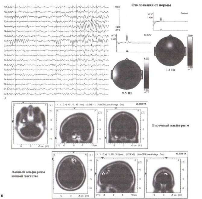
Figure 8. Case of abnormal localization of alpha-like rhythms
Top left: EEG fragment in the eyes-open state. Deviations from the norm of EEG spectra and corresponding topograms are presented. Below are sLORETA images of abnormal rhythm generators.
Our experience of working with healthy subjects and patients allows us to draw the following conclusion: if in an individual patient: 1) the maximum of rhythmic activity within the range of 7-13 Hz is localized in leads different from those mentioned for the norm, 2) the rhythm itself is so noticeable that there is a significant deviation from the norm in both absolute and relative power - then this rhythm can be considered abnormal. In our practice, the maximum distributions of abnormal alpha rhythms were found in the posterior temporal areas (for example, in connection with tinnitus or spinal injury), in the parietal areas of the left hemisphere (in connection with dyslexia), in the middle and anterior temporal areas (in connection with with age-related cerebrovascular disorders). Only in a few cases were abnormal alpha rhythms found in the frontal regions.
An example of the spectral characteristics of the EEG of a patient with abnormal alpha rhythms is presented in Fig. 8. The raw EEG recording shows two types of rhythms within the alpha frequencies: the first with a frequency of 9.5 Hz, located in the left middle temporal region, and the second with a frequency of 7.3 Hz, located in the Fz lead zone. The results of comparison with the normative database and sLORETA images are presented in Fig. 8 top right.
In general, changes in the normal functioning of thalamocortical pathways can result in a number of neurological disorders, such as epileptic seizures and tremor in Parkinson's disease, both of which have rhythmic components. Stimulation or destruction of the relevant part of the thalamus (for example, the ventral nucleus zones) is one of the accepted methods for alleviating tremor, apparently associated with the destruction of the rhythmic activity of thalamocortical networks. Abnormal rhythmic activity of thalamic cells (as shown in patients with implanted electrodes) can be observed in some neurological disorders associated with behavioral disorders that are not necessarily rhythmic in nature. For example, recordings of thalamic neuron activity in patients suffering from chronic pain as a result of sensory deafferentation (so-called phantom pain) demonstrate the presence of abnormal rhythmic bursts of action potentials. In this case, stereotactic lysis of the thalamic nuclei leads to a reduction in phantom pain.
Disadvantages of the method
Excessive wave stimulation can cause negative reactions in the body. So, with an excess of alpha activity, a decrease in concentration develops, the formation of attention deficit, depression, and a decrease in visual clarity. A person begins to need daytime sleep and gets tired faster. In addition, under the influence of alpha waves, suggestibility increases.
The downside of beta wave stimulation is increased anxiety, overexcitement and the development of obsessive-compulsive disorder. There are signs of stress and paranoia. Also, under the influence of such stimulation, muscle tension increases and insomnia occurs. The effect on the cerebellar amygdala and hypothalamus provokes an increase in blood pressure.
When exposed to theta waves, you can develop concentration disorders, depression, hyperactivity, or, conversely, apathy. Under the influence of such stimulation, drowsiness, indifference to life, and increased suggestibility occur.
Therefore, before resorting to any methods of stimulating brain wave activity, it is better to consult with specialists.
What to do?
Now there are a large number of methods for treating the disorder:
- medicinal;
- light therapy;
- psychotherapy.
The use of these techniques improves not only the mental, but also the physical condition of the patient. Biofeedback therapy is quite common and involves constant monitoring of physiological parameters of the body over time. During the treatment course, physiological indicators are adjusted, for example, the functioning of the muscular system, blood circulation, and brain activity. This treatment allows people to cope with fears, panic attacks, and excessive muscle tension.
This method is most often used for headaches, spastic torticollis, stuttering, constantly shaking hands, high blood pressure, impotence caused by a psychological problem, and epilepsy.
Dear readers! We strongly recommend that you consult a doctor before taking medications or self-medicating. There are contraindications.
NORMAL EEG IN ADULTS
Normally, in a person without any special health problems, who is in a relaxed state, the dominant impulse is the alpha rhythm. Its maximum activity is observed in the occipital part of the brain.
In 1/10 of the subjects, the oscillation amplitude is no more than 25 microvolts. Such fluctuations are called low-amplitude, but are considered as one of the normal variants.
In some people, normal α-waves are replaced by pulses with a frequency of 14-18 Hz and an amplitude of 50 μV. Like alpha waves, they are registered in the occipital zone and decrease towards the front (toward the temples and frontal lobe). This activity is also considered normal and is called the “fast ɑ-variant.”
In approximately 0.2% of studies, a “slow alpha variant” develops in the occipital zone - waves characterized by a frequency of 2.5-6 oscillations/sec. and amplitude 50-80 µV. They are not considered abnormal, but mark the border between normal and pathological.