Smell (olfactory bulb), structure and description
The receptors of the entire olfactory system are located in the area of the upper nasal passages. There is a vomeronasal system that includes the vomeronasal nerve, the terminal nerve, and the necessary accessory olfactory bulb in the forebrain.
It is considered as its own representative of the accessory olfactory system in the central nervous system.
According to electron microscopy, the following are structurally combined: olfactory club; supporting cell; central processes of olfactory cells; basal cell; basement membrane; olfactory hairs; microvilli of olfactory cells and microvilli of supporting cells.
The spherical thickening is the olfactory club, from which 12 hairs grow, about 10 microns each. The olfactory hairs are immersed in a liquid medium, which is produced by Bowman's glands.
The presence of these hairs increases the area of close contact of a particular receptor with molecules containing an odorant. An axon is located below the receptor cell. They correspond to the olfactory nerve, which runs at the base of the skull and is embedded in the olfactory bulb.
The sensitivity spectra of different cells overlap. Moreover, more than 50% of odorous substances are common to two of the many olfactory cells.
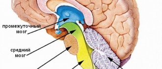
Rear
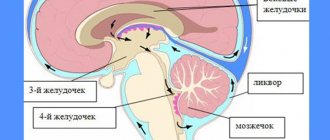
The structure of the hindbrain includes the pons and the cerebellum. The function of the bridge is very similar to its name, since it consists mainly of nerve fibers. The cerebral pons is essentially a “highway” through which signals travel from the body to the brain and impulses travel from the nerve center to the body. Along the ascending pathways, the brain bridge passes into the midbrain.
The cerebellum has a much wider range of capabilities. The functions of the cerebellum are to coordinate body movements and maintain balance. Moreover, the cerebellum not only regulates complex movements, but also contributes to the adaptation of the motor system to various disorders.
For example, experiments using an invertoscope (special glasses that invert the image of the surrounding world) have shown that it is the functions of the cerebellum that are responsible for the fact that when wearing the device for a long time, a person not only begins to navigate in space, but also sees the world correctly.
Anatomically, the cerebellum follows the structure of the cerebral hemispheres. The outside is covered with a layer of gray matter, under which there is an accumulation of white matter.
This department is responsible for the centers of the nervous system, somatic and autonomic reflexes: chewing, swallowing, moderation of salivation. The hindbrain has a complex structure and is divided into two parts: the cerebellum and the pons.
The pons has a cushion-like shape, is white in color and is located above the medulla oblongata. Responsible for muscle contractions and muscle memory: postures, stability, walking. The bridge consists of nerve fibers; it contains centers responsible for functions: chewing, facial expression, auditory and visual.
The cerebellum covers the posterior part of the pons, and the anterior one consists of multiple transverse fibers entering the middle cerebellar peduncle.
The cerebellum is responsible for certain functions:
- muscle tone, their memory;
- body position and coordination;
- motor function;
- implementation of signals in the cerebral cortex.
When pathologies of these sections occur, the following symptoms may occur: excess movements, paralysis, when walking, the legs are spread wide apart, the gait is uncertain and sways to the sides.
Coordination and balance during movements depend on the normal functioning of the hindbrain, and the main function is the connectivity of the forebrain and hindbrain.
Functions of the olfactory bulb (sense of smell)
The coding of odors, as well as their recognition in the very centers of the olfactory system, is based on the special properties of receptors. Each olfactory bulb registers its own electrical response, which is generated by an odorous substance: different odors create their own spatial mosaic from excited or inhibited areas of the tissue of the olfactory bulb.
The central projections that make up the olfactory system are sent to various parts of the brain: the anterior nucleus, the olfactory tubercle, the prepiriform cortex, the periamygdala cortex, and another part of the entire amygdala complex. This is how the olfactory bulb connects different parts of the “olfactory brain” in just a few switches.
And this ensures a close connection between the olfactory system and other sensory systems, which, in turn, affects human feeding, sexual and defensive behavior.
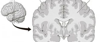
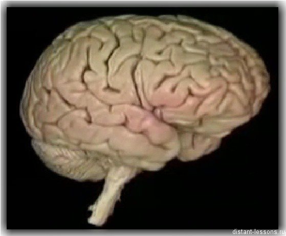
Large hemispheres
The right and left hemispheres make up 80% of the central nervous system; they are responsible for mental activity and consist of gray substance (cortex, basal ganglia) and white substance (fibers). In this part of the brain, the density of neurons is highest, they are closely interconnected. A longitudinal groove separates the hemispheric parts of the brain; in its deep layers there is a white substance - the corpus callosum, consisting of bundles of nerve tissue.
The inner layer of the substantia alba contains the basal ganglia, which regulate muscle tone. These structures also control reflexes, motor reactions, and the automaticity of actions, repeating cerebellar tasks.
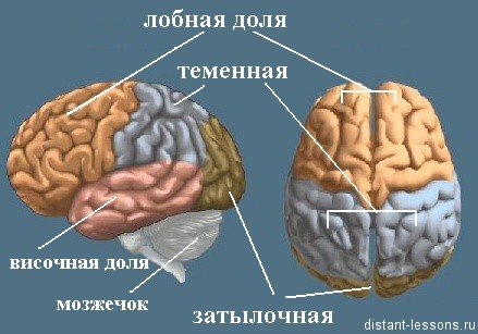
The lobes of the brain are responsible for specific tasks. The occipital lobe of the brain is responsible for vision, and underneath it is the cerebellum, which controls balance and coordination. The parietal lobe of the brain controls body sensations, the frontal lobe controls movement, and the temporal lobe controls hearing, memorization, and speech.
The thickness of the cortical layer ranges from 3 to 5 mm; the white substance of the right and left hemispheres is covered with the cortex. Nerve cells and woven filamentous processes, afferent, efferent types of neurons, neuroglia form the cerebral cortex.
It is formed by layers:
- granular and internal granular;
- pyramidal external and internal;
- spindle cells, molecular.
The cortex occupies approximately 50% of the volume of the hemispheric regions, the size is about 2200 cm². There are grooves on the surface cortical layer; a third of the total area of the cortex is localized in their depths. The furrows have different sizes and shapes.
The cortex is responsible for higher nervous activity and consists of new, old, ancient and intermediate cortex. The neocortex covers more than 95%, this is the outer part of the cortex. The archicortex covers approximately 2%. This old area controls instincts and the emotional sphere. The paleocortex covers 0.6%; this zone controls vegetative functions and the internal physiological balance of the body. The intermediate layer covers 1.6% of the bark.
Thanks to conditioned reflexes, the relationship of the GM with nerve tissues located in different parts of the body, the body functions fully. The cortex synchronizes mental activity, motor skills of internal organs, and the area that analyzes incoming impulses.
The tasks performed depend on the segment of localization of nerve fibers that receive one of the types of impulses (sensory, motor, associative), responsible for perception. The associative segment, responsible for acquiring and processing data from the sensory area, occupies more than 70% of the cortex. It coordinates the activities of sensory and motor areas.
The grooves of the hemispheric surfaces divide them into 5 segments. In front of the central sulcus, where pyramidal cell structures are localized, there is a frontal segment. This is the main motor area responsible for the precision of movement of the body, arms, and facial muscles.
The premotor cortical area is located above the motor area, which controls complex actions that depend on sensory feedback (grasping an object, moving over an obstacle).
Broca's center is localized in the lower part of the left dominant hemispheric zone, responsible for speech; with the activity of the right hemisphere, the emotional coloring of words is maintained. This segment takes part in short-term memorization of speech; thanks to the activity of this center, a person prefers to work with his left or right hand.
The optic lobe is a motor region that controls rapid movement of the eyes when observing a moving object.
The olfactory segment is located at the base of the frontal regions. Thanks to the activity of the olfactory region, a person perceives odors. This zone of the cortex is connected to the olfactory centers in the lower parts of the brain structures responsible for behavior, motivation, learning, memory, instincts (limbic system).
The prefrontal cortical area is a large area of the frontal lobe, responsible for memory and mental activity. Its activity shows how a person perceives the world, thinks abstractly, and how much control he has over himself.
https://www.youtube.com/watch?v=JX3OT2DFyeQ
The parietal lobe is responsible for how a person perceives different sensations through the skin (heat, cold, pain), taste. The correct functioning of this zone helps a person feel the size of his body.
The occipital region is responsible for vision. She perceives the received image from the retina into a meaningful image: a person recognizes words and faces. Below this zone is the cerebellar region responsible for the coordination of movements.
The auditory and vestibular areas are located in the temporal lobe: responsible for the perception and memorization of audio effects. There are centers responsible for speech, sound perception, emotions (fear, anger, joy, pleasure).
The extensive area responsible for speech function is represented by zones of speech formation and understanding.
The two hemispheres coordinate opposite parts of the body. For the majority of humanity (95%), the left hemisphere is responsible for understanding languages, mathematical and logical abilities. The right hemisphere controls visual perception: facial expressions, creative abilities (drawing, music), the emotional sphere.
The activity of the insular lobe appears when a person laughs, cries, listens to music, or empathizes. Here the feelings of aversion to bad smells and dirt are processed.
Basal ganglia
They lie in the deep layers of the white substance, interact with the cortex, and control motor activity. Thanks to this complex nervous structure, movements begin, stop, and their intensity is regulated. The selection of myofibers for movement occurs, and opposing muscle tissues are inhibited.
- caudal and red nucleus;
- shells;
- substantia nigra;
- globus pallidus;
- reticular formation.
Normal functioning of the autonomic nervous system is impossible with dysfunction of the basal ganglia. Thanks to them, a person performs automatic actions, while the central nervous system reserve is not consumed.
The main feature of the large brain is that it is divided into the right and left hemispheres. Each of them is responsible for different functions: for controlling one of the sides of the body, receiving signals from a certain side.
The right hemisphere is responsible for the following:
- the ability to perceive the situation as a whole;
- development of intuition;
- making decisions;
- recognition abilities: pictures, faces, images, melodies.
The left hemisphere is responsible for the functioning of the right side of the body, and also processes information coming from the right side. The left hemisphere is responsible for the following:
- speech development;
- analysis of the situation and related actions;
- ability to generalize;
- logical thinking.
The brain is a very complex organ with many sections. Even a minor injury or inflammation of one part of the brain can cause hearing, vision or memory loss.
Pathologies
A neurologist treats problems associated with dysfunction of the olfactory brain. First of all, neurodegenerative processes should be attributed to pathological problems with this area. They cause serious disturbances in the hippothalamus, as a result of which the patient is diagnosed with Alzheimer's disease. Other diseases include:
- increased feeling of anxiety, which entails damage to the tonsils of the brain;
- epileptic seizures. They occur in both adult patients and children. In this case, partial sclerosis of the hippocampus occurs;
- depression;
- in case of disruption of the tonsils and some olfactory gyri, patients are diagnosed with autism or Asperger syndrome;
- the presence of malignant tumors that pinch the nerve endings in this area;
- problems with the functioning of the circulatory system, which are accompanied by insufficient nutrition of blood vessels in the area of the olfactory brain;
- schizophrenia;
- increased aggressiveness and excitability;
- congenital pathologies;
- impairment of olfactory functions, which may be accompanied by loss of taste, vision or smell.
In the latter case, the patient experiences a delay in the functions of transporting odors to the neuroepithelium in the olfactory area. They can occur against the background of chronic inflammatory or infectious processes, injuries, or exposure to chemical substances on the mucous membranes that can impair the function of the sense of smell.
Article on the topic: Antibiotics for diarrhea in adults and children
To diagnose pathologies, MRI and CT examinations are used. They help to fully assess the state of all parts of the structure. Based on the research results, an effective treatment regimen is selected for the patient. For depression and increased excitability, tranquilizers and sedatives are used. Many disorders in the functioning of this area cannot be treated; the patient is given supportive therapy.
In older people, due to changes in this part of the brain, partial or complete memory loss occurs, tremors of the limbs occur, nerve cells die, etc. Unfortunately, such pathologies cannot be cured. In this case, the patient is constantly monitored by a specialized specialist.