Japanese encephalitis is an infectious disease with a transmissible transmission route. The disease is also called mosquito encephalitis.
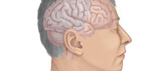
This name is not accidental: dangerous neurotropic viruses are carried by mosquitoes. When infected, the brain and its membranes are affected.
The disease is seasonal. Its outbreaks are observed during the period when new mosquitoes appear. The disease affects not only people, but also some animals: white mice, monkeys, horses, cows, goats, sheep.
History of discovery and study
The disease has a centuries-old history of origin. It was first identified in Japan in 1924, when an infectious outbreak killed a large number of people. Of the 7,000 infected, only 20% survived.
After research conducted by Japanese scientists, the diagnosis was confirmed - Japanese encephalitis. Later it was called summer encephalitis, mosquito encephalitis B, and autumn mosquito encephalitis.
The final name of the complex neuroinfection was fixed in the form of the term “Japanese encephalitis”.
After identifying the ability of encephalitis to occur in various natural centers, scientists concluded that it is spreading not only in Japan, but also in the Pacific countries.
Residents of Russia were also at risk: in 1938, it was described by Russian researchers in the territory of Southern Primorye.
Causes and sources of the disease
In the 30s of the 20th century, the causative agent of Japanese encephalitis was first identified. The virus is part of group B of filterable neurotropic arboviruses. It has special properties.
- despite its small size (no more than 22 mm), it has an increased degree of resistance to environmental influences;
- when boiling, the virus dies no earlier than after 2 hours;
- when exposed to alcohol, ether and acetone, the effect of suppressing the virus is observed after 3 days;
- cold has no effect, the virus can remain in a state of negative temperatures for up to 395 days.
Sources of infection include:
- waterfowl are the main source;
- rodents;
- pigs and horses - in anthropourgic foci;
- poultry;
- bats are a reservoir during the inter-epidemic period.
The main risk group for the disease are children under 10 years of age. The virus is especially dangerous for pregnant women. It can lead to miscarriage in the 1st and 2nd trimesters of pregnancy.

Encephalitis virus transmission
Japanese encephalitis is an infectious disease that affects brain structures - how to reduce the risks?
Japanese encephalitis (JE) is a flavivirus that causes yellow fever, dengue and West Nile fever. The infectious agent is transmitted by mosquitoes and provokes the development of viral encephalitis mainly in Asian countries. The mortality rate among those infected reaches 30%. Half of the patients may develop psychiatric and neurological disorders.
Classification
The disease is more common in certain climatic zones - Southeast Asia, Thailand, India, Primorsky Krai, and the south of the Far East. In warm countries, the activity of the virus is higher, but most local residents have stable immunity to the pathogen. Differences in symptoms among different people are explained by the characteristics of their body.
The following forms of JE are distinguished:
- asymptomatic – up to 3 weeks after infection;
- febrile form - lasts about a week;
- aseptic meningitis - characterized by inflammation of the meninges;
- encephalitis.
Each form is characterized by mild, moderate and severe course.
Seasonality of the disease
Outbreaks of JE incidence are typical for the subtropics and areas with a warm temperate climate. Such areas include some northeastern provinces of China, Japan, and the southern regions of the Far East and Primorye.
At an average temperature above +20 degrees, long-term epidemics (40-50 days) occur more often. The maximum number of cases is observed in August and September - during the hottest period.
In countries with warm climates, surges in incidence are recorded all year round.
Everyone is susceptible to JE. However, the virus poses varying degrees of danger to residents of different regions.
For example, in areas with an increased risk of infection (endemic), in most patients the infection develops latently. The ratio of overt to asymptomatic cases ranges from 1:25 to 1:1000.
Most of the population in these areas is immune to the disease. People who have had it develop lifelong immunity.
In most people who have moved, antibodies to the causative agent of Japanese encephalitis can easily be detected during examination after 5-6 years of living in a new place. During disease outbreaks, representatives of all groups of the population become ill, regardless of age.
The risk of infection is higher for residents of rural areas, especially for fishermen, hunters and farmers, who are often attacked by blood-sucking insects.
Transmission routes
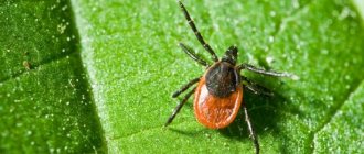
Japanese encephalitis is transmitted transmissibly through mosquitoes of the genus Culex and Aedes. The assumption of transmission of the virus through insects of the genus Anopheles is questionable. The route of transmission of the virus to humans is through mosquito saliva during blood sucking. People do not act as sources of infection, since the intensity of spread of the virus throughout the body is low.
A mosquito infected with a pathogenic microorganism retains the pathogen within itself for a long time. The pathogen accumulates in the insect's body, which makes the mosquito a more dangerous carrier. Females can live more than 3 months.
The virus does not persist in the body of overwintered mosquitoes. Typically, episodes of epidemic formation are associated with the introduction of the infectious agent by migratory birds. Despite the focal prevalence of the disease, it affects not only people who find themselves far from populated areas. Even in populated areas and cities there is a risk of becoming infected with JE.
Pathogenesis
With increased virulence of the pathogen (ability to infect) or with a large number of viral agents, some of them are destroyed at the point of entry into the body. The remaining particles end up in the blood and spread to different organs. The virus can spread along nerve trunks.
After spreading throughout the body, the pathogen ends up in the kidneys, lungs, liver and spleen. The disease is asymptomatic when most of the viral agents die in the blood. If surviving microorganisms multiply and accumulate in tissues, a second wave of disease occurs with pronounced clinical manifestations.
Description of the viral agent
The causative agent of Japanese encephalitis is Japanese encephalitis virus. JEV is common in Australia, Southeast Asia, America and Africa. Related viruses are found on most continents.
Japanese Encephalitis Virus (JEV)
The pathogen has a diameter of 40-50 nm. Many rodents, birds and monkeys are very sensitive to the virus. JEV is inactivated (neutralized) when heated to +60 degrees for half an hour and to +70 degrees for 10 minutes. When boiling, inactivation occurs after 2 minutes. The causative agent of the disease dies in acetone or alcohol after 3 days.
Pathogenesis of the disease
A person becomes infected with encephalitis when bitten by mosquitoes of the species Culex pipiens, Culex trithaeniorhynchus, Aedes togoi, Aedes japonicus. Insects can retain the virus in their bodies for about 3 months. An exacerbation of the disease occurs during the fertile period of female insects.
Article on the topic: Prevention of flat feet in adults - exercises and choice of shoes
Infection occurs mainly in places where there is high humidity. Most often these are swampy areas. Infection of the body is also observed in the evening in parks and open areas with large crowds of people. The development of the disease can be influenced by:
- general condition of a person;
- the rate of occurrence of the body's resistance reaction;
- the amount of virus that has entered the blood;
- virulence and strain properties of the virus.
The pathogen can spread through the hematogenous and neural route. When the body temperature is elevated, the virus can attack more strongly.
Crossing the blood-brain barrier, the viral infection affects the brain parenchyma. In this area, its active reproduction occurs.
During severe manifestations of the disease, the virus reproduces in the nervous system, as well as outside it. The moment it enters the brain, the peak of the disease is observed. During the lesion, neurons die, vascular edema and glial proliferation occur.
Pathogen
Japanese encephalitis virus belongs to the group of arboviruses. It contains RNA and measures 15-22 nm. The virus has a toxatonal form. Its surface consists of a large number of capsomeres.
Features of the pathogenesis of Japanese encephalitis are determined by the routes of introduction and circulation of the virus in the human body. The massive attack of mosquito vectors on humans and, consequently, the inoculation of a large amount of virus in a relatively short period of time lead to significant epidemic outbreaks and their severity.
A number of factors - overheating, malnutrition, chronic parasitic and microbial infections, unsatisfactory sanitary and hygienic living conditions and water sources, unhygienic working conditions, etc. - reduce the protective properties of the body and contribute to the manifestation of the pathogenicity of the neurotropic virus, as well as an acute disease with a short prodrome or without her. The virus inoculated into humans by mosquitoes is partially destroyed at the site of introduction. The undestroyed virus spreads hematogenously throughout the body. The first target of the virus in the human body is the endothelial walls of capillaries and precapillaries of parenchymal organs and the brain. The virus multiplies in the bloodstream and, due to a number of biological factors that contribute to a decrease in the protective properties of the blood-brain barrier, quickly reaches various formations of the brain and has a pathogenic effect on them, multiplying further in the brain parenchyma. Having reached the brain parenchyma and especially some neurons with specific biochemical properties, the virus, due to its neurotropism, begins to multiply rapidly and enters the bloodstream again.
The penetration of the virus through the endothelial walls of capillaries and precapillaries leads to a sharp increase in the permeability of brain vessels and vessels of other parenchymal organs for plasma and blood cells.
Symptoms and stages of disease development
The development of the disease involves several periods. During the incubation period, which lasts from 4 to 21 days, the presence of the virus in the body is detected. Further, the disease has several stages of progression.
From infection to brain injury 4 days
In the first 4 days, a person feels a sharp rise in body temperature (up to 39–41°C). The period of hyperthermia lasts about 10 days. Symptoms of acute development of the disease are also observed:
- chills;
- headache (especially in the frontal part);
- lumbar pain;
- pain in the abdomen, limbs;
- signs of nausea, vomiting.
There is hyperemia of the face, sclera, and upper chest. The person's sweating increases. The pulse may increase to 140 beats per minute. In this case, the pressure increases significantly, and peripheral capillaries may narrow.
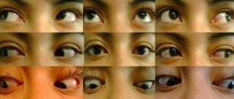
There is increased muscle tone throughout the body. The extraocular muscles work in a disturbed mode.
The height of the disease and exacerbation
At the height of the disease, on the 4th day, foci of brain damage are detected. A person is diagnosed with meningeal syndrome. Consciousness is impaired, and sometimes coma occurs. Mental disorders include delirium, delusions, and hallucinations.
During this period, the patient is forced to be in a supine position due to increased muscle tone, which is extrapyramidal and pyramidal in nature.
More often observed postures are on the back, on the side with the head thrown back, arms and legs bent. Muscle hypertension affects the masticatory and occipital regions.
If there is a deep lesion of the pyramidal system, then spastic hemiparesis, monoparesis, and also paralysis appear.
Choreiform hyperkinesis of the facial muscles and upper extremities, paresis of the facial nerve in the central part with an asymmetrical arrangement of nasolabial folds may appear.
The acute period is characterized by hyperemia of the optic discs. Sometimes bleeding and swelling may occur. The patient risks losing the ability to distinguish colors and react to light. Also, the field of vision is significantly narrowed. During this period, symptoms of bronchitis and pneumonia may appear.
Article on the topic: Treatment of nosebleeds in adults and children
Convalescence period
The convalescence period lasts from 4 to 7 weeks. The temperature is normalized or is in the low-grade stage. Residual brain damage may persist.
Among them are:
- hemiparesis;
- lack of coordination;
- muscle weakness;
- mental disorders.
Clinical picture[ | ]
The incubation period of the disease is from 5 to 15 days. The disease begins suddenly with rapidly increasing general infectious symptoms. Prodromal phenomena may be observed in the form of rapid fatigue, general weakness, drowsiness, and decreased performance. Sometimes diplopia, decreased visual acuity, speech disorders, and dysuric disorders occur. On the first day of illness, febrile fever occurs; from the second day, the illness is accompanied by a feeling of heat or tremendous chills, severe headache, vomiting, severe malaise, weakness, staggering, myalgia, facial hyperemia and conjunctivitis, bradycardia alternating with tachycardia, tachypnea. A coma and petechial exanthema often develop, which is accompanied by changes in consciousness. Frequent signs of the acute period are myoclonic fibrillary and fascicular twitching in various muscle groups, especially on the face and limbs, rough irregular tremor of the hands, which intensifies with movements. In the clinical picture of the disease, several syndromes are distinguished, which can be combined with each other.
Infectious-toxic syndrome
characterized by a predominance of symptoms of general intoxication with a minimum of neurological disorders. The picture of peripheral blood reveals an increase in ESR to 20-25 mm/h, an increase in the amount of hemoglobin and red blood cells, neutrophilic leukocytosis with a sharp shift in the leukocyte formula to the left, up to juvenile forms.
Meningeal syndrome
proceeds as serous meningitis. There are also convulsive, bulbar, comatose (90% mortality), lethargic, amentive-hyperkinetic and hemiparetic syndromes. The severity of the disease and the polymorphism of its manifestations are determined by the characteristics of brain damage. Symptoms of the disease reach their greatest intensity on the 3-5th day from the onset of the disease. Mortality is 40-70%, mostly in the first week of illness. Survivors recover very slowly, with prolonged asthenic complaints.[9]
Diagnosis methods
The first signal for diagnostics should be the presence of a person during an epidemic in an area that is a risk zone. If at this time a person was bitten by a mosquito, then diagnosis must be carried out immediately.
In Russia, cases of the disease are usually detected at the end of August and before the onset of cold weather in Primorye regions.
A reliable way to determine infection is a blood serum test. It notes the presence of specific antibodies. The most effective is the study of paired sera.
The first sample is taken at the very beginning of the onset of the disease, and the second - after 3-4 weeks. By the end of the first week, you can observe the body's reaction. Titles increase over time. A fourfold increase in titers is considered a positive result.
In addition to this type of diagnosis, hemagglutination suppression and neutralization reactions can be used. The PCR method is also used.
Japanese mosquito encephalitis affected area
Treatment of the disease
During specific therapy, which is carried out during the period of fever, the patient is administered immunoglobulin (3-6 ml 3 times a day) or reconvalescent blood serum (20-30 ml).
Positive dynamics are also observed with intramuscular or intravenous administration of hyperimmune horse serum (15–20 ml) in the first week after infection.
During pathogenetic therapy, the patient is prescribed diuretics and agents to remove toxins from the body. Treatment also involves taking corticosteroids, sedatives and drugs to eliminate seizures.
If the patient has a violation of respiratory function or cardiovascular activity, then resuscitation measures are carried out. Treatment is carried out under the supervision of a neurologist and psychiatrist.
Pathomorphology
In the deceased, the membranes and substance of the brain are swollen and congested; the substance of the brain contains hemorrhages and areas of softening. Histological examination reveals perivascular infiltrates, hemorrhages, neuronophagia and degeneration of nerve cells. In the internal organs - plethora, hemorrhage in the serous and mucous membranes, dystrophic changes, especially in the heart muscle, liver and kidneys. The immediate cause of death is damage to the brain stem, edema and swelling of the brain, ITS.
Vaccination of the population
The fight against the manifestations of Japanese encephalitis is possible through vaccination of the population. It is shown mainly to residents of endemic areas, as well as tourists traveling to regions where there is an outbreak of disease.
Vaccine
If an adult lives in a dangerous region, he is often not vaccinated. It is definitely recommended for children.
The vaccine remains effective for 3 years. The vaccination is given from the moment the child reaches one year of age. During vaccination, 3 doses are administered according to a 0–7–30 schedule.
In the accelerated mode, the last vaccination can be done a week after the second. Revaccination should be carried out after 2–3 years.
When traveling to dangerous endemic areas, you must take care of vaccination in advance. You need to get vaccinated no later than two weeks before departure. Vaccination is contraindicated for pregnant women and people with allergies.
Notes[ | ]
- Disease ontology database (English) - 2020.
- Monarch Disease Ontology release 2018-06-29sonu - 2018-06-29 - 2018.
- Uchaikin V.F., Shamsheva O.V.
Guide to clinical vaccinology // M. GEOTAR-Media. - 2006. - 592 p., ill. ISBN 5-9704-0189-7. (pp. 313-314). - Lobzin Yu. V., Belozerov E. S., Belyaeva T. V., Volzhanin V. M.
Human viral diseases // St. Petersburg: SpetsLit. — 2020. — 400 pp., ill. ISBN 978-5-299-00641-4. - Meningitis and Encephalitis Fact Sheet | National Institute of Neurological Disorders and Stroke (unspecified)
. www.ninds.nih.gov. Retrieved October 22, 2020. - ↑ 1 2 V. N. Timchenko, L. V. Bystryakova.
Infectious diseases in children. - St. Petersburg: SpetsLit, 2001. - 4,000 copies. — ISBN 5-299-00096-0. - Japanese encephalitis at www.adventus.info (unspecified)
(inaccessible link). Retrieved November 9, 2009. Archived November 28, 2009. - WHO - Japanese encephalitis (JE) in India (unspecified)
(unavailable link). Retrieved November 9, 2009. Archived July 24, 2009. - ↑ 12
Japanese encephalitis at health.centrmia.gov.ua (Russian) (inaccessible link). Retrieved August 14, 2009. Archived January 24, 2010. - E.P.
Shuvalova. Infectious diseases. — 5th edition. - Yaroslavl: Medicine, 2001. - P. 439-451. — 624 p. — 5000 copies. — ISBN 5-225-04578-2. - Patterns of persistence of the Japanese encephalitis flavivirus and experimental development of diagnostic and prophylactic drugs
- Japanese encephalitis on www.eurolab.ua (Russian) (inaccessible link). Retrieved August 14, 2009. Archived June 5, 2009.