- Treatment of chronic subdural hematomas
A hematoma on the head after a bruise or blow is a serious and potentially life-threatening condition that often requires immediate treatment. Brain damage can have a lasting impact on a person’s emotional, mental state and overall health.

Most often, a hematoma on the head appears as a result of injury.
The brain is a protected organ of the body, but in case of accidents this protection is often not enough. One of the most severe traumatic brain injuries is intracranial hematoma (bleeding).
Causes of pathology
The brain is located inside the skull, it is enclosed in soft membranes and washed with blood and brain fluid. They occupy a volume of 1.5–2 liters. These fluids are incompressible, so if blood begins to pour into the cranial cavity during a rupture of a vessel or an impact, a displacement of the brain occurs - dislocation.
Of the total number of head injuries, hematomas make up no more than 10%. However, they are the ones that account for the highest mortality rate. The most serious cases during hospitalization occur in unconscious victims.
In addition to traumatic etiology, hemorrhage can be vascular or iatrogenic in nature, occurring due to hemophilia, craniocerebral disproportions, sepsis, brain tumors, intoxication, etc.
Therapy
Treatment of intracerebral hematoma occurs in two ways: conservative or surgical. The neurosurgeon chooses which one should be used. If the size of the hematoma is less than three centimeters, it is treated medicinally, using drugs that reduce vascular permeability. At the same time, thromboembolism and high blood pressure are prevented. Surgical treatment is carried out when its diameter is larger, when a person’s consciousness is impaired and symptoms are pronounced. The neurosurgeon performs transcranial removal of the hematoma, endoscopic evacuation, or stereotactic aspiration. If there are multiple hematomas, only one of them, which is large, is removed.
Types of hematomas
The brain compressed by intracranial hemorrhage can move, as a rule, to the foramen magnum and the foramen of the tentorium cerebellum. At this level in the brain stem there is a respiratory center, sensory and motor pathways, as well as a single pathway for the outflow of cerebrospinal fluid from the brain.
The consequence of impaired outflow in the cranium will be an increase in excess volume of cerebrospinal fluid.
There are two types of hematomas on the head
There are two types of intracranial hemorrhage:
| Type of hematoma | Description |
| Epidural | Formed over the dura mater of the brain due to arterial bleeding (mainly due to damage to the middle meningeal artery), usually in the place where the blow occurred. Characterized by a progressive increase in the size of the lesion |
| Subdural | Formed under the dura mater of the brain due to rupture of the cortical bridging veins, usually on the side of the skull opposite to the impact |
In some cases, both types of pathology occur, the symptoms of which may be unexpressed.
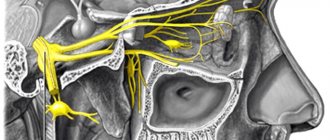
Epidural hematoma
This is an accumulation of blood between the inner surface of the skull and the periosteum (the outer layer of the dura mater). With this type of disorder, there is most often a history of trauma and an accompanying skull fracture.
Typically, the source of bleeding is the rupture of the branches of the meningeal artery (in most cases, the middle meningeal artery) by the sharp edges of a skull fracture.
Most often, the formations are limited by cranial sutures and have a biconvex shape. They can have a volumetric effect and cause herniation of brain tissue.
In a fairly large number of cases, this type of hemorrhage is unilateral, but bilateral and multiple epidural hemorrhages occur.
Subdural hematoma
This is an accumulation of blood in the space between the dura mater and the arachnoid membrane of the brain. Occurs in any age group of patients:
- children: hemorrhages usually occur as a result of accidental injury, most often they are bilateral; interhemispheric subdural lesions are predominantly observed in children with non-road traffic injuries;
- young people: the most common cause of hemorrhage is road traffic accidents; in adult patients, unilateral hematomas are more often formed;
- elderly people: the appearance of bruises on the head is usually associated with a fall.
Typical sites of localization of hemorrhages are the middle cranial fossa and the frontoparietal regions.
Chronic subdural hematoma
Chronic subdural hematoma is a volumetric polyetiological intracranial hemorrhage, which is located under the dura mater, causes general and/or local compression of the brain and has (unlike acute and subacute forms) a delimiting capsule.
The capsule usually appears two weeks after the hemorrhage. These characteristics determine all the features of cerebral pathophysiological reactions, clinical course and treatment tactics.
Intracerebral variety
An intracerebral hematoma of the brain is life-threatening. Its symptoms appear immediately after the blood vessels rupture. Most often detected in the frontal and temporal lobes. The shape of the hematoma is spherical.
The soft tissues of the brain are saturated with blood. Symptoms appear immediately, as this process affects nervous tissue. Speech disturbances appear, strength in the limbs is lost, the face becomes distorted. In some cases, the sensitivity of certain parts of the body is lost, coordination and vision are impaired. Mental disorders appear.
Even a small hemorrhage puts pressure on neighboring tissues. Intracranial pressure increases, and a displacement of brain structures appears. Depending on the location, trembling of the eyeballs, squinting, difficulty swallowing, and disturbances in respiratory and heart rhythm may occur. Body temperature may rise and loss of consciousness may occur. In some cases, such a violation leads to death.
Symptoms
Symptoms of hemorrhages on the head may not be expressed, most often it manifests itself in the form of a headache. You may also experience an unpleasant sensation such as slight dizziness when changing your body position or moving vehicles in front of your eyes.
One of the main symptoms of the pathology is headache
Other possible signs of pathology:
- vomiting, nausea;
- drowsiness;
- confusion;
- loss of speech or slow speech;
- weakness in the limbs on one side of the body;
- difference in pupil size.
In the early stages of injury, an accurate diagnosis can often only be made through special studies. Therefore, in case of any head injury, it is recommended to immediately contact a neurologist or neurosurgeon.
Diagnostics
Before starting to treat the pathology, a diagnosis is carried out. The physical examination of the patient includes:
- general examination: consists of assessing the location and area of damage, as well as the color of the injury area;
- palpation: assessment of pain in the area of damage, tension of the subcutaneous formation, determination of signs of fluctuation.
Computed tomography has important diagnostic value
At the outpatient level and during emergency hospitalization of the patient, the following diagnostic examinations are indicated:
- radiography of the skull in two projections;
- computer and/or magnetic resonance imaging of the brain.
Additional diagnostic tests:
- general blood analysis;
- X-ray of the cheekbones and nasal bones.
Complications and consequences
Sometimes internal hematomas lead to compression of the brain and slow loss of consciousness. As the lump diverges, it may begin to fester if there is an infection. During this, the person’s body temperature rises, the pain of the bruise increases and a change in its color is noted. Instead of becoming lighter, it becomes brighter and more saturated. Extensive and deep injuries can lead to a coma and loss of vital human reflexes.
A person who has suffered a head injury must be examined and have a magnetic resonance computed tomography scan, even if there are no external signs of a hematoma. Deeply located damage to the blood vessels of the brain, which is not visible from the outside, can pose a real threat to life.
Source
A hematoma on the head after a bruise or blow is a serious and potentially life-threatening condition that often requires immediate treatment. Brain damage can have a lasting impact on a person’s emotional, mental state and overall health.
Most often, a hematoma on the head appears as a result of injury.
The brain is a protected organ of the body, but in case of accidents this protection is often not enough. One of the most severe traumatic brain injuries is intracranial hematoma (bleeding).
Treatment
The goal of treatment is to reduce systemic manifestations of the inflammatory response.
If necessary, apply a bandage to the site of impact
In mild cases, with subcutaneous bruises, a tight bandage is applied. In the presence of intense subcutaneous hemorrhage, it is necessary to open it, which is performed under local anesthesia.
Typically, the hematoma is semi-liquid and can be removed through a small hole using a flexible endoscope. The endoscope is equipped with a video camera, which allows you to take photos if necessary.
Treatment of chronic subdural hematomas
With this type of pathology, spontaneous resorption is often observed, which allows for conservative therapy. This is only possible in the absence of neurological deficit and with periodic monitoring using computed tomography.
Main indications for conservative treatment:
- small amount of damage with the patient remaining in the clinical phases of subcompensation and compensation;
- spontaneous reduction in the volume of damage during repeated studies, if there is a positive trend in neurological symptoms;
- patient's refusal to undergo surgery.
Treatment at home
At home, treatment of a hematoma on the head after a bruise can be carried out only in mild cases. Therapy consists of applying cold to the injury site in the first hours after the injury. In the future, it is necessary to apply external agents to the area of impact to promote the resorption of the resulting bruise.
It must be remembered that the symptoms of the pathology may not appear immediately, so you should carefully monitor changes in your physical, emotional and mental state.
It is also important to tell family members about the injury, as hemorrhage may be accompanied by further memory loss.
The adequacy of the therapy is assessed by stabilization of the general condition and regression of external manifestations of the injury.
Complications and consequences
A hematoma in the head can provoke various complications. It can cause dangerous brain damage. The main symptoms of its presence are pain, unconsciousness, and focal signs. Focal symptoms include speech and visual disorders, motor dysfunction.
After a head blow, even when there is no bleeding or external damage, it is necessary to undergo an examination and a CT scan. This pathology, which forms as a result of vascular damage, becomes a threat to life and can provoke various complications and adverse consequences.
A hematoma can provoke motor dysfunction, frequent headaches, inadequate perception of what is happening, speech disorders, and memory impairment. The duration of rehabilitation is individual and depends on various factors. To prevent the occurrence of adverse consequences, it is necessary to promptly seek help from doctors.
Hemorrhage in the head can be caused by various reasons: accident, accident, difficult childbirth. As a result, various adverse consequences often appear.
A timely examination, therapy under medical supervision, and a course of rehabilitation make it possible to return to your previous lifestyle and restore the normal functioning of the body.
Source
It is very easy to get a hematoma, you just have to hit something hard. And if subcutaneous hemorrhages on the arms and legs do not pose any threat to life, then a hematoma on the head is dangerous because it compresses the brain and negatively affects its functioning.
Head hematomas occur due to trauma. This could be a bruise, blow, pinching or birth injury. The treatment tactics for such hemorrhages depend on how severely the vessels are injured, where exactly the bruise is localized, and also what its size is.
Forecast
With the epidural form of the pathology, in case of timely treatment, the prognosis is good. The prognosis of subdural hemorrhage varies widely and depends on the size of the affected area and the duration of the injury.
It is also important to consider that intracranial hematomas in most cases are accompanied by other serious injuries: subarachnoid hemorrhage, diffuse axonal damage to the brain, contralateral contusions, etc.
After any significant impact to the head resulting in loss of consciousness, or in the event of any signs that may indicate the presence of a hematoma, you should consult a doctor.
Subdural variety
The most common of these injuries is subdural hematoma of the brain. The consequences of this hemorrhage can be fatal. Such hematomas can spread widely throughout the tissues. The cavity has a crescent shape.
In the acute form, a person may not regain consciousness after an injury. All vital signs are gradually deteriorating. Consciousness is impaired. Deviations affect the respiratory and cardiovascular systems. If the patient does not lose consciousness after the blow, he may have a severe headache, speech disorders, pyramidal insufficiency, and convulsions.
If the hematoma is a subacute form, at the time of hemorrhage the person loses consciousness for a couple of minutes. Then he comes to his senses. A person remains conscious for up to 14 days. At this time, a moderate headache appears. The person feels a little weak and tired. The pressure may rise slightly. After some time, convulsions and loss of consciousness appear. The person may fall into a coma. If breathing and heartbeat are impaired, the patient may die.
Chronic varieties differ only in the duration of the development period. A few weeks after the injury, symptoms of cerebrovascular accident appear.