Forms the posteroinferior part of the brain skull. It consists of a basilar (main) part, lateral parts and occipital scales. All these parts surround the foramen magnum, foramen magnum, through which the cranial cavity communicates with the spinal canal.
Basilar part
located in front of the foramen magnum. By the age of 18-20 years of life, it fuses with the body of the sphenoid bone into one whole. The medullary surface of the basilar part has the shape of a groove and, together with the body of the sphenoid bone, forms a platform inclined towards the foramen magnum - the slope. The groove of the inferior petrosal sinus runs along the lateral edge of the basilar part. On the lower surface of the basilar part there is a well-defined pharyngeal tubercle.
Lateral part
the steam room has an irregular shape and, gradually expanding, passes posteriorly into the occipital scales. On the lower surface of each lateral part there is a well-defined ellipsoidal occipital condyle. The condyles, with their convex surfaces, connect with the superior articular fossae of the atlas. Through each lateral part, above the condyle, passes the hypoglossal canal, which contains the hypoglossal nerve. Immediately behind the occipital condyle is the condylar fossa. At its bottom there is a hole for the venous outlet - the condylar canal. Lateral to the occipital condyle there is a jugular notch. Posteriorly, this notch is limited by the jugular process directed upward. Next to the process on the cerebral surface of the lateral part there is a well-defined groove of the sigmoid sinus.
Occipital scales
It is a wide plate with a concave inner surface and a convex outer surface. In the center of the outer surface there is an external occipital protrusion (tubercle), from which the external nuchal crest descends down the midline to the posterior edge of the foramen magnum. From the occipital protuberance to the right and left there is a downward curved superior nuchal line. Parallel to the latter, approximately at the level of the middle of the external occipital crest, the lower nuchal line extends from it in both directions. In addition, above the external occipital protrusion there is a less noticeable highest nuchal line.
On the inner, cerebral, surface of the occipital scales there is a cruciform elevation formed by grooves that divide the cerebral surface of the scales into 4 pits. The center of the cruciform eminence projects forward and forms the internal occipital protrusion. At the level of the protrusion, to the right and left there is a groove of the transverse sinus, which passes into the groove of the sigmoid sinus. Upward from the internal occipital protrusion there is a groove of the superior sagittal sinus, which continues into the groove of the same name in the parietal bone. Inferiorly, the internal occipital protuberance narrows and continues as the internal nuchal crest, which reaches the foramen magnum. The edges (lambdoid and mastoid) of the upper and lateral parts of the occipital scales are highly jagged; in these places the occipital bone is connected to the parietal and temporal bones.
Hello. My skull has been deformed since birth. Diagnosis of trigonocephaly. The forehead looks very strange and repulsive. I have never seen anything like this in other people in my life. Now I am 36 years old, and I still find it very difficult to communicate with people. Please tell me, are there methods for correcting this problem in adults? And, in general, are there any precedents in this area? Thanks in advance for your answer.
Dear Oleg Sergeevich! Trigonocephaly is a form of craniostenosis. Surgical treatment of the disease is carried out in early childhood, this ensures normal development of the brain and prevents secondary deformations of the skull. As for adult patients, the possibilities of neurosurgery in this case are limited. Contact a neurosurgeon for a face-to-face consultation; after examination and the necessary examination, the doctor will be able to answer your questions.
Hello, dear doctor. Tell me how to find out whether a deformation of the skull interferes with the normal functioning of the brain? I have a lot of abnormalities in my skull. The forehead looks like a square cap. And dents on both sides in the temporal lobe of the head. What is this? And is it possible to remove this Andrey ↓↓ 0 Aprelkov Andrey Ivanovich (0 / 1) 04 Dec 2017 17:04 ““ #55 Reply
Hello, I am 15 years old and have had hydrocephalus since childhood, because of this my head is slightly uneven, the left side of the head is larger than the right, very noticeable, even the ear on the left side is different from the right, can I somehow fix this?
Dear Oleg! Patients under the age of majority can be provided with medical care only with the consent of their legal representatives - parents or guardians. Surgical correction of the shape and size of the ears is possible in some cases. As for changes in the shape and size of the head associated with hydrocephalus, due to objective reasons, surgical correction is not performed. You need to be under the supervision of your attending neurologist.
Hello! I am 33 years old. The problem is that when I look at myself, I understand that the right side of my face is slightly different from the left. It’s as if the left one looks a little better in terms of health than the right one. In the photographs it is very noticeable that the face seems to be crooked. I’ve noticed this before for about 25 years + - several years. But something has been bothering me lately
Dear Stepan Alexandrovich! Slight asymmetry of the right and left halves of the body is not a pathology. At your age, the growth of skeletal bones is complete, and it is impossible to say in absentia what is causing your symptoms. Consult a maxillofacial surgeon; after an examination and, possibly, the necessary examination, the doctor will be able to answer your questions.
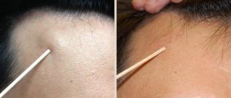
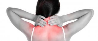
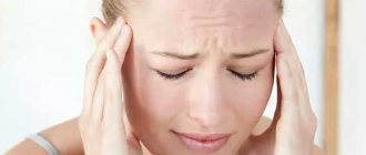
Sometimes, when talking about pain in the back of the head, patients say that their occipital protuberances hurt and draw the doctor’s attention to this. What can this condition mean, and which doctor should you contact if your occipital protuberances hurt? Most often, complex causes are involved: otogenic, vascular, associated with meningeal symptoms.
A little anatomy
In order to know why the occipital protuberances hurt, you need to remember their anatomical structure. The occipital protuberances themselves are usually simply convexities of the lateral surfaces of the unpaired occipital bone; below and on the sides they are delimited by the mastoid processes of the temporal bones.
Occipital bone internal view
It is these processes that are responsible for the development of many types of pain in the back of the head, especially if the occipital protuberance hurts on one side. This most often indicates inflammation, which is caused by a “problem” in the temporal bone.
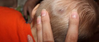
Sometimes the pain occurs higher, in the very back of the head, which has an aching character. It is then that patients, in order to show the nature of diffuse and aching pain, simply put their hand on the back of their head. Thus, the pain is diffuse, and this is typical, for example, for tension headaches, and if patients show the place of pain with one finger, then this most often indicates otogenic causes.
Definition of the word “Bump” according to TSB:
Shishka - Shishka (&Scaron.i&scaron.ka) Karol (b. 19.3.1906, Serenča, Yugoslavia), Czechoslovak surgeon, academician of the Academy of Sciences of Czechoslovakia (1965). Member of the Communist Party of Czechoslovakia since 1945. In 1932 he graduated from the Faculty of Medicine at Karpov University (Prague). Participant of the Slovak National Uprising of 1944. Since 1950, head of the surgical clinic of the University. Komensky and at the same time (since 1955) director of the Institute of Experimental Surgery of the Slovak Academy of Sciences. Main works on the problems of experimental and clinical surgery of the heart and lungs, arts. blood circulation Foreign member of the USSR Academy of Sciences (1971) and the USSR Academy of Medical Sciences (1969), honorary member of the Society named after. J. Purkyne, Society of Polish Surgeons. Vice-president (1961-65), president (1970-74) of the Slovak Academy of Sciences, vice-president (1970-74) of the Academy of Sciences of the Czechoslovak Socialist Republic. Member of the Central Committee of the Communist Party of Slovakia (1958-62), member of the Central Committee of the Communist Party of Czechoslovakia (1962-1976). Member of the Slovak National Council (since 1969). Awarded 5 orders and medals.
Cone - Cone (biol.) a collection of spore-bearing spikelets (strobili) at the ends of the shoots of coniferous plants. often the term "Sh." used as a synonym for strobila.
Occiptalgia of otogenic origin
This tricky term is simply translated, namely: pain in the back of the head, which developed as a result of a sore ear. As a rule, the patient knows that he has chronic otitis media, which has a habit of worsening in cold, damp, and windy weather. Therefore, if the occipital protuberances hurt due to the weather, this is one of the signs of chronic otitis media, as well as mastoiditis.
The thing is that in the mastoid process, both on the right and on the left, there are cavities filled with air, which can “belatedly” respond to changes in atmospheric pressure. Irradiation from these zones of chronic inflammation may radiate posteriorly, and in this case, the subcutaneous nerve trunks may be involved in this process.
The mastoid process is highlighted in the photo.
Thus, pain may occur in the occipital protuberances, which intensify when pressed. This can be a means of differential diagnosis, distinguishing painful sensations arising from the ENT organs from neuralgia. What are the signs of neuralgia?
Notes
- Pine cone jam - photo recipe for preparing for the winter (Russian). nazimu.info. Retrieved June 7, 2020.
- Baturitskaya N.V., Fenchuk T.D.
Amazing experiments with plants // Biology, No. 42, 44-47/1999; 2/2000 - Plant life. In 6 volumes / Ch. ed. Al. A. Fedorov. - M.: Education, 1978. - T. 4. Mosses. Moss mosses. Horsetails. Ferns. Gymnosperms. Ed. I. V. Grushvitsky and S. G. Zhilin. — P. 280.
- Geraldika.ru. Coat of arms of Krasnochikoysky district
. Retrieved March 10, 2013. Archived March 11, 2013.
Vascular occiptalgia
In addition to neuralgia, the source of pain impulses, for example, when the occipital protuberances of one or both sides hurt, are blood vessels. And not venous, but arterial, which can change their lumen. These vessels have the property of sharply undergoing spasm, which is relatively asymptomatic and painless, and then expand compensatoryly, more than it should. This is an expansion and can cause pain, usually in one side of the head. This attack is characteristic of migraine, or hemicrania.
True, in this case, most often there is a one-sided localization, but there is a situation where attacks of occipital migraine occur, which can be symmetrical in nature.
If the occipital protuberances hurt, and this pain is of vascular origin, then what to do? In this case, drugs that help narrow the blood vessels again usually help. Such drugs include, for example, caffeine sodium benzoate, Coficil, Askofen, Caffetamine, Caffetin, Citramon, that is, all drugs that contain caffeine.
Finally, the occipital protuberances hurt and during an attack of high blood pressure, this may be a symptom of a crisis. Often, a hypertensive crisis begins with pain in the back of the head. If the occipital protuberances hurt, especially at the end of the working day, then this may be a signal to measure your blood pressure level.
What if intracranial pressure rises?
This reason can also be quite significant. But if hypertensive-hydrocephalic syndrome is the cause of pain, then this pain soon spreads throughout the head. After all, an increase in cerebrospinal fluid pressure causes the receptors of the meninges to react, and as a result we get a diffuse headache, the characteristic companions of which are usually photophobia, or photophobia, intolerance to loud sounds and, in general, all irritants.
In conclusion, we can list several more reasons. Thus, headache and heaviness in the back of the head are characteristic of tension headaches, of infectious intoxication syndrome if the pathogen has a pronounced neurotropic effect, for example, with influenza. And sometimes patients who have a sedentary job may simply tell doctors, for example, that their “occipital protuberances are swollen and painful.” As a rule, this secondary symptom occurs against the background of exacerbation of cervical osteochondrosis. But if you complete a course of massage, go swimming, and undergo several sessions of physiotherapy, then this pain recedes.
Diagnosis and treatment
For such pain, only a qualified specialist can prescribe the correct treatment. If these pains are caused by neuralgia, then self-medication is contraindicated. In this case, the following diagnostic methods are prescribed:
- computed and magnetic resonance imaging;
- radiography.
Depending on the causes of the disease, the doctor determines what to do if the occipital protuberances hurt, then there may be a nerve block method, drug treatment, antidepressants, massage and other popular methods of treating the affected areas of the nerve and surrounding soft and bone tissues.
There is no need to worry too much if you notice a lump or ball on the back of your head. Most of them, as a rule, do not pose any danger. However, it is recommended to visit a doctor after the appearance of such a formation, especially if it causes concern, in order to establish a correct diagnosis.
Painful balls or bumps on the back of the head that appear suddenly within a day or two can be caused by infection or injury. If the formation was caused by an infection, the area around it will be red and warm to the touch. You can always consult a doctor for advice. A lump in the neck or back of the head most often represents:
- Injury.
You may, for example, hit something and unconsciously not notice it or soon forget it - Enlarged lymph nodes
. Usually indicates that you have an infection. - Skin tumors
are harmless soft and less often hard growths on the skin that are a cosmetic defect. - Cysts are
fluid-filled bumps that may disappear on their own. They usually do not require any medical treatment.
But there are other reasons, which are discussed in more detail below.
Types of tumors on the head and their signs
A lump on the back of the head may be small in size and not cause discomfort, but at the same time pose a danger to human health. On the Internet you can find a lot of information about what kinds of soft tissue tumors there are, how to deal with them at home with the help of medications or traditional medicine. The use of such approaches poses serious dangers. Even if the problem can be resolved, there is no guarantee that it will not return. Also, health care workers often send tumors for additional study to rule out the possibility of cancer.
Hemangioma
One of the most dangerous types of neoplasms. It can occur on any part of the head, including the back of the head. Appears against the background of a malfunction of the blood vessels. It looks like a bright crimson or purple bulge with clear edges; on its surface you can see the vascular network. The conglomerate can cause disruption of the trophism of the tissues surrounding it. It also tends to grow to impressive sizes and degenerate into a malignant form. Treatment is carried out strictly under the supervision of a surgeon or oncologist. Most often, surgical removal of the hemangioma is required, after which the biomaterial is sent for histology.
Allergic reaction
Swelling on the skin of the back of the head can occur as a result of the influence of an allergen on the body.
The danger is posed by aggressive cosmetics, household chemicals, hats and synthetic bedding. In this case, the formations are usually small in size, numerous, do not hurt, but itch. Therapy consists of eliminating exposure to the allergen on the body, using antihistamines, and strengthening the patient’s immunity. A special place in this group of causes is occupied by an insect bite. Bees, flies, and mosquitoes can provoke a violent reaction of the body in the form of severe swelling of soft tissues. Typically, these bumps are painful, hot to the touch, and itchy. When pressing on the bite site, a clear liquid or exudate may be released. It is better not to self-medicate, especially if it is not known who bit. We cannot exclude the possibility of the presence of a tick that has crawled under the skin, and only a doctor can get rid of it.
Fibroma and sarcofibroma
Fibroma is a benign tumor, the basis of which is connective tissue. Its danger lies in its ability to grow to impressive volumes and turn into a malignant analogue - sarcofibroma. Initially, formations appear against the background of endocrine disorders, metabolic disruptions, and hormonal changes in the body. The lump itself is hard to the touch and does not cause any discomfort. It only hurts when there is mechanical damage. Treatment of fibroids involves monitoring its growth by an oncologist. If the tumor grows quickly or is in the way due to its location, it is removed.
Lipoma (fat) on the head
A benign tumor often appears in women after 30 years of age. This is facilitated by frequent changes or disturbances in hormonal levels and problems with fat metabolism. The formation is soft, mobile, round in shape, does not hurt when pressed and rarely bothers you. When located at the back of the head, it can cause discomfort due to frequent use of a comb, wearing a headdress, or simply the presence of an aesthetic defect. The treatment method is selected by the doctor depending on the size of the lump and its growth rate. This can be excision of the mass, its removal with a laser, or the introduction of a conglomerate of special anti-lipid drugs into the body.
Pimples and bites
Such bumps are rarely isolated. Most often, they appear unexpectedly, do not reach significant volumes, and go away on their own within 4-7 days. Usually the formations are itchy - scratching leads to infection and inflammation. In this case, the discomfort increases, and the local temperature may rise. In the absence of care or weak immunity, an abscess forms. You can try to deal with such bumps yourself by regularly treating the affected area with an antiseptic. If the condition worsens or there is no effect within 2-3 days, it is recommended to consult a doctor.
Bruise or injury
A blow to the head is the most common cause of a bump. Typically, such consequences of injury do not cause serious problems and do not require special treatment. It is enough to apply cold to the sore spot as soon as possible and leave the compress for a quarter of an hour.
A doctor's help is necessary if:
- nausea and vomiting occurred;
- the child has a problem;
- after the blow there was loss of consciousness;
- there is dizziness or signs of incoordination;
- the temperature has risen.
You will learn about methods for eliminating headaches after a blow from this article.
A small hematoma will go away on its own in 3-5 days. To speed up the process, you can use “Troxevasin”, “Rescuer”, “Troxerutin”, and heparin ointment. All these products are good at relieving swelling, stimulating the resorption of bruises, restoring blood vessels, and eliminating pain.
Wart on the head
The most common benign tumor, which is equally often diagnosed in people of different ages, gender, and social status. Most often it occurs in people with impaired metabolism, against a background of stress, when tissues are damaged by an inflammatory process or their frequent mechanical irritation. A wart on the head is rarely dangerous, but is an aesthetic defect. The bumps can take on different appearances, reach significant sizes, and spread over the surface, occupying a large area of the skin. Treatment must be coordinated with a dermatologist.
Atheroma on the head
The formation looks like a lipoma, but has a yellowish tint. It appears due to disruption of the sebaceous glands and blockage of their ducts. A dense lump with a smooth surface begins to cause pain and significant discomfort as it grows. The tumor must be removed surgically or using a laser. The resulting material is sent for histology, but it rarely contains cancer cells.
Causes
If a lump under the skin in the back of the head causes pain, then it is difficult not to notice it and not think about it. People can sometimes live with painless growths for years, paying attention to them only occasionally, for example, when combing, styling their hair or cutting their hair. Moreover, often the same reason may be accompanied by unpleasant sensations, or may not physically bother you at all.
Main reasons include:
Lipoma
Lipoma (fat)
This is a tumor that mainly consists of fat cells. It is a capsule that usually feels like a soft and mobile lump. The reasons for its occurrence are unknown, although it is believed that they may be genetic.
Symptoms include:
- A small bump that may become larger over time
- Feels like a soft and doughy ball
- Pain may occur if it continues to grow
Atheroma
Atheroma
This is a small cyst that appears when the sebaceous gland is blocked. They are usually harmless. However, it is important to have a doctor examine these growths, especially when they begin to change in appearance, size, or shape.
Symptoms of atheroma include:
- They may ooze, leaving a dry crust over time.
- The scalp is flaky in this area
- Sometimes they may encyst and turn into hard lumps
- May become inflamed, painful, swollen and red
Hair (follicular) cyst
Pilar cyst
This is a type of cyst that forms in the area of the hair follicles. It is a type of the above-mentioned atheroma, along with an epidermal cyst.
The pilar cyst is filled with keratin, which helps your hair grow. These lesions mainly affect the scalp, but can also be found in any other part of the body where hair grows.
May develop into cancer in rare cases. Therefore, many doctors prefer to remove them to be on the safe side.
Symptoms include:
- Presence of a hard lump on the back of the head
- Inflammation
Ingrown hairs
Lumps due to ingrown hairs
The cause of the appearance of a hard ball can be as simple as. These growths can appear after shaving, cutting or damage to the hair as a result of the cut hair continuing to grow inside your skin next to the follicle.
The problem is common among people who regularly shave their hair, in this case the back of their head. Ingrown hairs should be examined for signs of infection, which can cause them to grow to the size of a noticeable bump. Common symptoms of its presence include:
- Redness
- Painful bump that looks like a pimple
Gout
Tophi with gout on the head most often form on the auricle.
But there are also atypical cases involving other areas of the head and neck () This disease is characterized by an increase in the level of uric acid in the blood. This is a form of arthritis that also causes joint pain. Uric acid forms crystals, which then accumulate in your body.
Clusters of such crystals are called tophi or gouty nodes. They can form in areas such as the head, fingers, joints, and also on the earlobes. Tophi are known to be extremely painful, but the pain usually goes away when the level of uric acid in the blood begins to decrease.
Common symptoms include:
- Joint pain
- Swelling and redness in the affected area
- Fever
- Problems walking
Keloid acne
The condition is also called papillary dermatitis. But the term “keloid acne” is not entirely correct, since it is not acne, but a severe form of folliculitis. Moreover, histologically the lesions are not keloid scars. They form almost exclusively on the back of the head, and are found only in black men who serve in the army and shave the back of their heads. For example, in the USA, this is a massive problem for African-American military personnel, so even protocols have been developed to prevent it. In the early stages, keloid acne can be treated well, but in advanced stages the prognosis is not so favorable.
Attached tick or Lyme disease
After the bite of an infected tick, a small papule appears, around which redness spreads more and more every day - this is the beginning of Lyme disease.
A tick can also transmit Lyme disease, one of the first symptoms of which is sometimes the formation of a small nodule (papule), although not always. Its appearance usually takes about three days from the moment of the bite. Other symptoms of this serious infection include:
- Headache
- Fever
- Nausea
- Joint pain
- Rash at the site of the bite
Types and causes of hair bumps on the head
Bumps on the head in the hair are pronounced or unnoticeable compactions on the back of the head, crown, and in the crown area. They can be painful when touched, but more often their formation is not accompanied by discomfort.
In any case, the reason for the formation of the seal must be clarified. It is this that becomes the determining factor when choosing therapeutic tactics. Various methods are used in treatment - conservative and surgical.
Causes and provoking factors
Bumps on the back of the head always form for pathological reasons. Large, small, hard or soft swellings indicate that at some stage a malfunction occurred in the body. Perhaps the sebaceous gland is simply clogged and a common pimple appears. But in case of unfavorable development of events, the formation of a benign or malignant tumor cannot be ruled out.
Often a person discovers a lump when it grows to a size of 1-2 cm. This is a signal of the progression of the disease, especially if the general state of health deteriorates. Seals in the scalp are dangerous and require careful examination of adults and children.
Injury
If a lump appears under the skin on the head after a bruise, then the relationship is easy to establish. This is the name of a closed traumatic injury, which occurs quite rarely in everyday life. Usually a person hits himself when he falls at home or on the street.
Seals are formed due to sports and work injuries. New growths are especially dangerous after blows with a blunt object, traffic accidents and falls from a height.
Indeed, in such cases they are often accompanied by skull injuries and (or) concussion.
Unlike other parts of the body, the scalp has almost no fatty tissue, tightly adjacent to the bone. Therefore, after an impact, the blood is not distributed evenly, but accumulates, forming a compaction. Symptoms characteristic of this type of injury:
- severe pain when pressed;
- the appearance of extensive hematoma or pinpoint hemorrhages;
- puffiness, swelling, redness of the skin.
As the lesion heals, the color of the dermis around the lump changes from dark purple to yellow-green. Its size decreases, and then it dissolves and disappears without a trace. If a person has a bump, then an ice pack becomes an excellent protection against hematoma and swelling.
You should consult a doctor immediately if, after the blow, in addition to the bump, you are bothered by attacks of nausea and vomiting, dizziness and headaches. These are specific signs of a concussion that do not go away on their own without consequences.
Allergy to insect bites
Allergies are another common reason why a lump appears on the back of the head.
Only it does not develop through the consumption of food or certain medications, or the penetration of pollen or animal hair into the body.
A lump in the scalp is the result of an insect attack. These could be mosquitoes, gadflies, horseflies, midges, human fleas living in basements. What indicates the development of an allergy:
- the seal does not hurt even with strong pressure. During a bite, many insects inject bioactive substances into the blood that have anesthetic properties;
- Itching occurs, which makes a person scratch constantly. At the same time, it involuntarily microtraumas the skin, provoking the development of a bacterial infection.
In people who are not predisposed to allergies, only a slight swelling forms after bites. It does not require treatment and disappears within a day. And allergy sufferers often develop large itchy bumps, which resolve after taking antihistamines.
Insects often attack an adult or child in their sleep. They prefer to bite through the thin, delicate skin behind the ears, in the temples and back of the head.
Treatment methods
Methods for eliminating soft or hard bumps on the head depend on their origin, the severity of symptoms, and the age of the patient. If it does not increase in size and does not interfere with a person’s normal lifestyle, then doctors adhere to a wait-and-see approach. Only with its sudden growth will conservative or surgical treatment be carried out.
Ointments, gels, creams, balms
Immediately after a head injury, it is advisable to use a cold compress. A plastic bag should be filled with ice cubes and then wrapped in a thick cloth. Apply the compress every hour for 10 minutes. If a lump does form, external remedies will help. What to do to eliminate pain, what medications to use:
- non-steroidal anti-inflammatory drugs - Diclofenac, Nimesulide, Ketorol, Nise, Voltaren. Ointments and gels relieve pain, relieve inflammation, and prevent the formation of edema;
- angioprotectors, venoprotectors - Troxevasin, Troxerutin, Lyoton, heparin ointment. They restore the integrity of damaged capillaries and eliminate bleeding.
Time-tested remedies have proven themselves well in resolving bumps on the head - balsamic liniment according to Vishnevsky, Naftaderm, balms with comfrey or cinquefoil. They are characterized by complex effects. The components remove excess fluid so that it does not compress sensitive nerve endings and reduce the severity of pain.
Tablets, capsules, dragees
The formation of bumps on the head, large and painful, often signals infection of the tissues.
A similar situation usually arises when a doctor’s prohibition is violated - do not touch the lump, be careful when washing your hair. Staphylococcus aureus and epidermal staphylococci and opportunistic fungi penetrate into microcracks.
An infectious-inflammatory process starts, which can only be stopped by taking antibiotics or antimycotics:
- macrolides Clarithromycin or Azithromycin;
- semisynthetic penicillins Amoxiclav, Augmentin;
- cephalosporins Ceftriaxone, Cefazolin;
- antimycotic agents Flucostat, Nystatin.
When the allergic nature of the neoplasm is identified, antihistamines are included in the treatment regimen - Suprastin, Tavegil, Loratadine. A persistent headache may indicate compression of the nerve endings by the lump. Ketorol, Maksigan, Spazgan are used as analgesics.
Surgery
Surgical removal of cones is the most commonly used treatment method with many advantages. Within 30-60 minutes, the patient gets rid of the tumor, which causes both physical and psychological discomfort.
The likelihood of compression of blood vessels and nerves, the formation of ulcers and malignant degeneration of the tumor is completely eliminated.
Surgical therapy is performed by doctors in government institutions and private medical centers.
How the operation is performed:
- the neoplasm is completely excised with a scalpel or a certain part of it is removed;
- the surgeon makes a puncture at the appointed point and removes the contents of the formed cavity;
- Laser excision is practiced for small tumors, localized mainly in the occipital zone, on the top of the head.
The operation is performed under local anesthesia in a hospital setting. The patient's condition is monitored by medical staff for several days, and then he is discharged home.
Folk remedies
Doctors allow traditional medicine to be used only to eliminate hard or soft lumps formed as a result of a bruise. Their use is also possible at the final stage of allergy therapy. In all other cases, folk remedies will not help, but will only harm.
As long as a person uses ointments and infusions with mild analgesic properties and does not consult a doctor, the pathology rapidly progresses. If the tumor is malignant, then both health and life are at risk.
The earlier cancer is diagnosed, the significantly greater the chances of a complete cure.
For injuries that lead to bruises, swelling and bumps, the following homemade ointments help well:
- Grind a tablespoon of aloe juice and full-fat sour cream. Without ceasing to stir, add 150 g of Vaseline, 2 drops of juniper essential oil;
- In a mortar, mix a teaspoon of tar and honey. Add a drop of essential oils of thyme, spruce and fir, 100 baby cream.
Folk remedies for oral administration are recommended for use in case of insect bites. They will speed up the removal of allergens from the body, relieve pain, and relieve swelling.
To prepare the infusion, pour a glass of boiling water over a teaspoon of dry plant material, cool and filter after 20 minutes. Drink 1/2 glass 2 times a day.
Sage, birch leaves, linden blossom, and nettle are used as raw materials.
Source: https://vsepromozg.ru/nedomoganiya/shishki-na-golove-v-volosah
Painful lump on the back of the head
There are several different reasons why painful lumps may form under the skin on the back of your head. This may be inflammation of the lymph nodes or enlargement of the salivary glands, as well as atheroma or lipoma, which sometimes begin to hurt.
An inflamed lymph node is one of the causes of painful lumps
The reasons associated with enlarged lymphatic nodes or salivary glands can be as minor as an allergic reaction or quite serious, such as oral cancer.
The reason why the lymph nodes in the cervical region are inflamed may be bacteria. But there are also cases when viruses are to blame, which can be a symptom of the following diseases:
- Rubella
- Herpes
- Infectious mononucleosis
- Viral pharyngitis
Additional causes that may contribute to swelling in this area include:
- Allergic reactions
- Hodgkin's lymphoma
- Allergy to medications
- Oral cancer
- Non-Hodgkin's lymphomas
- Food allergies
- Leukemia
- Thyroid disease
A soft skin formation that appears on the back of the head may be an atheroma or a lipoma. Although most often they are not painful, this can change over time. A medical examination is mandatory to determine the correct diagnosis.
A lump on the back of the head can also be painful or not, depending on whether it is benign or malignant. The actual cause can only be determined through a medical examination.
You can always try to use proven home remedies to get rid of bumps that are not dangerous to your health. However, it is important that the doctor conduct a preliminary examination of this formation.
Causes of lumps
Bumps appear on the head not only after blows. Experts identify dozens of reasons for the formation of conglomerates of various sizes, consistency, and appearance. Their occipital localization deserves special attention due to the abundance of blood vessels and nerve endings in the area and the increased risk of injury to the area.
The main reasons for the appearance of bumps on the back of the head:
- a skin reaction is the result of an insect bite or the use of inappropriate hair cosmetics. It can be a small bump or a large swelling, which depends on the cause and the reactivity of the body;
- trauma – a consequence of rupture of blood vessels in a small area and the formation of a hematoma;
- tumor - a benign or malignant formation of subcutaneous or intradermal localization. Such cones can have different appearance, character, shape, and consistency. Each has its own specifics and requires specific treatment;
- enlarged lymph nodes – when an infection enters the body or inflammation develops, the lymph nodes begin to work more actively, which is why they increase in size. Sometimes they become so swollen that they rise above the surface of the skin;
- inflammatory diseases - boils on the head are not uncommon for people who do not care about their health and neglect the rules of personal hygiene. Purulent-necrotic lesions of the soft tissues around the hair follicle can take on the appearance of a large and painful swelling.
Even a hard lump that appears on the back of the head after a blow deserves a specialist’s assessment. You should not wait for the formation to resolve on its own. It is better to immediately visit a doctor, make sure there is no danger to health, and begin therapy.
Lump on the back of the neck
The appearance of a lump in this area can also be a sign of malignancy or infection. The lump on the back of the neck can be soft and tender or, on the contrary, hard and tough. It can originate just under the skin or much deeper.
Many tissues, organs and muscles are located near the back of your head and neck. They include:
- Arteries and veins
- Neck muscles
- Cervical vertebrae
- The lymph nodes
- Salivary glands
Enlarged lymph nodes are considered the main cause of the appearance of balls and bumps on the back of the neck. Lymph nodes are made up of cells that help your body fight infections and also prevent cancer cells from spreading. These protective organs enlarge when you are sick to help your body fight infections.
Additional causes of swollen lymph nodes may include:
- Pharyngitis
- Sinus infection
- Dental infections
- Ear infections
Features of bumps on children's heads
In childhood, bumps on the back of the head in most cases are the result of injury. Even newborn babies are not immune from them - the soft tissues of the head are damaged during the process of passing through the birth canal. Typically, such bumps do not require special treatment, but it is better to consult a doctor.
Also, the appearance of swelling on the back of a child’s head may indicate inflammation and enlargement of the lymph nodes. Children are not immune from wen. They occur in childhood due to disruption of the sebaceous glands or ignorance of basic rules of personal hygiene. The likelihood of a malignant tumor or precancerous condition forming in a child is small, but it cannot be excluded.
Lump on the back of the head behind the ear
There are a number of different reasons for the appearance. They also tend to come in a variety of shapes. Lumps in this area may be harmless, while others are a sign that something serious is going on in your body.
A bump on the back of the head behind the ear can often be caused by swollen lymph nodes. They are an essential part of the immune system, acting as a biological filter and usually enlarge when an infection enters the body.
Enlarged lymph nodes should return to normal on their own after treatment of the underlying disease. Ear or skin infections are the main reason for their enlargement. It is recommended to consult a doctor if the formation does not go away for more than fourteen days or if additional symptoms appear.
Other causes of a lump in the back of the ear include:
- Acne
Pimples behind the ear can occur when pores or hair follicles become clogged. Although it is rare, the possibility that acne can appear behind the ears cannot be ruled out.
- Infections
Infections that can cause swelling in the throat can also cause a lump. Mononucleosis is one of the main diseases, which is accompanied by inflammation of the lymph nodes behind the ears, and the throat also swells.
- Abscesses and cysts
Abscesses (boils) and cysts are very similar in that they are both fluid-filled structures. The distinguishing factor is that an abscess is filled with pus, while a cyst most often is not. Abscesses can develop due to an infection or foreign body in the ear.
- Atheromas
They are also known as epidermoid cysts. Atheromas are known to form from clogged sebaceous glands and are the most common type of skin cyst. Atheroma can also appear in rare cases behind the ear.
- Mastoiditis
There are mastoid processes of the temporal bone, which are located directly in the area behind the ears. An ear infection can spread to this bone in some cases. The result will be swelling and a lump behind the ear.
Why bumps appear on the head: reasons
Often people suffer soft tissue injuries or are exposed to a variety of diseases. This may cause a lump to form on the back of the head or another area of the head. In most cases, a hard or soft ball under the skin is a consequence of a bruise, an insect bite, or a benign tumor. To avoid health problems in the future, you should inquire in advance about the symptoms of bumps on the back of the head of various origins.
A bite of an insect
If a lump on the head hurts when pressed, itches, itches, then this is the result of an insect bite. To get rid of the tumor, you can use special antihistamine medications. An antiseptic, wound-healing agent will also cope well with this problem. If the swelling does not go away for a long time, it is better to consult a doctor.
Injury
One of the most common causes of a lump on the back of the head is a blow, bruise, or any other soft tissue injury. You can fall unsuccessfully and get injured on the left or right. In this situation, swelling and growth appear. Sometimes when pressed, painful sensations occur. As a rule, such skin damage goes away on its own, but you can speed up healing by using a regular cold compress.
Atheroma
A lump on the head under the skin is sometimes an atheroma - a hard formation that forms due to blockage of the ducts of the sebaceous glands. Such a tumor does not cause pain, but can grow quickly and become inflamed due to infection. The result is a painful tumor. To eliminate a tumor on the head in the form of a lump, you need to visit a doctor who will prescribe treatment (laser therapy or standard surgery).
Hemangioma
When the lump on the back of the head is red and round in shape, it is probably a hemangioma - a benign vascular tumor (as in the photo). Often it is formed due to failures in the development of blood vessels. This growth is very painful when injured and can bleed. It is recommended to remove the hemangioma, otherwise there will be a risk of a number of complications. A tumor on the back of the head is eliminated using various methods: surgery, laser surgery, cryodestruction.
Lipoma
A mobile, soft lump on the head of an adult without a bruise is a lipoma (fat). This tumor refers to benign connective tissue formations that form under the skin. In most cases, this bump does not threaten the health and life of the owner. The wen increases in size very slowly and does not cause discomfort. However, it is worth undergoing a medical examination.
Fibroma on the head
A hard or soft lump in the occipital region, which may have a stalk - fibroma. This neoplasm consists of fibrous and connective tissue. Sometimes such a noticeable growth under the skin causes pain if it is accidentally touched or injured. Fibroma is always eliminated; this happens through cryodestruction, laser therapy, electrocoagulation and other techniques.
Lump on the back of the head made of hard tissue
Some hard lumps may appear on the back of the head and remain there for several months or even years. Those that form in this area are usually inactive. Although they are benign in many cases, they can also be malignant.
If you notice a hard lump under the skin on your head that is not a regular bruise, it was not soft before and does not go away over time, then it may be:
- Osteoma
is a benign bone tumor that is known to occur in this area - A dermoid cyst
is a rare tumor consisting simultaneously of various hard tissues, among which hard tissues may predominate. It can be diagnosed in a child. It is usually benign and does not hurt, although it can become infected.
Osteoma is a hard formation of bone tissue.
It is very important to consult a doctor to establish the correct diagnosis. Surgery may be necessary, depending on the cause and the problems created.
The occipital lymph nodes are located on the back of the neck and are part of the human lymphatic system, consisting of a network of lymph nodes and vessels. Lymph moving through the vessels is cleansed by the lymph nodes from pathogenic bacteria, pathogenic viruses and degenerated cancer cells and microorganisms. Which provides immune protection to human health.
In normal condition, the lymph nodes on the back of the head are inconspicuous, practically not palpable and are characterized by a round shape, small size, soft structure, elasticity and mobility. Usually the size of the lymph nodes on the back of the head does not exceed 10 mm. However, in some cases, in normal conditions they can be larger.
If the occipital lymph nodes are enlarged, this is a sign that an inflammatory process caused by an infection has begun in the body.
A lump on the head: determining the diagnosis and causes
A tumor on the head in the form of a lump appears at least once in almost every person’s life. When a bump appears on the head, as a rule, it brings with it not only inconvenience, but also a lot of worry about the cause of its appearance.
And the worries are not in vain, because a bump can be not only a cosmetic defect, but also a symptom of more serious diseases.
The development of the disease can continue for several years without symptoms such as itching, dizziness and nausea.
The bulge can appear both on the head in the hair and under the skin, causing significant inconvenience with its presence. A lump in the head can have various reasons for its appearance, some of which have not yet been precisely studied. As a rule, growths on the scalp, which also itch, appear in the back of the head.
Reasons for appearance
Many people are interested in the question: “What causes a lump on a person’s head?” In the course of various studies, experts have identified the main factors that can cause the formation of a bump on the head:
- Various types of blows and injuries.
- Insect bites.
- Atheroma.
- Osteoma.
- A lump on the head can form as a result of various inflammatory diseases.
- Lipoma.
- Wart.
- Allergic reaction.
Now it’s worth understanding each point in more detail.
Bumps and bruises
During the course of his life, a person repeatedly receives injuries of varying degrees of complexity. From hitting your head on any hard object, a person can get a lump-hematoma in any part of the head.
The color of the hematoma can be different: either red or light blue. When pressing on the lump, a rather sharp pain will occur. In order for the bump to go away, you need to apply something cold to your head.
This type of injury goes away quickly, and most often there is no need to see a doctor.
However, in addition to causing a painful bulge on the head, symptoms of a traumatic brain injury may occur. A person experiences dizziness, nausea, and headaches. In this case, only a doctor should treat the sick person. In addition, an x-ray examination of the damaged area should be carried out.
Hemangioma
This is the name of a lump that is formed as a result of a congenital anomaly of vascular development. Some types of this vascular tumor resemble a red growth. There are cases when through the tubercle on the head you can see the same irregular interweaving of blood vessels.
This type of disease poses a significant threat to human health, since the tumor can grow rapidly and become malignant. It can appear on such areas of the head as the forehead, ears, cheeks. Such a bump on the head can appear in representatives of both sexes and at any age.
Warts
They can be located in the form of bumps on the forehead above the eyebrows and not only, causing unpleasant sensations to the person. However, they do not cause any pain. Quite a lot of warts form over a person’s entire life, and they do not have to be removed.
The exception is the occurrence of various infections. The reason why warts appear is infection with the human papillomavirus. They are capable of growing and reaching a size of 0.5 cm. For warts on the head under the hair, treatment will consist of several stages, the implementation of which guarantees getting rid of the bumps on the head.
First, a sick person should consult a dermatologist, who will prescribe a histological examination, as well as dermatoscopy. Then a medicinal course of treatment will be prescribed, including antiviral drugs of general and local action. In some cases, laser wart removal is used.
Osteoma
This type of disease is another type of tumor. If you look at medical photographs (x-rays), you will see that the tumor forms on the bone.
The swelling is very dense to the touch. However, no matter how frightening it may look, it does not pose any danger to the patient.
When formed, it does not itch or hurt, and a person does not feel any discomfort on the head.
As a rule, these types of tumors on the head appear in children and adolescents in the age group from 4 years to 21 years. There are two development options:
- When you press on this growth, painful sensations will occur.
- An option with a painless development of the situation is also possible.
However, this disease does not go away on its own, and surgical intervention is necessary, which involves removing the affected part of the bone tissue of the skull.
It is worth remembering that if the types of lumps that appear cause even minor pain, then you need to consult a doctor
Results
There are many types of tumors located on the head, which can be either malignant or benign.
If any growths “pop out” on your head, you should not try to diagnose yourself.
Moreover, you should not resort to self-medication, as this can end sadly.
Growths on the head are a very serious problem, so in such cases you should immediately seek medical help.
But which doctor should I contact?
First you need to visit a therapist who will refer you to a dermatologist, surgeon or oncologist.
Timely treatment will allow you to quickly and permanently get rid of the problem.
Source: https://golovnie-boli.com/bolezni-golovy/na-golove-shishka.html
Inflammation of the lymph nodes in the back of the head in adults
Symptoms of inflammation of the occipital lymph nodes
In itself, an enlarged lymph node in the back of the head is not evidence of the presence of any serious disease. This condition is not accompanied by the symptoms described below. It only states that a particular lymph node is working more actively compared to the others.
Inflammation of the lymph nodes can be general and local, acute and purulent, and non-purulent.
The main symptoms of inflammation of the lymph nodes on the head are their enlargement and pain when palpated. And pain in the back of the head, often radiating to the throat area.
In the initial stage, inflammation of the occipital lymph nodes manifests itself in slight discomfort in the back of the neck. At the same time, changes in the appearance and temperature of the skin surface are almost invisible.
Over time, a person notices that his occipital lymph node is enlarged on the right or left. In some cases, inflammation of the occipital lymph nodes occurs on both sides.
The skin around the inflamed lymph node swells. There is a local increase in temperature and pain when touched. When palpated, you can clearly feel a swollen lymph node on the back of your head. Note that an inflamed lymph node on the back of the head in an adult is diagnosed later than an enlarged lymph node on the back of the head in a child. This is associated with the habit that an adult develops of not paying attention to minor ailments.
At the next stage, the enlargement of the lymph nodes becomes obvious, rounded hard tubercles form under the skin on the neck, redness of the skin is observed around, and an increase in local temperature. Even without pressing on the affected area, the occipital lymph nodes hurt.
In the acute stage, lymphadenitis of the occipital lymph nodes is characterized by throbbing pain, dizziness, nausea, lack of appetite, fever, chills and fever. The affected area feels hot and loose to the touch.
Causes of enlarged occipital lymph nodes and treatment
Inflammation of the lymph nodes signals a problem in the body. And its intensity characterizes the intensity of the immune system. If the lymph node in the back of the head hurts, you should not try to diagnose yourself. Since inflammation of the occipital lymph node is a symptom and not an independent disease.
The lymph nodes at the back of the head become inflamed in order to neutralize the vital activity and reproduction of pathogens that enter them and remove them from the human body.
That is why ignoring the condition in the initial stages of its onset can lead to death due to untimely provision of medical care due to the patient’s carelessness.
As soon as you feel that the lymph node on your head is inflamed, do not wait for the right moment to go to see a specialist. In case of a sharp deterioration in health and an increase in general temperature, call a doctor at home or emergency care.
While waiting for the doctor, you can apply cold compresses to the affected areas, and in case of severe pain and/or fever, you can take a pain reliever and/or antipyretic. Let us remind you that inflamed areas cannot be heated under any circumstances, since this can only accelerate the occurrence of suppuration and sepsis.
If the occipital lymph node is enlarged in an adult, this may generally indicate the presence of diseases such as:
- Allergy
- Angina
- Brucellosis
- Chickenpox
- Herpes
- Flu
- Dermatitis
- Diphtheria
- Upper respiratory tract diseases
- Oral diseases
- Rubella
- Leukemia
- ENT diseases
- Meningitis
- Mononucleosis
- Oncological diseases
- Rheumatoid arthritis
- Rash of various origins
- Toxoplasmosis
- Trophic ulcers
- Tuberculosis
- Pharyngitis
- Phlegmon
- Furunculosis of various origins
In addition, inflammation of the lymph nodes can be caused by vaccinations and other local damage to the skin.
If the occipital lymph nodes are enlarged, timely diagnosis will help determine the causes of their inflammation, eliminating the risk of developing serious conditions.
Diagnosis of a patient’s condition with inflammation of the lymph nodes includes the following steps:
- Visual inspection
- Identification of medical history
- Instrumental studies, including ultrasound, fluorography and x-rays
- Cytological studies, including blood tests, tissue biopsy of the affected area.
Based on the results of diagnostic studies, it is established why the lymph nodes on the head are inflamed. What to do to eliminate the disease, the strategy and treatment methods are determined.
How and with what to treat lymph nodes on the back of the head
Since inflamed suboccipital lymph nodes are a sign of a certain disease, to treat lymphadenitis, first of all, effective treatment of the disease that caused the painful condition of the lymph nodes is necessary.
Depending on the type and severity of the underlying disease, treatment is carried out on an outpatient or inpatient basis - in order to avoid the spread of the underlying disease and timely provision of professional assistance.
In this case, anti-inflammatory and antipyretic drugs, antiseptics and antibiotics, as well as antihistamines, immunomodulators and other drugs, including herbal ones, can be used. Medicines are used for both internal and external use. In some cases, surgical intervention and surgical removal of lymph nodes are indicated.
The use of local homemade remedies is possible only after receiving the appropriate recommendations from a medical specialist.
Diagnosis of the disease
Often, the clinical picture alone is sufficient to make a diagnosis and determine a treatment regimen. In some cases, the use of additional diagnostic techniques is still required. In particular, they are necessary to clarify the nature of the tumor and exclude its malignant nature.
To determine the nature of the lump, you may need to undergo the following procedures:
- general blood and urine tests - aimed at identifying an infectious or inflammatory process;
- blood biochemistry - allows you to identify problems with metabolism and suspect oncology;
- the use of tumor markers is an informative method for detecting cancer;
- X-ray or CT scan of the skull - help assess the condition of the bones of the skull, exclude their fractures, the presence of cracks;
- Ultrasound – aimed at studying the structure of the lump, its composition and the depth of penetration into the tissue;
- histology – studies the cellular structure of the biomaterial, which allows you to quickly make a diagnosis;
- A biopsy is another way to examine tissue for cancer.
Based on the diagnostic results, the optimal treatment method is selected. If necessary, the procedures are repeated several times, which makes it possible to evaluate the effectiveness of the treatment.
Inflammation of the lymph nodes in the back of the head in children
Due to the increased susceptibility to infections and restrictions on the use of types of diagnostics and drug therapy, the lymph nodes on the back of the head in pregnant women and children require special attention, since their enlargement or soreness indicates increased activity of the node, including due to the onset of any inflammatory process in body.
Most often, inflamed occipital lymph nodes in a child are accompanied by tearfulness and general depression, discomfort in the area of the affected nodes, and in more severe cases, fever, headache, nausea and vomiting.
Children often cannot accurately characterize their condition, so the adults responsible for them must especially closely monitor the condition of their lymph nodes in order to promptly identify whether hard lymph nodes have appeared on the back of the child’s head and immediately seek help from a pediatrician. As in the case of an adult’s disease, when the temperature reaches a threshold of 39 degrees, the child can be given an antipyretic.
The most acute inflammation of the lymph nodes was observed in children aged five to eight years. After this age, in general, the child’s body is able to independently eliminate the cause of lymphadenitis, and acute situations are observed less and less often.
Diagnostic methods
Ultrasound is one of the diagnostic methods for the formation of a lump in the solar plexus
To find out the causes of compaction in the solar plexus area in men and women, consultation with a surgeon or therapist is required. The doctor prescribes the following procedures:
- Ultrasound of the chest and abdominal cavity;
- Ultrasound of the thyroid gland;
- x-ray of the marked area;
- if gastrointestinal diseases are suspected, FGDS may be required.
Blood tests are also taken to determine inflammatory agents. If the doctor finds out the underlying causes, additional tests and examinations, including hormones, may be required.