Magnetic resonance imaging, better known to everyone as MRI, is one of the effective and informative forms of patient examination. With its help, the doctor can accurately make a diagnosis, because during the examination both soft tissues and blood vessels, as well as internal organs, are checked. This technique is considered the best for identifying diseases of the spine, brain and spinal cord. It has no analogues, the procedure is absolutely unique. Sometimes it can complement or even replace other forms of diagnostics. For example, thanks to the possibility of 3D modeling of the objects being studied, it is possible to avoid angiography (contrast examination of blood vessels based on X-ray techniques) or to narrow the search area for pathologies.
Among the undoubted advantages of MRI are painlessness and lack of radiation exposure. But there is also a big drawback - it is impossible to carry out in the presence of contraindications, so in such a situation you will either have to refuse the examination or look for ways to still carry it out.
The question of conducting an MRI is especially acute when the patient is claustrophobic, since the study takes place when the patient is placed in a closed tube. About 20 percent of the population suffers from fear of enclosed spaces. Such people refuse diagnosis, believing that claustrophobia and MRI are incompatible concepts. However, there is still a way out.
Claustrophobia as one of the contraindications
As mentioned above, one of the advantages of MRI is safety. However, the procedure has a number of contraindications, in the presence of which the study is prohibited, since there are threats to the patient’s health and failure of diagnostic equipment. These include:
- electronic implants in the body, such as a pacemaker or hearing aid,
- pins or metal structures,
- clipping of cerebral vessels in the medical history.
Claustrophobia is not an absolute contraindication to MRI.
Let us immediately make a reservation that true claustrophobia - a mental disorder characterized by a pronounced fear of being in a closed space, with accompanying clinical symptoms - is very rare. As a rule, such a diagnosis is established by a psychiatrist. At the same time, the development of a panic attack when entering a narrow, confined space is a frequent occurrence. Patients have different degrees of severity of such manifestations.
In people suffering from a fear of closed spaces, this pathology manifests itself in different ways. Some begin to experience tachycardia, a feeling of fear when entering a locked or cramped room, others experience a panic attack and hysteria. The causes of claustrophobia have not yet been precisely determined. Experts believe that the matter lies in psychological trauma or severe stress.
The pathological syndrome can manifest itself in any confined space from which it is impossible to leave: from the elevator to the board of the aircraft.
Claustrophobia has the following symptoms:
- increased level of anxiety - this is where the disease begins,
- tachycardia – caused by the release of a special hormone,
- dizziness, loss of orientation,
- rapid breathing,
- increased sweating,
- dry throat,
- tinnitus,
- shiver.
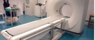
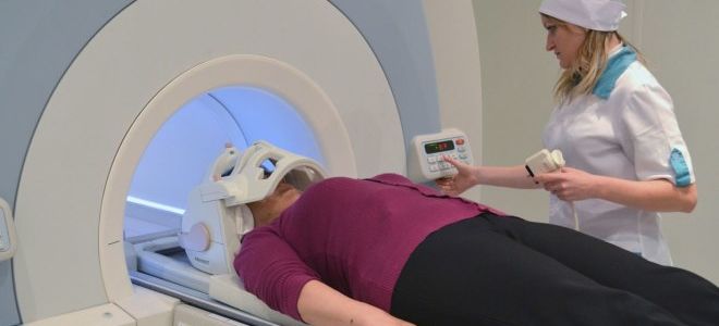
The MRI procedure lasts from 20 minutes to several hours, depending on the area being examined and the pathology. For a patient with claustrophobia, even 5 minutes in a capsule is a difficult ordeal. However, it is not necessary to refuse the examination; the problem can be solved. MRI for claustrophobia can be performed because, in addition to classic closed devices, there are open-type devices.
Advantages of open MRI
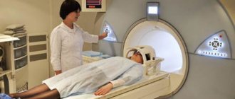
In a closed-type device (it is a large capsule), the patient is completely immersed in the tube for the duration of the procedure. For those suffering from claustrophobia, this is a difficult experience. There is a good alternative for them - an open-type MRI machine. There are no walls on the sides; only a special canvas is placed on top.
Carrying out an examination with such a tomograph completely eliminates being in a confined space, so the procedure will take place without fears or problems.
AFTER A PATIENT'S ADMISSION TO THE MRI CENTER
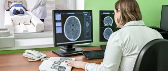
When a patient arrives at an MRI center, it is necessary to explain in detail all issues related to the procedure: the position of the magnet on the table in the tunnel the sound that occurs when creating a gradient the need for administration of contrast agents approximate examination time
If methods of cardiac synchronization and respiratory compensation are used, it is necessary to explain why this is necessary and what the patient may feel.
Before the MRI session begins, the patient should be explained in detail that during the MRI examination he will have to hold his breath for a while, open his mouth or not blink. If the patient does not understand why this is necessary, the entire explanation will be useless. It is preferable for the MRI procedure to be explained by the physician because it establishes contact between the physician and the patient and provides direct feedback to the physician regarding the patient's specific concerns.
When a patient enters the scanning room, the sight of the magnet tunnel and the unfamiliar surroundings usually increase their anxiety. You must be prepared to again explain the MRI procedure and answer any questions the patient may have.
The most common difficulty with MRI is claustrophobia. The closed area inside the magnet and the presence of equipment such as the head coil contribute to the appearance of claustrophobic disorders in patients and their increased nervousness.
Tips to help reduce claustrophobia in a patient:
- If the tomograph is equipped with a mirror, it is necessary to check that it allows the patient to see what is happening outside the magnet tunnel
- If possible, the patient should be examined in a prone position, as this will often allow them to see what is happening outside the magnet tunnel. This patient position is used for MRI of the pelvis, abdomen, chest, and areas of the upper and lower extremities such as the hip and wrist
- Remove the pillow from under the patient’s head. At the same time, the distance from his face to the inner wall of the magnet tunnel increases.
- Ask the patient to close his eyes or place a tissue over his eyes. Some patients don't like this, but others feel more comfortable knowing that if they accidentally open their eyes during an MRI exam, they won't see the enclosed space inside the magnet.
- Turn on the lighting and fan inside the magnet; At the same time, the atmosphere will become brighter and the air will be fresher
- Explain to the patient that the magnet tunnel is open on both sides. Often these few words are enough to calm him down
- Explain to the patient that he can exit the magnet tunnel at any time and that the examination can be paused without harm to the tomographer or personnel. This allows the patient to feel that he is being monitored
- Ask the person accompanying the patient to stay with him in the scanning room
- Inform the patient via intercom about the progress of the MRI examination and the duration of each sequence
- Warn the patient about the existence of an emergency button, which he can press if necessary, giving the doctor a signal during the MRI examination
- If the proposed measures are ineffective, then a successful examination is possible only after prescribing sedatives to the patient
How to get an MRI if you have claustrophobia
To reduce the risk of unexpected stressful emotions during MRI, you should:
- try to find a clinic where the procedure is performed using open-type equipment,
- lie on your side or stomach, but not on your back,
- study in detail the features of the procedure and its order in order to know what happens during your stay in the capsule,
- Ask a relative or friend to be present during the MRI.
Proper preparation for the examination plays an important role. We are talking, first of all, about the psychological mood. It will be useful to communicate with those who have already undergone the procedure. They will tell you how they felt and how effective this method is.
How to do a closed MRI for claustrophobia
Finding a clinic with an open MRI machine can be difficult, and sometimes impossible, so you have to prepare for the examination in a classic capsule. It will pass without problems if you listen to some recommendations:
- you need to inspect the device to understand that it is not completely closed, there is a way out of it,
- to increase the space inside the capsule, just remove the pillow,
- To provide the space with fresh air, you can turn on ventilation,
- you can close your eyes during the procedure or use a blindfold,
- turn on the lighting to maximum to assess the situation around you,
- ask the doctor to talk throughout the procedure,
- turn on relaxing pleasant music,
- take a sedative.
If the measures described do not help, and fear remains, then it is better to undergo an MRI under anesthesia. By the way, for children this is a mandatory condition, since the child is unlikely to be able to remain motionless for a long time, and this is important for obtaining results. The use of anesthesia has significant advantages:
- obtaining images of maximum quality,
- eliminating the possibility of movements during the examination,
- speeding up the procedure time,
- reducing the likelihood of obtaining an incorrect result.
Whatever procedure is performed - a spinal examination or an MRI of the brain - psychological preparation is extremely important for claustrophobia. If the patient is sure that he will not be able to stay in the capsule for a long time, then it is better to decide on the use of sedatives or immersion in medicated sleep.
Special equipment
The MRI machine is equipped with special equipment that will help cope with fears. This includes:
- panic button – in case of discomfort, the patient can always press it to interrupt the diagnosis,
- headphones - they are needed to communicate with the doctor, in them the patient will hear what he should do - for example, hold his breath, you can play pleasant music in the headphones to reduce stress,
- internal communication - the patient can always tell the doctor about his condition,
- mirror - with its help the patient will see what is happening outside,
- light and ventilation - to create comfortable conditions inside the device.
What doctors can do
The technologists servicing the tomograph and the doctor must tell the patient in detail what awaits him, how the examination is carried out, and explain that it is safe and will not cause any harm. They will show the device so that the patient understands that the capsule has an entrance and an exit, it is not completely sealed.
During the scan, doctors can constantly talk to the person being examined, saying that nothing bad will happen, no one will forget about him and will help if he becomes ill.
The specialist will offer a sedative if necessary, for example, Corvalol.
The doctor is obliged to inform the patient about the presence of a panic button. The most important thing is to explain to the patient that MRI for claustrophobia is possible and will not cause harm.
What can the patient do?
The patient should tell the doctor from the very beginning that he suffers from claustrophobia. The most important thing is to conduct psychological preparation in order to tune in positively to the procedure.
You need to decide in advance whether lighting and ventilation are required. You can ask a friend or relative to accompany you. Experts such as psychologists and psychotherapists will tell you how to deal with the fear of closed spaces.
Services and prices
An examination in a closed tomograph costs on average 4-8 thousand rubles, in an open one - 2-10 thousand.
Prices for computed tomography start from 4 thousand rubles.
Prices depend on the device, as well as the area that needs to be examined.
Are there any risks with an MRI scan?
There are currently no generally accepted risks associated with undergoing MRI. Magnetic resonance imaging is painless, passes quickly and does not entail any lasting effects. The scanner does not touch your child during the scan. MRI in children does not cause any harm to the child’s health
However, MRI is not suitable for people who have metal objects in their body (metal implants, surgical clips, etc.), as the scanner creates a strong magnetic field. Your child should wear clothes without zippers or metal fasteners during the MRI scan.
Before the scan begins, the nurse or MRI doctor will check that your child does not have a pacemaker, metal implants, a history of metal objects getting into the eye, or any allergies.
You will also need to leave behind metal objects such as jewelry, watches, keys, pens, or credit cards with metal strips. Your child will need to lie still during the MRI scan and may be sedated.
Some studies involve administering a contrast agent to improve the accuracy of the images. Before the injection, your child will be given a topical anesthetic cream.
Alternative options
If the listed methods do not help, and layer-by-layer scanning of the organ is the only way to make a correct diagnosis, you need to look for other options.
One alternative has already been indicated - an open apparatus. But it's not easy to find. Therefore, we have to select other options. And there is a way out - computed tomography. This technique is not as accurate as MRI for studying soft tissue, but for patients with claustrophobia it can be a real lifesaver.
MRI is a modern and accurate diagnostic method. Patients with claustrophobia do not need to give it up. Proper preparation is the key to success.
Adviсe
You can partially get rid of claustrophobia before an MRI using psychological influence. You yourself must verify the safety and harmlessness of the method. Read the information about how the procedure goes, and you will come to the conclusion that there is nothing to be afraid of. We ourselves create panic and provoke a phobia.
A detailed self-analysis or a visit to a psychologist can overcome the fear of an MRI. The right attitude will help cope with claustrophobia. Think about what you are more afraid of: examination due to a closed space or test results.
Why don't electronics “love” MRI?
Tomograph for MRI
A tomograph is a device used to perform MRI; it is a huge magnet under a plastic shell. It creates a powerful magnetic field - 10 times stronger than the Earth's magnetic field.
For humans, this is completely harmless and gives a good diagnostic result, which greatly helps different doctors in making a diagnosis and prescribing treatment for patients.
But electronic gadgets suffer in a magnetic field. The most expensive parts of hearing aids can be damaged: microcircuits, amplifiers, microphones.