Tachycardia during panic attacks
A panic attack is an increased feeling of fear and discomfort that appears in certain situations.
This condition is often uncontrollable, so a person needs the help of a professional. A specialist will determine the causes of the feeling of panic and help you cope with it using different methods. Attacks occur due to a stressful situation, emotional overload, social factor, or illness. For example, withdrawal syndrome from vasoconstrictor drops, which provokes tachycardia and panic attack due to psychological dependence on the drug. The patient cannot stop using the drug. He thinks he will stop breathing.
The reasons for the appearance of the condition are the psychological factor from the experience:
- pregnancy;
- childbirth;
- stroke;
- heart diseases;
- depression;
- difficult life situation.
The crisis can be caused by excessive consumption of coffee, medications, and central nervous system stimulants.
The most common signs of a panic attack:
- Increased heart rate . What is the heart rate when a panic attack occurs? During an attack, a person’s heart rate accelerates to 180 beats per minute. In this way, the body reacts to changes in its overall well-being, saturating the blood with oxygen and mobilizing its resources. At this moment, the person feels as if he is on the verge of a heart attack. Rapid heartbeat during a panic attack is its main and main symptom.
- Feeling of weakness, dizziness. The appearance of this symptom sometimes gives rise to the manifestation of other signs of this condition. A person feels the approach of fainting and, as a result, a deterioration in the clarity of vision occurs and instability appears. The reason for this sensation is a change in the level of oxygen in the body.
- Symptom of cottony legs. Muscle tension during an attack creates an inversely proportional effect and the person feels as if his legs cannot support him. However, this effect is deceptive; despite the tremors in the muscles, a sufferer of this disease is unlikely to fall.
- Heat. Due to increased blood circulation, a feeling of heat occurs.
- Lack of air. With active hyperventilation, a person’s breathing becomes shallow. The lungs are filled with oxygen and when taking a deep breath, the person loses the feeling of fullness. Tight chest and abdominal muscles also create a feeling of tightness.
- Feeling of derealization, depersonalization. At the moment of crisis, a person seems to lose the reality of the events happening around him. He feels like he is no longer in control of the situation.
Nonspecific depression of st heart what is it
- Reduced concentration and attention are manifested in difficulty memorizing and low academic performance. Physical activity is also significantly reduced, to the point of stupor, which may be considered laziness. Adolescent and childhood depression is often accompanied by aggressive attacks and increased conflict, which hide self-hatred.
- The mood becomes better in the evening. Self-confidence disappears and self-esteem decreases.
Due to these feelings, the patient moves away from society and strengthens his emerging feeling of inferiority. Long periods of depression in patients over 50 years of age are accompanied by deprivation and a clinical picture that is similar to dementia.Constant gloomy thoughts, a pessimistic attitude, an increasing sense of guilt, self-deprecation - a familiar state? It is this that is most often shown in all films, associating it with the depression of the Art. segment. And the patient, just like in all such films, thinks about causing harm to himself, or even comes to thoughts of suicide.
- The patient begins to sleep poorly, may have nightmares, and finds it very difficult to get up in the morning. Appetite worsens, and there is a frequent preference for carbohydrate foods over protein foods. The desire to eat may appear in the evening. A person in a state of depression has a distorted sense of time: for him it lasts a very long time.
- Another important sign is a reluctance to take care of oneself, which leads to an extremely sloppy appearance, at a minimum.
- Communication with such a person often comes down to discussing his past problems. The patient’s speech itself is slowed down, and the formulation of ideas becomes a difficult task for him.
- During the examination, patients look at the light or out the window. Gestures are directed towards oneself, hands are pressed to the chest. During anxious depression, the hands are pressed to the throat, a Veragut fold is observed in facial expressions, and the corners of the mouth are lowered. When manipulating objects, actions will be fussy. The voice becomes lower and quieter, long pauses appear between each word, and low directiveness is noted.
Such reasons can indirectly confirm the diagnosis of st interval depression:
- Dilated pupils.
- Tachycardia.
- Constipation.
- Reduced elasticity of the skin, it becomes flabby.
- The brittleness of nails and hair is significantly increased.
- The patient seems much older than his years.
- Due to cravings for foods rich in carbohydrates, weight may increase uncontrollably.
- Sexual desire increases, because this reduces the level of anxiety.
What can cause depression?
- At the genetic level, depression ct is caused by a pathology of the eleventh chromosome.
- With the biochemical path of development of this diagnosis, the exchange of catecholamines and serotonin is complicated.
- Neuroendocrine development manifests itself when the rhythm of the pituitary gland, hypothalamus and limbic system, as well as the pineal gland is disrupted, which is why the level of production of releasing hormones and melatonin is reduced. Daylight is involved in the creation of these hormones - the less of it, the worse the production.
- Between the ages of twenty and forty, there are increased spikes in depression.
- A sharp decline in a person's social class.
- Presence of suicide in the family.
- Loss of loved ones and relatives in adolescents over eleven years of age.
- The risk group includes people with increased conscientiousness, diligence and anxiety.
- Naturally, stressful events and problems with satisfying sexual desires also lead to depression.
- Some doctors add homosexuality and the period after childbirth to this.
What does CT segment depression mean?
Depression is a condition in which the segment is located below the isoelectric line by more than 0.5 mm. The condition can be caused both by physiological characteristics and by the presence of pathological processes and diseases of the cardiovascular system.
https://www.youtube.com/watch?v=-Ru4g_VXwko
Possible development due to vegetative-vascular dystonia. Pathology of the nervous system causes the development of arterial hypertension and affects the functioning of the heart muscle.
Depression may occur with excessively low potassium levels. A lack of this microelement leads to heart contraction disorders.
With tachycardia, ST segment depression is also possible. This type of deviation is called oblique; it is necessary to distinguish it from ischemic for timely and competent treatment of the disease.
May appear with hyperventilation. If the patient breathes frequently, shallowly, the tissues will become too saturated with oxygen; the carbon dioxide content will decrease. Most often diagnosed with fear, a stressful situation, or strong emotional arousal.
Possible development in pregnant women. Due to increased load on the heart and blood vessels, tachycardia may develop.
How does depression develop?
Recent research in the field of st segment depression has helped to compose three options for the development of anxiety and arterial hypertension:
- Due to somatovegetative disorders, depression begins and hypertension additionally develops. Due to increased nerve impulses, pressure increases in the smooth muscles of peripheral vessels. In this option, neurocircular dystonia or hypertension is treated, but the initial alarming factor remains unknown.
- Arterial hypertension develops, and only then anxiety depression is added. This disease is considered a more dangerous form for treatment. Using electrocardiography, the brain component can be identified, which will allow the diagnosis of the disease.
- In the third and final version, depression manifests itself as a complication of arterial hypertension. Due to increased symptoms, hypertension and depression, unique clinical pathologies arise, which allows for accurate diagnosis.
The National Heart Center conducted a number of studies. In patients with arterial hypertension, an increased degree of anxiety was observed and there was a high risk of depression when the patient changed his group from the first to the third.
After analyzing the medical records of inpatients, we found that doctors could have made mistakes when prescribing treatment for patients with hypertension. Due to the fact that attention was paid to the patient's anxiety extremely rarely, the ability of antihypertensive drugs to resist the disease fell further and further.
When making a diagnosis, the doctor is based on the reasons given by the patient. But you should always check for possible mental disorders. With such violations, the clinical picture will be disrupted.
In current realities, st depression and arterial hypertension should be monitored by both a psychiatrist and a cardiologist. Naturally, it is important that the patient himself participates in the course of treatment, because it is he who uses the drugs and follows the regimen that the doctor prescribed for him.
Features of treatment and prognosis
To confirm the diagnosis, additional examinations are necessary. You will have to tell your doctor about any medications you are taking: medications can affect heart function. Blood samples must be provided for analysis. If there are blood clotting disorders, you should also tell your doctor about them.
Treatment depends on the cause. If myocardial ischemia is noted, antiplatelet agents, nitrates, adrenergic blockers, and statins are prescribed.
If medications are not effective enough, surgical methods of intervention are used: coronary artery stenting, coronary artery bypass grafting.
You should reduce body weight if you are overweight, walk in the fresh air more often, exclude foods with a lot of fat and sugar, and give preference to natural foods.
With VSD, it is necessary to normalize the state of the nervous system. Prescribe the amino acid Glycine to get rid of the neurotic component. In addition, doctors often prescribe nootropic and sedatives.
Corvalol and potassium supplements may be prescribed.
In addition, you should give up bad habits (smoking, drinking alcohol), normalize your diet, follow a drinking regime, avoid stress, and get enough rest.
Prevention methods include a healthy lifestyle. In addition, you should regularly visit doctors to promptly detect emerging pathologies and treat them. The prognosis is more favorable if there is a mild form of pathology.
How to analyze the causes of depression?
First, let's recap the possible symptoms of ST segment depression:
- Excess oxygen in the lungs.
- Low potassium levels.
- Long-term use of antiarrhythmic drugs.
- Increased concentration of adrenal hormones due to frequent stress.
- Fibrosis, subendocardial ischemia.
Potassium deficiency is detected on the cardiogram by a pronounced U wave with ST segment depression.
Atrial repolarization is noted in leads avf, 3, 2 with a decrease in st. The same situation can be seen with pulmonary emphysema.
Let's explain the rules that doctors use when observing the electrocardiogram of a patient suffering from coronary artery disease:
- The traditional method involves considering the shift in QRS cycles that are above the isoline.
- The bias level itself is found by comparing it with PQ. If you forget about this point, you can mistakenly establish segment elevation.
- The starting point of measurement is located after the end of the QRS for sixty to seventy seconds. This is a general standard. In case of ventricular repolarization or suspicion of this, the PQ level is taken as the point.
- Leads AVR and V1 do not make it possible to understand whether the segment has increased or not.
- With a heart rate exceeding one hundred and thirty beats per minute, pathologies can be seen, which incorrectly signals false elevation due to the hard work of the myocardium.
What are the symptoms of ischemic segment depression?
It is not always possible to see such a disease based on clinical symptoms. It is rarely possible to detect pathology during a medical examination. A symptom can be called pain, the source of which is located behind the sternum.
If it is present, the doctor carefully examines the source of pain, using the Metelitsa classification:
- No pain in the pit of my stomach.
- Physical activity is accompanied by pain in the chest.
- Pain in the pit of the stomach, which makes physical activity impossible.
- Pain that dissipates with Nitroglycerin.
Additional visual characteristics of the diagnosis are cold sweat and skin, its blueness, rapid breathing, and muscle fatigue.
To assess the ability of the heart muscle to respond to an increase in contraction frequency, tests using physical activity must be performed.
A healthy person has no pathologies, because his heart responds adequately to increased load. With physical activity, arterial hypertension decreases, in rare cases increasing systolic pressure.
In the presence of a previous myocardial infarction, myocardial ischemia is called an important reason for low blood pressure. With pathologically frequent contractions of the heart, reduced functional cardiac capabilities indicate ventricular dysfunction. This situation occurs when using cardiotropic drugs.
Source: https://remson58.ru/nespetsificheskaya-depressiya-serdtsa/
Treatment of tachycardia during panic attacks
Tachycardia during panic attacks can be cured by the following methods:
- working with a psychologist - this includes the use of special relaxation techniques based on systematic breathing controls;
- the practice of yoga, meditation, an example can be seen here;
- systematic exercise (walking, jogging in the fresh air, swimming);
- art therapy, mastering new skills;
- a healthy lifestyle, including proper nutrition, good sleep, maintaining the required level of vitamins and minerals in the body, and the absence of bad habits (abuse of alcohol, cigarettes).
Important! Treatment of tachycardia during a panic attack using tranquilizers and antidepressants should only be selected by the attending physician.
A panic attack without the occurrence of tachycardia indicates that the patient has vegetative-vascular dystonia of the hypotonic type.
Diagnostics
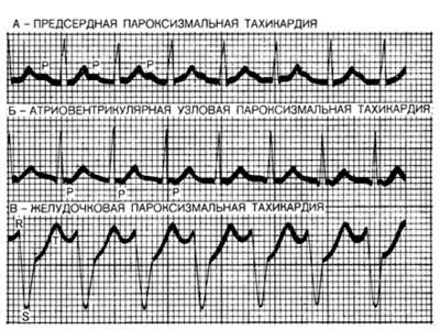
If paroxysmal tachycardia is suspected, the patient should consult a general practitioner or cardiologist to confirm or refute the diagnosis. The main research method is electrocardiography (ECG). Not all attacks are felt by the patient, so the doctor may prescribe daily
Additionally, the patient may be prescribed the following diagnostic methods:
- magnetic resonance imaging of the heart;
- layer-by-layer scanning of the heart muscle (MSCT);
- Ultrasound diagnostics of the heart.
How panic attacks affect the heart
The heart is more involved than other organs during a crisis. Due to an attack, a disturbance in cardiac function occurs. When pain occurs, a person's feeling of anxiety increases. In medicine, this phenomenon is called a panic attack of the heart. A dependence is created in which disruptions in the functioning of the heart give rise to and prolong a feeling of anxiety, which, in turn, leads to other disorders of the human body.
The reaction of the heart muscle is directly related to the increased level of adrenaline in the blood. Physically, the following happens:
- Acceleration of heart contractions - a person feels booming beats.
- Prolonged exposure to adrenaline provokes the appearance of arrhythmia.
- Nearby muscles begin to “suffer” - the person experiences heart pain due to the resulting pressure in the thoracic region.
Important! A low pulse during panic attacks can cause a person to have cardiovascular dystonia of the hypotonic type.
The disease in one form or another is common among 5% of the population. It is treatable and often does not require medication. By contacting a qualified psychologist, a person can easily solve this problem.
Tachycardia during VSD. The reason is in the head
“I haven’t slept well for many nights. The heart is pounding, as if wound up. Although there is no good reason to be nervous, I cannot sleep. I feel panicked, feeling like my heart might stop.
The family had to call an ambulance. When the doctor came into the room and examined me, he said everything was fine. Pulse is normal. The pressure is also fine.
But he couldn’t explain why his heart was pounding so much.”
What diagnosis would you make?
Currently, you can often find the diagnosis “tachycardia with vegetative-vascular dystonia” in the patient’s medical documents. Women are especially susceptible to this disease. This can be explained by a more acute reaction of women to emotional stress, which leads to changes in the endocrine profile.
Women's lives are always full of events. Sometimes the burden of the daily hut is so heavy that they do not have enough time to rest. This can lead to health problems. In addition to women, men and teenagers may also periodically complain of low pulse and rapid heartbeat.
Is tachycardia based on dystonia so dangerous?
Vegetative-vascular dystonia is a problem that simultaneously affects several systems in the body. VSD and extrasystole may be accompanied by the following heart rhythm disturbances:
- Tachycardia;
- Cardialgia;
- Bradycardia;
- Arrhythmia.
Symptoms of tachycardia based on dystonia
Cardiodynia is the most common problem associated with hyperhidrosis. If you feel a sharp pain in the chest, which has nothing to do with actual overload, this is the result of a disorder of the sympathetic part of the nervous system. Osteopathy is prescribed as a treatment in such cases, helping to cope with the disorder.
With prolonged internal tension and stress, bodily manifestations arise in the form of tachycardia, depression, neurosis, and fears. THIS is VSD. Tachycardia with VSD is an unconscious excitement during neurosis, provided that doctors do not find heart pathologies
Tachycardia after sleep is also the most common sign of a parasympathetic nervous system disorder.
If a patient has osteochondrosis, the disease usually manifests itself as VSD and arrhythmia, which can be easily confused with myocarditis.
Bradycardia occurs when the heart rate drops to 40-60 beats per second, and the person is in a subconscious state. Other signs include sudden mood swings and extremely cold hands and feet.
Sweating of the hands and feet is possible with bradycardia. But excessive sweating of the armpits for no reason (worries or physical work) already indicates the presence of dystonia.
A slightly elevated temperature - 37.1 - 37.5, rising in the evenings, is an alarm bell. The best way out in this case is a full medical examination. Your goal is to rule out a more dangerous disease.
When should you see a doctor?
The following warning signs are reasons to make an appointment with a neurologist or psychotherapist:
- Inexplicable outbursts and paranoia;
- Dizziness and headaches;
- Sudden mood swings and drowsiness;
- Excessive sweating and weakness;
- Shortness of breath and a feeling of heaviness in the chest, a seeming lack of air;
- Suspicion and anxiety;
- Buzzing and ringing in the ears;
- Inexplicable panic
Hidden signs and acute states of chronic diseases make diagnosis difficult. Unfortunately, doctors do not have all the necessary measures to fully determine vegetative-vascular dystonia. To do this, the patient must undergo a thorough examination by the following specialists:
- Neurologist;
- Ophthalmologist;
- Psychotherapist;
- Otolaryngologist;
- Endocrinologist.
Is it possible to cure arrhythmia caused by nerves?
It is almost impossible to completely get rid of vegetative-vascular dystonia. However, the patient can and should take steps to reduce his symptoms. Atrial fibrillation often returns as a result of each intense nervous breakdown. Effective therapy in this case includes psychotherapy and antidepressants.
What is priority: traditional methods or pharmaceutical drugs?
Anaprilin for tachycardia is recommended only in cases where the pulse exceeds the dangerous mark - from 90 beats per minute, and the pressure is significantly “off scale”.
Anaprilin reduces heart rate, but is also addictive. Therefore, such drugs should not be overused. It is better to take a validol tablet or drink 15 drops of Corvalol.
These drugs will relieve shortness of breath, blood pressure and lower the pulse.
Folk remedies and herbs cope well with sympathoadrenal crisis. For example, you can brew dill seeds and drink them at night. Decoctions of chamomile, mint and lemon balm, hawthorn reduce blood pressure and soothe.
Experienced specialists can help patients cope with problems and teach them how to manage temporary setbacks that are easy to overcome!
A healthy lifestyle, good sleep, healthy eating habits and exercise can help prevent the negative consequences of extrasystoles in VSD, even with a hereditary background.
Timely and quality rest
Don't ignore this question. You should rest regularly, even if you are too busy. The ideal rest regimen for “dystonics” is to go to bed early at night (before 10) and get up early in the morning (before 8 o’clock). Irregular sleep or lack of sleep makes it difficult to get rid of tachycardia.
The quality of sleep also affects heart failure during VSD.
Maintaining a healthy diet
Maintaining a healthy diet will act as a firewall against such an unpleasant symptom as VSD tachycardia. Thus, regular healthy eating should contribute to the normal functioning of all organs, including the heart.
Correct daily routine
You must stick to a regular daily routine. Tachycardia with VSD is dangerous only when a person is often nervous.
After all, the more often we are exposed to stress, the greater the risk of having a stroke, heart attack and other troubles associated with the nervous system.
Although arrhythmia with VSD is more a symptom than a disease, there are still risks with real interruptions in the functioning of the heart muscle. With a stable lifestyle, a person is less nervous and, accordingly, reduces anxiety symptoms.
Avoid drugs and alcohol
Drugs and alcohol can ruin anyone's life. Moreover, people who are worried about bradycardia syndrome risk developing real bradycardia.
Keep your blood pressure under control
Do not do any unnecessary or unnecessary work. Don’t take everything “to heart,” that is, take very simple questions so seriously.
Avoid anything that can create psychological pressure on you. This will help control your pulse during VSD. Follow the rules of a healthy lifestyle.
It wouldn’t hurt to discuss questions about your mental health with a good neurologist or psychotherapist.
Tachycardia and panic attacks
Panic attacks, according to modern medicine, can provoke tachycardia. Moreover, paroxysmal tachycardia as a form of arrhythmia is one of the most striking symptoms of panic, accompanying each attack. Tachycardia during panic attacks lasts, with short intervals, from ten minutes to half an hour.
In some cases, a patient who experiences a panic attack for the first time and has no idea about this phenomenon experiences severe fear, considering the arrhythmia to be a consequence of cardiovascular disease. His first instinct is to seek medical help. However, the electrocardiogram does not show any abnormalities in the heart. The patient becomes more confident that the doctors are not qualified enough and that they did not refuse him help, although he is sick. Anxiety and fear for your life provoke the next series of attacks. This is how the self-starting panic mechanism begins to operate.
Are panic and anxiety conditions really dangerous for the heart?
On the one hand, the heart is an organ that is involved in all significant processes occurring in the body. The consequences of a neurotic disorder for the heart and cardiac muscle cannot be underestimated. Psychiatrists and neurologists note that progressive neurosis negatively affects the patient’s heart, head and stomach, contributing to the development of psychosomatic diseases affecting these organs.

Panic states most often begin with vague anxiety, and already then the first heart rhythm disturbances occur. As fear and anxiety increase, disruptions intensify. This activates a mechanism that informs all systems and subsystems of the body about a problem that has arisen.
The blood flow increases, the so-called emergency system is mobilized. A panicked person, like an astronaut before going into outer space, is prepared by caring organ assistants for action in extreme conditions.
Law enforcement officers who have worked for a certain period in an intensive regime are given rest to recover. A similar recovery period is also needed for the heart, the brain, the stomach, the circulatory system... and the patient himself who has experienced panic.
However, no one gives him, as a hero, additional vacation or even a day off. After an attack of panic, normal life returns to normal, and the victim of panic attacks returns to his family or work, where no one takes into account the fact that he “worked a shift in heavy duty.”
On the contrary, if his problems are already known to others, everyone considers him almost a parasite. After all, the cardiologist, to whom the patient turned with complaints of heart ailments, probably also pronounced a verdict that he was healthy.
However, one can hardly call paroxysmal tachycardia, accompanying anxiety and panic, a healthy and normal phenomenon. Especially if the tachycardia lasts ten, twenty, or even forty minutes - almost the entire period of a panic attack!
We invite you to familiarize yourself with: Dukan Diet “Attack” - menu for the week

All these unpleasant sensations are caused by an imbalance of oxygen, increased blood supply, and an imbalance in the functioning of a system that is usually harmonious.
This type of arrhythmia, paroxysmal tachycardia, is the most common type of cardiac ailment associated with vegetative-vascular dystonia and panic. Arrhythmia causes fear in the patient, indicates that everything is clearly not all right with him, encourages him to think that malfunctions in the heart are not the consequences of a disorder in the body, but, on the contrary, the cause.
And a medical examination, during which no pathology is detected, not only increases fear, but also generates distrust in doctors. The patient “finds himself alone with his illness,” no one believes him, and, therefore, no one will help.
A vicious self-starting mechanism comes into play: growing fear gives rise to a new panic attack with symptoms of “heart pathology”, then another one... Panic attacks become chronic. And the manifestations of arrhythmia also become chronic, increasingly frightening and depressing the patient.
How to distinguish tachycardia from panic?
If tachycardia is a consequence of fatigue, vegetative-vascular dystonia or the initial stage of cardiovascular disease, then it goes away with medications. Drugs such as Corvalol, Valocardine, as well as sedatives - valerian root, glycine, novopassit - help stop tachycardia. In the case of a panic attack, if they help the patient, it will be a placebo effect: the patient must believe that his condition will improve from these drugs. If he convinces himself of the severity of the disease, these measures will not be enough.
Just as a panic attack provokes tachycardia, tachycardia in a patient suffering from panic attacks can cause another attack.
Everything is complicated by the fact that paroxysmal tachycardia, associated with a series of too frequent heart contractions, feels very similar to the extrasystole that accompanies atrial fibrillation - a serious disease that requires immediate treatment, or even surgical intervention.
Treatment of atrial fibrillation
Before starting treatment, it is necessary to determine the source of the disorder, namely with the interruption of which valve or ventricle it is associated with. It is necessary to completely examine the entire cardiovascular system. The course of treatment consists of lifelong observation by a cardiologist, drug treatment, and, in some cases, surgical procedures. Most often, arrhythmia is classified as a chronic pathology, and surgical intervention is rarely required. It is necessary for people with congenital heart disease and is performed in children under 14 years of age when drug treatment is ineffective.
Tachycardia and panic
Fear is a state when a person wants to run and hide from current circumstances. Tachycardia during panic attacks is a natural phenomenon, since adrenaline immediately enters the blood. A stressful situation gives rise to thoughts of a bad outcome and even death, a person panics. The fear hormone is released at a subconscious level to make the body more active. As a result, the heart rate increases.

Diet food
When paroxysmal tachycardia is diagnosed, the patient needs to constantly monitor blood sugar and cholesterol levels. Gaining excess weight is not allowed. Overeating can trigger the onset of an attack of tachycardia. Therefore, it is important to eat small portions 4-5 times a day.
The last meal should be no later than 2 hours before going to bed. It is important to exclude “provoking” foods and drinks:
- coffee Tea;
- confectionery sweets and buns;
- foods containing a lot of sugar and starch;
- butter, lard and mayonnaise.
Causes and symptoms of a panic attack
Panic attacks occur against the background of any stressful situation, be it heart or lung disease, emotional stress or social factor.
The main reasons that contribute to panic include:
- myocardial infarction;
- cardiac ischemia;
- beginning of sexual activity;
- pregnancy;
- childbirth;
- menopause;
- adrenal diseases;
- taking medications.
The symptoms are most pronounced during the first attack. A person’s blood pressure rises sharply, their heart rate increases greatly, and they may lose consciousness. The duration of the attack can be from 15 minutes to 1 hour. After this, the person feels depressed and defeated. Typical symptoms of a panic attack include:
- cardiopalmus;
- increased sweating;
- feeling of heat;
- lump in the throat;
- headache;
- numbness of the limbs.
Return to contents
Is there tachycardia?
The activity of the heart is closely related to the psychological state of a person. This is explained by the presence of the fear hormone - adrenaline - in the body. It enters the bloodstream every time an extreme situation occurs and a person needs to make an urgent decision. In life, such circumstances rarely occur and pass quickly. During a panic attack, a person does not stop thinking about bad things for a long time. A large amount of adrenaline enters the blood, provoking attacks of tachycardia. Adrenaline increases the force of heart contractions and speeds up the rhythm. A person feels like his heart is “jumping out of his chest.”
ST segment depression: causes and treatment methods
Forms of depression
26.12.2017
23.3 thousand
15.6 thousand
6 min.
A downward displacement of the ST segment relative to the isoelectric line (depression) is a reason for a more detailed examination of the patient, since the presence of such a change allows one to suspect ischemia of the heart muscle.
It should be remembered that analysis of this segment alone in isolation from the overall picture of the electrocardiogram is not informative enough. A correct conclusion is possible only after a comprehensive detailed analysis of the recording in all leads.
https://www.youtube.com/watch?v=q9ddORshGYo
A segment on a cardiogram is a section of the curve located between adjacent teeth. The ST segment is located between the negative S wave and the T wave.
The ST segment is a fragment of the electrocardiogram waveform that reflects the period during which both ventricles of the heart are fully involved in the excitation process.
The duration of the ST segment on the ECG depends on the heart rate and changes with it (the higher the heart rate, the shorter the duration of this section on the cardiogram).
Each section of the electrocardiographic curve has its own diagnostic value:
| Element | Meaning |
| P wave | The same shape and size of a positive P wave and its presence before each QRS complex is an indicator of normal sinus rhythm, the source of excitation in which is localized in the atrio-sinus node. With a pathological rhythm, the P wave is modified or absent |
| Q waves | Determined by the process of excitation of the interventricular septum (depolarization of the interventricular septum) |
| R wave | Reflects the excitation of the apex of the heart and adjacent areas of the heart muscle (depolarization of the main part of the ventricular myocardium) in leads v 4, 5, 6, and in leads v1 and v2 - reflects the process of excitation of the interventricular septum |
| S wave | It is a reflection of the excitation of the interventricular septum adjacent to the atria (basal) (depolarization of the base of the heart). On a normal electrocardiogram it is negative, its depth and duration increase with complete blockade of the left bundle branch, as well as the anterior branch of the left bundle branch. |
| T wave | Is a manifestation of the processes of repolarization of the ventricular myocardium |
| U wave | An unstable element of the electrocardiographic curve, recorded after the T wave and appearing due to short-term hyperexcitability of the ventricular myocardium after their repolarization |
| PQ segment | The duration of this interval indicates the speed of electrical impulse transmission from the atrial myocardium to the cardiac muscle of the ventricles of the heart. |
| QRS complex | Displays the progress of the process of distribution of excitation throughout the ventricular myocardium. Lengthens with right bundle branch block |
| ST segment | Reflects the saturation of myocardial cells with oxygen. Changes in the ST segment indicate oxygen starvation (hypoxia, ischemia) of the myocardium |
| PQ interval | Conducting electrical impulses; an increase in the duration of the segment indicates a disruption in the conduction of impulses along the atrioventricular pathway |
| QT interval | This interval reflects the process of excitation of all parts of the ventricles of the heart; it is commonly called electrical ventricular systole. Prolongation of this interval indicates a slowdown in impulse conduction through the atrioventricular junction |
On a normal cardiogram in the limb leads, the ST segment has a horizontal direction and is located on the isoelectric line. However, its position is also recognized as a variant of the norm, slightly above the isoelectric line (one and a half to two cells). This picture on the electrocardiogram is often combined with an increase in the amplitude of the positive T wave.
When analyzing an electrocardiogram, the greatest attention is paid to this segment when coronary heart disease is suspected and when diagnosing this disease, since this section of the curve is a reflection of oxygen deficiency in the heart muscle. Thus, this segment reflects the degree of myocardial ischemia.
The conclusion about ST segment depression is made when it is located below the isoelectric line.
The descent of the ST segment below the isoline (its depression) can also be recorded on the cardiogram of a healthy person; in this case, the position of the electrocardiogram curve in the ST segment does not fall below half a millimeter of the isoelectric line.
When analyzing an electrocardiogram, it is necessary to take into account that modifications of some of its elements can be caused by medications that the patient is taking, as well as deviations in the electrolyte composition of the blood.
Downward displacement of the ST segment relative to the isoelectric line is a nonspecific sign. This electrocardiographic phenomenon is observed in various leads in a number of conditions:
- Subendocardial or acute transmural ischemia (in acute myocardial infarction).
- Acute myocardial ischemia of the anterior wall of the left ventricle. This may also be indicated by ST elevation in the precordial leads.
- Acute ischemia of the lower wall.
- The result of exposure to drugs of the cardiac glycoside class.
- Hyperventilation of the lungs (excess oxygen in them).
- Reduced potassium content in the peripheral blood (hypokalemia) - in this case, there is a possibility of an additional U wave.
- Hypertrophic changes in the left ventricle, which in some cases can be interpreted as a sign of its overload.
- The horizontal displacement of this segment downwards is specific for the chronic course of coronary circulatory failure with myocardial ischemia.
- Vegetovascular dystonia.
- Pregnancy. During this period, a shift of the ST segment below the isoelectric line may be recorded against the background of tachycardia; the degree of depression in these cases does not exceed 0.5 mm.
A change in the ST-T complex in the form of its displacement down relative to the isoelectric line can be caused by a complex of reasons. For example, in a patient with myocardial hypertrophy (of any origin) and receiving therapy in the form of cardiac glycosides, there is a possibility of acute subendocardial ischemia.
Detection of ST segment depression is the reason for a thorough analysis of the electrocardiogram recording in all leads for a more accurate diagnosis of the location of the lesion.
In typical cases, myocardial ischemia (hypoxia) is manifested by pressing pain, discomfort, and burning in the chest area. Irradiation of pain to the back and left upper limb is typical. A painless form of myocardial ischemia is also possible, manifested by feelings of discomfort in the chest space, tachycardia, a decrease or increase in blood pressure, heartburn, and shortness of breath.
In the differential diagnosis of ischemic myocardial damage with VSD, the features of the clinical picture are taken into account: vegetative-vascular dystonia is characterized by ST depression in a young patient, more often in women, against the background of an increase in heart rate, in the absence of symptoms typical of angina pectoris. In this case, changes in the electrocardiogram are regarded as “nonspecific” or as “signs of increased influence of the sympathetic nervous system.”
With transient ischemia, Holter monitoring (recording an ECG during the day) helps make a diagnosis. The Holter displays all episodes of oxygen starvation of the heart muscle of patients that occurred during the day.
Application of Holter
In order for treatment to be effective, it is necessary to act directly on the cause of hypoxia, which is determined using special examination methods. Possible reasons are:
- atherosclerotic vascular lesions;
- unbalanced diet containing excessive amounts of cholesterol;
- emotional stress;
- presence of bad habits;
- sedentary lifestyle;
- excessive physical activity when the body is unprepared;
- metabolic disorders in the body leading to obesity;
- diabetes.
When treating myocardial ischemia, complex therapeutic regimens are used, consisting of the following drugs described in the table:
| Group | Drug names | Effect |
| Antiplatelet agents | Acetylsalicylic acid, Thrombo ACC, Cardiomagnyl | Prevents the aggregation of blood cells and improves its rheological properties |
| Nitrates | Nitroglycerin, Nitrosorbide, Nitrospray, Nitromint, Isoket | Dilate coronary vessels and improve blood supply to the myocardium |
| Adrenergic blockers | Metoprolol, Atenolol, Propranolol | Normalizes blood pressure and heart rate |
| Statins | Simvastatin, Atorvastatin | Reduce blood cholesterol levels to prevent atherosclerotic vascular disease |
If conservative therapy is insufficiently effective, surgical treatment methods are used:
- stenting of coronary arteries and (or) their branches;
- coronary artery bypass grafting.
In the treatment of vegetative-vascular dystonia, the main role belongs to the normalization of the excitability of the nervous system. The amino acid Glycine is capable of normalizing the metabolism of nervous tissue. The beneficial effect of this substance on nervous tissue helps to reduce the astheno-neurotic component.
It is also advisable to use nootropic drugs with an additional sedative effect.
If tachycardia or tachyarrhythmia is present in vegetative-vascular dystonia, the use of Corvaldin, Corvalol, and potassium preparations is indicated.
For effective treatment of vegetative-vascular dystonia, it is necessary to adhere to a protective regime: giving up bad habits, a balanced diet, combating physical inactivity, and eliminating stress. Massage, physiotherapy and acupuncture show high effectiveness, especially as part of complex therapy.
Source: https://neurofob.com/mood-disorders/depression-forms/depressiya-segmenta-st.html
How to stop a panic attack?
If a person is seized by panic and notices a rapid heartbeat, you need to pull yourself together and calm down. Positive thoughts will help him with this. He must remember that this is just a malfunction of the body, which does not pose any danger to life or health. Breathing exercises will relax you and speed up the end of the attack. A person must concentrate on breathing: you need to breathe with your stomach, while the inhalation is shorter than the exhalation. If the patient is at home, a contrast shower will help him. To do this, turn on either cold or warm water every 20 seconds. At the same time, massage your fingers and face, concentrating on the sensations. You can use a distraction and start counting people passing by or outside the window. To quickly overcome an attack, you can drink 10 drops of peony or valerian tincture, after diluting them in a glass of water.
If there is a loved one nearby, then he is obliged to support and help the patient. His emotional support is important. He should simultaneously perform breathing exercises, massage the neck and shoulders with a contrast shower, brew herbal soothing tea, offer to sing a song, take up drawing or watch a positive film.
Treatment of tachycardia during panic attacks
Treatment of tachycardia is aimed at removing the root cause of the attacks, that is, panic attacks. It is carried out at home using methods such as relaxation of the body through daily breathing exercises, meditation or yoga, sports activities in the form of cycling, morning jogging or swimming. Art therapy, humorous programs, and learning new skills help a lot. Decent self-esteem, good sleep, proper and timely nutrition are important. It is worth eating foods rich in vitamin C, magnesium, calcium and zinc. It is advisable to exclude alcoholic drinks and coffee, replacing them with herbal teas. In case of severe attacks, the psychiatrist prescribes medications to the patient. These may be medications from the group of tranquilizers or antidepressants.
PAROXYSMAL TACHYCARDIA OR PANIC ATTACK?
Hello! I have already contacted you. Here is the previous correspondence No. 705677 I constantly feel extrasytols Julia Female, 21 years old. Ukraine Chernovtsi Registered user has been tormented by heart problems for over a year now... I must say they happened before, but very rarely, when I stood up and sat down abruptly... I noticed that they began after suffering from a sore throat, the high temperature was only for one or two days. .but about 4 years have passed since then..frequent extrasitols began after getting married and moving to a new apartment..there was a lot of stress, because of this I have been living in constant stress for more than a year..I recently decided to get a holter, I want find out your opinion, is this scary, especially regarding the blockade, and what should I do about it, because I have no strength anymore? Here is the Holter conclusion: Average heart rate 92, during the day 100, at night 75 Minimum during sleep 60 Maximum 166, when climbing the stairs Types of contractions Normal 127952, 99.96% Superventricular 1 Ventricular 53 Single 53 Sinus rhythm. Predisposition to tachycardia in the daytime against the background of light loads. At night, a 1st degree block is recorded with an interval of PQ = 0.22-0.24, at 9:03 an episode of 2nd degree AV block with an atypical Wenckebach periodicity of 6:5 is recorded. Ventricular extrasystoles of the trigeminy type were recorded during excitement (she passed the exam). There are no diagnostically significant ST-T dynamics. HRV indicators are normal.
I should also say that I have stage 1 mitral valve prolapse with stage 1 regurgitation 06/17/2013 15:42 Complain about this message to the site administrator Replies: 2;
Direct specialty Kuklina Elena Nikolaevna. cardiologist Rating. Kuklina Elena Nikolaevna Complain about this message to the site administrator
Elena Nikolaevna Kuklina, cardiologist You’re not describing anything terrible, it doesn’t interfere with life. Stress and neuroses can and should be overcome; rational physical education will help you with this. If you can’t cope on your own, seek help from a psychotherapist. Creation time: June 17, 2013 21:19
Ratings: 1 Direct specialty Google Eduard Romanovich. Cardiologist. Pensioner. www.guglin.ru; [email protected] Rating. Guglin Eduard Romanovich Complain about this message to the site administrator
Guglin Eduard Romanovich Cardiologist. Pensioner. www.guglin.ru; Here it is not extrasystoles that need to be treated, but neurosis. This is mainly done by psychotherapists. Creation time: June 17, 2013 21:52
With this in mind, I went to a cardiologist; in conclusion, she wrote me metabolic cardiomyopathy, as I understood, cardiomyopathy and ventricular extrasytolia. I found on the Internet that you can die from ILC. Now I'm even more afraid of everything. They prescribed me ATP-long 20 mg, Magnerot and A-Diston (herbal tincture for calming). Eduard Romanovich, I’m only 21 years old, but I’m already like a senile old woman. There was an exam, so I couldn’t leave for a week, the extrasitols didn’t let me sleep, and on top of all this, there was something wrong with my stomach, as if there was always food in my throat. I have been suffering with all this for over a year now, I also have vegetative crises, I hope that this is them and not something serious. Heat rushes to the head from the back, the heart jumps out of the chest, the hands become cold and very wet, and not only the hands. And on top of all this, I’m afraid that I have myocarditis, or something from this opera. It's scary with all this. And until recently I was active, even too much, there were a lot of me everywhere. She was involved in swimming, volleyball and dancing. Nothing ever bothered me. Advise something, or say that yes, I am sick, go get examined and treated. Because no one believes me anymore that something hurts me, and my family and husband constantly laugh at me.
My question is this. Three times this month something incomprehensible has happened to me. I'm afraid this is paroxysmal tachycardia. The attack begins at rest, sometimes during passive communication, today at night I woke up at about 4 o’clock and it also began. Your heart begins to pound, heat gradually creeps up from your back into your head, it feels like you are being poured over with boiling water, your heart begins to beat quickly, it makes you sweat, your hands and arms melt like ice, and then you feel chills. When this all starts, I try to stretch, it seems to recede. but then it comes again. It can roll like this three or four times in a row. All this lasts about an hour, although after this I walk around all day in fear that the attack might happen again. Once I called an ambulance, they injected me with magnesium, because my blood pressure jumped to 15090, and they gave me valerian and told me to cross-stitch. calm your nerves. but when they arrived, the heart rate was normal 80. but there was a chill after they left. somewhere there were two more waves, but I somehow stretched and tried not to pay attention. Dear cardiologists, what does this look like, and should I go get examined again? Now I am undergoing treatment from a psychotherapist, my general condition is normal, but these attacks unsettle me. It must also be said that there is a certain dependence between these attacks and my emotional state
What does ST segment depression mean in an electrocardiogram?
- July 23, 2018
- Psychiatry
- Evdokimova Irina
Sometimes in the transcript of the electrocardiogram the doctor writes about ST-segment depression. In some cases, this is a sign of pathology, but it can also be a normal variant. Patients do not always understand this term, so you should understand in more detail the reasons for this ECG result.
What is the ST segment?
An ECG shows the electrical processes that occur in the heart muscle during contraction and relaxation. If you look at the result of the study, you can see a line with many teeth. A straight segment is called an isoline, and the distance between two adjacent teeth is called a segment.
The ST segment represents the interval from the end of the S wave to the beginning of the T wave. This segment shows the state of the heart muscle at the time of contraction of both ventricles. Normally, the segment lies completely on the isoline and does not deviate from it. If the segment is located below the isoline, then doctors talk about depression of the ST segment.
Does this indicate a dangerous heart pathology? It all depends on the degree and type of segment decline. The electrocardiograph records the examination results on checkered paper.
If the ST segment is located no more than half a cell below the isoline, then this is a variant of the norm and occurs in healthy people. This result is considered acceptable in both precordial and limb leads.
A stronger decrease in the segment may indicate cardiac pathology.
Why is the ST segment reduced?
The causes of ST segment depression are divided into coronary and non-coronary. Coronary causes include conditions associated with insufficient blood supply (ischemia) to the heart muscle. These are different types of coronary heart disease and myocardial infarction. Non-coronary causes include:
- lack of potassium in the body (hypokalemia);
- secondary myocardial lesions in non-cardiac pathology;
- paroxysmal supraventricular tachycardia (ST segment depression can be up to 8 mm);
- taking certain medications (cardiac glycosides, antiarrhythmics, phenothiazines);
- hypertrophy of the left ventricle of the heart;
- vegetative dystonia;
- mitral valve prolapse;
- emotional stress;
- intense breathing (hyperventilation).
Types of segment reduction
When making a diagnosis based on electrocardiogram results, the type of ST segment depression must be taken into account. In cardiology, there are several types of such deviations:
- oblique;
- oblique;
- horizontal.
Oblique and horizontal depressions may indicate the presence of cardiac pathology. An oblique decline sometimes occurs in healthy people.
Oblique and horizontal type of decline
If the segment between the teeth is an oblique line directed downwards, then in this case they speak of oblique depression of the ST segment. Such electrocardiogram readings are considered pathological. This indicates myocardial ischemia. Another reason for this result may be left ventricular failure.
https://www.youtube.com/watch?v=gQeri1wD8ME
A sign of insufficient blood supply to the heart muscle is horizontal depression of the ST segment. What it is? The segment between the S and T teeth is parallel to the isoline. This ECG result is also a marker of ischemia.
The status of the ST segment is checked in two adjacent leads. That is, the electrodes of the cardiograph are connected to two points located nearby, on the chest or on the limbs. And if a decrease in the segment was detected twice, then this, as a rule, indicates ischemia.
Oblique descent type
Oblique ST-segment depression is a deviation in the electrocardiogram when the line between the teeth is directed upward. This usually happens with tachycardia. This phenomenon may be temporary, for example when your heart rate increases after exercise. In this case, changes in the electrocardiogram do not indicate pathology.
But if a high T wave is observed on the electrocardiogram along with oblique ST segment depression, this may indicate a disease. This ECG result occurs in the acute stage of myocardial infarction, with left ventricular hypertrophy, hyperkalemia.
Nonspecific depression
A decrease in the distance between the S and T waves is not always associated with coronary heart disease. This can be observed both normally and in conditions where the blood supply to the myocardium is not impaired. Typically, such a decrease is caused by non-coronary causes. In this case, doctors talk about nonspecific ST-segment depression.
Such changes in the electrocardiogram can be observed under the following conditions:
- mitral valve prolapse;
- taking cardiac glycosides, diuretics, psychotropic drugs (the ST segment has a trough-shaped shape);
- vegetative-vascular dystonia;
- left ventricular hypertrophy;
- hypokalemia;
- tachycardia;
- cardiac conduction disorders;
- hyperventilation;
- inflammation of the pancreas;
- water and electrolyte disturbances;
- Wolff-Parkinson-White syndrome (a disease with periodic attacks of tachycardia).
In some cases, there are mixed causes of ST segment depression. For example, a patient may suffer from left ventricular hypertrophy and at the same time use cardiac glycosides. This can lead to the development of myocardial ischemia.
The photo shows an ECG of a patient who has been taking one of the potent heart medications for a long time. There is a noticeable decrease and trough-shaped shape of the ST segment.
It is difficult for a patient who does not have special medical knowledge to understand the results of an electrocardiogram. Sometimes it is necessary to prescribe additional research methods. The ECG interpretation must be shown to the attending cardiologist; only he will be able to make an accurate diagnosis.
Source: https://SamMedic.ru/338079a-chto-oznachaet-depressiya-st-segmenta-v-rasshifrovke-elektrokardiogrammyi
| When interpreting an ECG the isoelectric line is usually taken to be the PQ interval. The TP segment represents a true contour line, but is practically not used in most routine clinical measurements. The appearance of J-point depression > 0.10 mV (1 mm) from the PQ transition level with a relatively flat ST segment (eg, 0.10 mV and duration 80 ms after the J-point (ST 80) for three consecutive cardiac cycles with stable isolines are considered as a pathological reaction. As a general rule, ST 60 should be measured whenever the heart rate is > 130 beats/min. ST segment depression can be observed at rest. In this case, point J and ST 60 or ST 80 must further decrease by >0.10 mV to be considered abnormal. Most laboratories print ECGs in 3x4 or 2.5 sec format for a group of three leads. To increase compliance with the above criteria, some laboratories have adopted a strategy of recording ECGs in leads II, aVF, and V5 for 10 seconds after changes below the ischemic threshold occur. This is a 10 second recording increases the likelihood of identifying several consecutive pathological cardiac cycles with a stable isoline. When the level of ST segment depression > 0.1 mV, the stress ECG becomes less specific and imaging should be considered. In patients with early repolarization and ST segment elevation at rest, a return to the PQ transition level is normal, so the degree of FN-induced ST segment depression in patients with early renolarization should be determined from the level of the PQ interval, and not from the elevated position of the J point before the test. ST segment depression , induced by load, not only does not determine the localization of myocardial ischemia, but does not even indicate which coronary artery it is associated with. Unlike depression, exercise-induced ST-segment elevation is relatively specific in identifying the area of ischemia and the coronary artery that may be causing that ischemia. |
Panic attack or heart problems? How to distinguish one from the other and how to treat it
What is a panic attack
Panic attacks can occur in a completely healthy person - healthy both physically and mentally. However, when this symptom appears, the sufferer is terrified that he is seriously ill and will either die from cardiac arrest/suffocation or go crazy. Let’s make a reservation right away: neither one nor the other will happen.
A panic attack is an intense fear that results in increased production of adrenaline. We have already written in more detail about the causes of panic attacks.
A typical scenario: during the first attack of fear, a person calls an ambulance, then the attacks are repeated, he is examined, but no somatic diseases are found. In a favorable scenario, one of the doctors examining the patient refers him to a psychotherapist, and the person finally learns that he is not alone, he has panic attacks, a fairly common problem. It is worse if the attacks become regular, and the person begins to limit himself in activities or movements in order to avoid repeated attacks (some are afraid to ride the subway, others are afraid to leave the apartment altogether). Panic disorder affects up to 5% of the population.
Treatment
The choice of treatment depends on the form and type of disease. The next attack requires immediate hospitalization. The patient is admitted to the cardiology department if there are noticeable attacks more than 2-3 times a month.
The exception is short-term attacks with a smooth course that do not cause complications. In this case, the doctor may administer an antiarrhythmic drug intravenously (Propranolol, Quinidine, Cordarone). If the attack does not stop, then electrical impulse therapy may be prescribed.
An experienced doctor can stop paroxysm of tachycardia. A mechanical method of influencing the vagus nerve is used. For example, the following methods are used:
- applying gentle pressure on the upper corner of the eyeball from the inside;
- straining;
- inducing the gag reflex by irritating the root of the tongue;
- Pinch your nose and mouth and exhale forcefully in this state.
If an attack of tachycardia occurs several times a month, then the doctor may prescribe the following drugs to reduce the risk of relapse: Celanide, Kinylentin, Verapomil.
In particularly severe cases, doctors resort to surgical intervention:
- installation of a pacemaker;
- destruction of the impulse pathways and the source of tachycardia by mechanical, laser or electrical action;
- using an electric defibrillator.