“Depressive” genes and the “sad” brain: where, how and why depression occurs
Depression is a disease that has been known since antiquity, but has become especially widespread in our time.
Every tenth person over 40 years of age suffers from it, and two thirds of patients are women. This disease is quite mysterious - until recently, neither the genetic nor the physiological causes of its occurrence were known. True, doctors and biologists have long assumed that depression may be hereditary.
And in 2020, this hypothesis was finally confirmed - researchers from the USA managed to discover the genes responsible for this disease. And scientists from China and Great Britain have discovered the physical roots of depression.
In general, we are serious people. The granite of science crunches on our teeth. We illuminate such harsh, complex corners of biological knowledge that the lights of other popular science sites have not reached.
But sometimes we just want to fool around. And talk about science in a fun way, show it from a different angle. Draw funny pictures, write light and funny text.
That’s why we opened a new section - “12 biological news in pictures.”
The intellectual partner of these illustrated stories is RVC JSC.
Depression is a mental disorder in which a person’s mood declines, thinking is impaired, and a person’s motor activity decreases. Finding out the causes of this disease is a very important task.
After all, clinical depression significantly reduces the patient’s quality of life, and, even worse, is one of the important causes of morbidity and mortality in people. Therefore, it is important and necessary to fight depression, but it is difficult to do this if the causes of the disease are unknown.
When scientists from Massachusetts General Hospital in the USA discovered regions of the genome associated with depression, it became clear that this was a breakthrough discovery [1, 2]. How did they do this?
The researchers turned to 23andMe, a private biotech firm that provides genetic testing. The company provided them with biomaterial from hundreds of thousands of people, some of whom had a confirmed diagnosis of depression, and some did not report this diagnosis.
Scientists analyzed the data obtained using a GWAS study (Genome-Wide Association Studies). GWAS includes methods that compare the genomes of a group of sick people with the genomes of healthy people (a control group).
If a study can identify genomic variants that are significantly more common in people with a given disease, then the variant is said to be associated, or associated, with the disease. As a result of GWAS testing, scientists discovered two interesting genomic regions.
One of them contained a poorly studied gene, about which all that is known is that it encodes a protein synthesized in the brain. Another area has discovered a gene that is associated with epilepsy and mental retardation, factors that increase the risk of depression.
To see the picture in full size, click on it.
Then we carried out a similar analysis of two other large groups with controls. As a result of these large-scale studies, scientists were able to identify 15 genomic regions that contain 17 independent single nucleotide substitutions (SNPs) likely to be directly associated with depression. Some of these SNPs turned out to be located in genes (or near genes) involved in brain development.
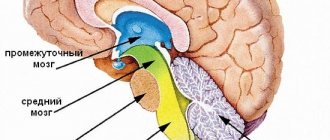
Soon after this discovery, another group of scientists, from China and Great Britain, discovered a new, no less important fact [7, 8]. It has become known in which part of the brain depression “hides”.
To do this, almost a thousand people were examined using high-precision magnetic resonance imaging - this method allows you to obtain images of the brain of a living person and understand which areas are involved in certain mental processes.
MRI studies have shown that depression affects the orbitofrontal cortex, an area of the brain in the prefrontal cortex that is responsible for signaling expected rewards or punishments in certain situations.
The medial, or middle, part of the prefrontal cortex is responsible for reward, while the lateral, or lateral, part is responsible for non-reward.
In people suffering from depression, the work of the “positive” medial region is slightly reduced, while the work of the “negative” lateral region remains at the same level.
Therefore, it is likely that a depressed person feels a sense of loss and disappointment associated with the lack of reward - this explains the depressed and depressed state of the patient.
The orbitofrontal cortex is also connected to the precuneus, a part of the brain that, among other things, is responsible for the sense of self and self.
If changes are observed in the orbitofrontal cortex, then they may also negatively affect the functioning of the precuneus, which could potentially lead to thoughts of personal loss and low self-esteem. It was also found that during depression, the connection between the “positive” medial region and memory systems in the brain is weakened, which explains the decrease in joyful feelings from pleasant memories in patients.
To see the picture in full size, click on it.
Fresh research will definitely help in the fight against depression. After all, they open up broad opportunities for finding new strategies for treating this disease.
But what conclusions can each of us draw based on these studies? Of course, genetically determined depression cannot be cured at home, and if you discover suspicious, depressing symptoms, you should consult a specialist doctor.
But everyone can bring themselves little joys every day! Reward yourself for your successes and for your mistakes too (after all, those who do nothing make no mistakes). Remember more often the joyful and pleasant things that happened to you in life (and there were a lot of them, right?).
And be attentive to your loved ones who suddenly feel sad for some reason - maybe right now they need your help and support. And then there is a high probability that depression will pass by!
- Craig L Hyde, Michael W Nagle, Chao Tian, Xing Chen, Sara A Paciga, Jens R Wendland, et al.. (2016). Identification of 15 genetic loci associated with risk of major depression in individuals of European descent. Nat Genet
.
48 , 1031-1036; - Novel study method identifies 15 genomic regions associated with depression. (2016). Science Daily;
- Code of Life: Reading does not mean understanding;
- Genetics of psoriasis: immunity, skin barrier function and GWAS;
- GWAS and psychogenetics: consortia in search of associations;
- The mysterious genetics of the “mysterious skin disease” - vitiligo;
- Wei Cheng, Edmund T. Rolls, Jiang Qiu, Wei Liu, Yanqing Tang, Chu-Chung Huang, et al.. (2016). Medial reward and lateral non-reward orbitofrontal cortex circuits change in opposite directions in depression. Brain
.
139 , 3296-3309; - Depression's physical source discovered. (2016). Science Daily...
Source: https://biomolecula.ru/articles/depressivnye-geny-i-grustnyi-mozg-gde-kak-i-pochemu-voznikaet-depressiia
Functions
Brain centers within the speech system are functionally connected and interact with such departments as areas of the associative cortex, areas of motor and articulatory activity, and subcortical structures. Participation of numerous brain regions in the speech system is associated with a large number of variants of the course of aphasia (for example, cortical, subcortical, dynamic, motor, acoustic-mnestic, optical-mnestic forms).
On the other hand, the presence of a large number of structural components of the speech system contributes to the restoration of speech function according to a compensatory type in cases where a certain area of the brain is damaged. Expanding the ability to restore functions is associated with the theory of neuroplasticity - the ability of normal nerve cells to take on the tasks of damaged ones.
Broca's area is responsible for the reproduction of verbal stimuli, Wernicke's area is responsible for the perception of verbal stimuli, both areas are located in the left hemisphere. The structures of the auditory cortex located in the left hemisphere take part in the perception of audible verbal signs. Neurons in this region perceive and identify phonemes—the simple sounds that make up words.
Phonemic hearing (the ability to distinguish phonemes) is impaired when designated areas of the cortical layer are damaged. The speech apparatus contains departments - regulating and performing. The regulatory department lies in the left hemisphere of the brain, which, to a greater extent than the right, is responsible for speech production and hearing. The regulatory apparatus includes pathways and nuclei of the brain stem (mainly located in the medulla oblongata).
Nerve endings belonging to the regulatory part of the brain extend in the direction of the muscles responsible for articulatory skills (tongue, lips, cheeks). Nerve endings as part of the regulatory department extend towards the muscles of the respiratory system and the vocal apparatus. Some areas of the cortical layer play a decisive role in the formation of verbal stimuli:
- Frontal gyri. The motor speech center is located in this area of the brain. Thanks to the activity of the designated structures, a person pronounces words and phrases.
- Temporal gyri. They perceive auditory stimuli - someone else's speech.
- Parietal lobe of the cortex. Participates in the process of understanding other people's words.
- Occipital lobe. Participates in the perception and identification of visual stimuli and, accordingly, written text.
- Subcortical nuclei. They regulate the rhythm, tempo, and expressiveness of reproduced words and phrases.
The pathways connect the cortical areas of the brain with the organs of the articulatory and vocal apparatus, which allows you to control muscle contractions and create the desired sounds using the tongue, lips, and cheeks. The pathways of the speech system originate in Broca's area.
Area 25 in the brain causes depression
The father of medicine, Hippocrates, told his students that the origins of all our diseases must be sought in the brain. This is not a new theory, but Hippocrates was right. When we are depressed, our brain works differently.
From this article you will learn what changes occur in the brain during depression.
Have you ever wondered what happens in your brain when you are depressed? And have you ever wondered how exactly antidepressants make a difference? It's time to find out how your brain works during depression, what changes occur in it.
Brain function in depression
Very often, people suffering from depression are given rather stupid analogies. Have you, too, been told that your batteries are running low and you need a recharge, or that your depression is like a broken leg that no one else can see? Nonsense.
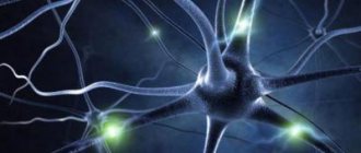
Your brain is a colony of 10 billion interconnected cells exchanging information with each other. Between cells there are zones called synapses. A synapse is a kind of junction point between two brain cells where they almost touch each other. Imagine that you and a friend are traveling on a bus.
You only need to turn around to speak to him. But out of habit, you grab your mobile phone and dial your friend’s number. Brain cells use the equivalent of your cell phone to communicate with each other through a synapse. Electrical impulses are used to make contact between cells.
But the impulse does not go beyond the synapse, just like your voice does not go beyond the bus. While you are talking on the phone, your brain cells release chemical messengers (transmitters).
These chemical transmitters are usually contained in small capsules called vesicles, and when needed, the transmitters are released and conduct nerve impulses across the synapse, transmitting information to the neighboring cell.
You may have heard of these chemical communicators before. They are called mediators or neurotransmitters (chemical transmitters of impulses between nerve cells). No one knows exactly how many of these neurotransmitters there are, but at last count there are more than forty. They carry different information.
Very important changes occur in the brain during depression. Some neurotransmitters play a role in stimulating the brain, others, on the contrary, in calming it. We won’t stuff you with scientific information, so we’ll only tell you about a couple of transmitters, and leave the remaining thirty-eight for the long winter evenings.
Serotonin, norepinephrine, neurotransmitter
Serotonin regulates sleep and wake cycles. You've probably heard that people suffering from depression have low serotonin levels. Instead of transmitting impulses across the synapse, allowing contact between cells, serotonin is resorbed, so information about mood, sleep, appetite and sexual desire is lost.
Norepinephrine can be compared to an organizer that controls brain activity. But when you're depressed, norepinephrine is released from brain cells very slowly, so brain activity decreases.
Brain cells are equipped with receptors for these chemical messengers.
Just as the key to your house fits only one door on your street, the shape of the receptors is such that it will only fit one type of neurotransmitter.
An attempt to connect another transmitter with an inappropriate receptor can be compared to the attempt of Cinderella's stepsisters to squeeze their thick knives into her glass slipper.
Your brain is a great recycler. Once the neurotransmitters have done their job, the brain cell from which they were released takes them back. Experts call this process reuptake.
Many antidepressants work by stopping reuptake.
For example, selective reuptake inhibitors help increase serotonin levels, allowing important brain signals to complete their journey.