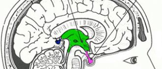
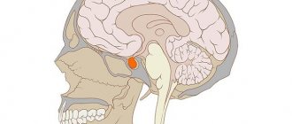
Empty sella turcica – this is a figurative name for sagging of the pituitary diaphragm. It covers the entrance to the pituitary gland - the sella turcica. Normally it is stretched over its processes and has an opening for the passage of the leg. For it to penetrate into the location of the pituitary gland, two conditions are necessary - low strength of the dura mater of the brain, of which it is part, and increased pressure in the cranial cavity.
The sella turcica is actually not empty; it contains cerebrospinal fluid (CSF), pituitary tissue, and often the optic nerves sag into it. This pathology is congenital and in about 50% of cases does not manifest symptoms until some time or throughout life. Most often, acquired forms are the result of treatment of pituitary tumors.
Reasons for formation:
- genetic inferiority of connective tissue;
- high cerebrospinal fluid pressure due to heart failure, pulmonary failure, hypertension, skull trauma, neoplasms inside the skull, sinus thrombosis;
- hemorrhage or infarction of the pituitary gland;
- destruction of adenoma;
- infection - meningitis, encephalitis, cyst suppuration, arachnoiditis;
- autoimmune Hashimoto's thyroiditis, hypophysitis (inflammation of the pituitary gland);
- long-term use of hormones for contraception, prednisolone and analogues;
- pregnancy, especially multiple births, menopause, repeat births, abortions;
- surgery for pituitary adenoma, radiation therapy;
- greater than usual need for pituitary hormones.
If, during treatment or after natural normalization of hormonal levels, the pituitary gland returns to normal, then the diaphragm sags into its cavity. High intracranial pressure causes crushing of the tissue of the organ, as if it spreads along the walls and bottom of the sella turcica. When visualized during this period, the sella appears empty.
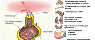
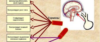
Functioning cells, even in this state, continue to synthesize hormones , but the control of their activity by the hypothalamus is disrupted. This causes the development of endocrine symptoms.
Neurological signs:
- headache of varying strength and circumstances of occurrence, does not have a constant localization, paroxysmal or monotonous aching;
- fluctuations in blood pressure;
- sudden episodes of muscle tremors;
- difficulty breathing, feeling of lack of air;
- anxiety, inexplicable fear, irritability, fluctuations in emotional background;
- loss of interest in the environment, depression;
- periodic increase in body temperature to 37.3-37.5 degrees;
- muscle spasms of the limbs;
- unsteadiness when walking, poor coordination of movements;
- memory impairment, fatigue;
- dizziness, fainting.
Endocrine disorders:
- excessive secretion of prolactin with sexual dysfunction in women and men;
- increased formation of growth hormone with manifestations of acromegaly;
- Itsenko-Cushing's disease;
- diabetes insipidus;
- metabolic syndrome, manifested by obesity, arterial hypertension and decreased tissue response to insulin, prediabetes mellitus;
- decreased production of pituitary tropic hormones.
Visual symptoms:
- pain in the eyes,
- tearfulness,
- doubling the contours of objects,
- fog before the eyes,
- flashes of light, sparks,
- dark spots,
- loss of fields,
- deterioration in vision clarity.
Decreased vision clarity
The risk of complications is higher if more than 90% of the pituitary gland is damaged due to its compression . Against the background of a stressful situation, patients may experience:
- severe vegetative crises;
- fainting conditions;
- personality changes;
- hypothyroid and thyrotoxic crises;
- sexual dysfunctions, infertility;
- progression of diabetes insipidus, Itsenko-Cushing's disease, acromegaly.
At the first stage, patients are referred for laboratory diagnostics of pituitary hormones, target organs - adrenal cortisol, thyroid thyroxine, testosterone and estrogen. The MRI method is recognized as the most informative.
Drug therapy is prescribed only if an imbalance of pituitary, thyroid, reproductive or adrenal hormones is detected. In case of disturbances in the functioning of the autonomic system, sedatives and adrenergic blockers are indicated. Intracranial hypertension requires discovery of the cause of its development and elimination.
Indications for surgery include : compression of the optic nerves due to their sagging into the opening of the diaphragm, blurred vision, leakage of cerebrospinal fluid through the nasal passages.
Read more in our article about empty sella syndrome.
What does the term “empty sella turcica” mean?
The name of this pathology misleads patients who have been diagnosed with this disease. They mistakenly believe that there is a void in their brain. In fact, this is a figurative name for sagging of the pituitary diaphragm. It covers the entrance to the pituitary gland - the sella turcica. Normally it is stretched over its processes and has an opening for the passage of the leg.
The fixation of the diaphragm, its density and opening parameters may have anatomical differences. In order for it to penetrate into the location of the pituitary gland, two conditions are necessary - low strength of the dura mater of the brain, of which it is a part, and increased pressure in the cranial cavity.
The sella turcica is actually not empty, because it contains cerebrospinal fluid (CSF), pituitary tissue, and often the optic nerves sag into it. This pathology can be congenital and approximately 50% of cases do not manifest symptoms until some time or throughout life. Acquired forms are most often the result of treatment of pituitary tumors.
We recommend reading the article about x-rays of the pituitary gland. From it you will learn about the indications for x-rays of the pituitary gland, the place of x-rays among imaging methods, as well as the diagnostic results.
And here is more information about MRI of the pituitary gland.
Probable circumstances of the onset of the disease
First of all, people who have a hereditary predisposition to this problem should be included in the risk group. Children often receive from biological parents an immature diaphragm, which is an underdeveloped, incomplete membrane.
Basically, even in the prenatal period, the brain of the fetus in the womb can be formed with the presence of defects under the negative influence of environmental factors. The most dangerous elements are considered to be radiation, environmental unsafety, modified food products, stress experienced by the expectant mother, poor quality of drinking water and much more.
Reasons for formation
Sagging of the diaphragm with crushing of the contents of the pituitary bed is promoted by:
- genetic inferiority of connective tissue;
- high cerebrospinal fluid pressure due to heart failure, pulmonary failure, hypertension, skull trauma, neoplasms inside the skull, sinus thrombosis;
- hemorrhage or infarction of the pituitary gland (hemorrhagic or ischemic stroke);
- destruction of adenoma;
- infection - meningitis, encephalitis, cyst suppuration, arachnoiditis (inflammation of the arachnoid membrane of the brain);
- autoimmune Hashimoto's thyroiditis, hypophysitis (inflammation of the pituitary gland);
- long-term use of hormones for contraception, prednisolone and similar drugs;
- pregnancy, especially multiple births, menopause, repeat births, abortions;
- surgery for pituitary adenoma, radiation therapy.
In case of primary insufficiency of the thyroid gland, adrenal glands, as well as in all cases when there is a greater than usual need for pituitary hormones (adolescents, pregnant women, menopause), the tissue of the organ increases. Such functional hyperplasia (proliferation) of the lobes and legs leads to protrusion and stretching of the diaphragm, its openings, and the membrane becomes thinner.
If, during treatment or after natural normalization of hormonal levels, the pituitary gland returns to normal, then the diaphragm sags into its cavity. High intracranial pressure causes crushing of the tissue of the organ, as if it spreads along the walls and bottom of the sella turcica. When visualized during this period, the sella appears empty.
Empty sella syndrome
Functioning cells, even in this state, continue to synthesize hormones, but the control of their activity by the hypothalamus is disrupted. This causes the development of endocrine symptoms. Eye manifestations are caused by tension in the nerves and disruption of their nutrition.
Treatment tactics
The goal of treatment is to correct disorders of the nervous, endocrine systems and organ of vision. Depending on the characteristics of the pathology in a particular patient, drug treatment or surgical intervention may be recommended.
If sella turcica syndrome is discovered completely by accident, does not manifest itself with any symptoms, does not cause discomfort to the patient, and in this case it does not require treatment. Such patients should be under medical supervision and undergo periodic examinations so that the doctor can promptly detect a possible deterioration in their condition.
- In case of laboratory-confirmed hormonal disorders in the form of a lack of certain hormones in the blood, hormone replacement therapy is carried out - the missing substance is introduced into the body from the outside.
- If symptoms of autonomic disorders occur, the patient is prescribed symptomatic therapy (sedatives, blood pressure lowerers, painkillers and other drugs).
Intracranial hypertension cannot be corrected with medications; it will go away on its own after the cause that caused it is eliminated.
In some clinical situations, it is unfortunately impossible to do without the intervention of a neurosurgeon. Indications for surgery are:
- sagging of the optic chiasm into the enlarged opening of the diaphragm, accompanied by their compression;
- leakage of cerebrospinal fluid (CSF) through the thinned bottom of the sella turcica; this symptom is called “liquorrhea” and is manifested clinically by the outflow of colorless liquid (the same cerebrospinal fluid) from the patient’s nasal passages.
In the first case, transsphenoidal fixation of the optic chiasm is performed - in this way, its compression and sagging are eliminated.
To eliminate liquorrhea, tamponade of the sella turcica is performed with a muscle.
Symptoms of empty sella syndrome
The clinical picture is characterized by an alternation of symptoms, periods of relative remission and progression of symptoms.
Neurological signs
The following disorders are detected in patients:
- headache of varying strength and circumstances of occurrence, does not have a constant localization, paroxysmal or monotonous aching;
- fluctuations in blood pressure;
- sudden episodes of muscle tremors;
- difficulty breathing, feeling of lack of air;
- anxiety, inexplicable fear, irritability, fluctuations in emotional background;
- loss of interest in the environment, depression;
- periodic increase in body temperature to 37.3-37.5 degrees;
- muscle spasms of the limbs;
- unsteadiness when walking, poor coordination of movements;
- memory impairment, fatigue;
- dizziness, fainting.
Watch the video about empty sella syndrome:
Endocrine disorders
The second group of symptoms relates to hormonal disorders:
- excessive secretion of prolactin with sexual dysfunction in women and men;
- increased formation of growth hormone with manifestations of acromegaly (accelerated body growth, skeletal deformation);
- Itsenko-Cushing's disease due to increased secretion of adrenocorticotropic hormone (obesity, moon-shaped face, hair growth on the face and torso, purple-purple stretch marks);
- diabetes insipidus – increased thirst and urination. Appears when there is a deficiency of vasopressin produced by the pituitary gland or due to a change in its release by the posterior lobe of the pituitary gland into the blood;
- metabolic syndrome is a consequence of dysfunction of the hypothalamus. Manifested by obesity, arterial hypertension and decreased tissue response to insulin, prediabetes;
- decreased production of pituitary tropic hormones – partial or complete pituitary insufficiency (panhypopituitarism).
Visual symptoms
Their intensity depends on the severity of circulatory disorders in the area of passage of the optic nerves and the circulation of cerebrospinal fluid in the subarachnoid space. Patients may experience:
- pain in the eyes,
- tearfulness,
- doubling the contours of objects,
- fog before the eyes,
- flashes of light, sparks,
- dark spots,
- loss of fields,
- deterioration in vision clarity.
Possible combination of empty sella syndrome and glaucoma
Poor circulation in the area of the optic nerves
Forecast
If you follow all the doctors' recommendations, the prognosis is usually good. However, just as there is no identical manifestation of the disease, no identical treatment, there is no unambiguous prognosis. Some patients will need to take medications regularly to manage symptoms. The most important thing for a person with disorders of the sella turcica is healthy habits, adequate sleep, and exclusion of smoked meats and other unhealthy foods from the diet. Regular physical activity is recommended, but a sense of proportion is important: you should not bring the body to overwork.
If close relatives have had cases of this disease, it is worth visiting a doctor and undergoing an examination. On the other hand, although you need to be careful about your health, you also cannot live in constant fear of illness. After all, according to statistics, the primary syndrome is diagnosed not so rarely - in 10% of newborns, while it interferes with the life of no more than 3% of patients.
How dangerous it is for the patient
The risk of complications is higher if more than 90% of the pituitary gland is damaged due to its compression. Against the background of a stressful situation, patients may experience:
- severe vegetative crises leading to loss of ability to work;
- fainting conditions;
- personality changes;
- hypothyroid and thyrotoxic crises;
- sexual dysfunctions, infertility;
- progression of diabetes insipidus, Itsenko-Cushing's disease, acromegaly.
Visual impairment and the threat of blindness are indications for surgical treatment
Treatment of other diseases starting with the letter - s
| Treatment of salmonellosis |
| Treatment of cutaneous sarcoidosis |
| Treatment of pulmonary sarcoidosis |
| Treatment of Kaposi's sarcoma |
| Treatment of uterine sarcoma |
| Treatment of Ewing's sarcoma |
| Treatment of type 2 diabetes mellitus |
| Treatment of type 1 diabetes mellitus |
| Treatment of seborrhea |
| Treatment of sepsis |
| Treatment of heart failure |
| Treatment of anthrax |
| Treatment of Budd-Chiari syndrome |
| Treatment of Guillain-Barré syndrome |
| Treatment of Gilbert's syndrome |
| Treatment of Parhon's syndrome |
| Treatment of polycystic ovary syndrome |
| Treatment of irritable bowel syndrome |
| Treatment of Sjögren's syndrome |
| Treatment of sinusitis |
| Treatment of systemic lupus erythematosus |
| Treatment of systemic scleroderma |
| Treatment of systemic sclerosis |
| Treatment of syphilis |
| Treatment of scoliosis |
| Treatment of somatoform disorders |
| Treatment of spinal stroke |
| Treatment of spondylosis and spondyloarthrosis |
| Treatment of laryngeal stenosis |
| Treatment of tracheal stenosis |
| Treatment of stenosing ligamentitis |
| Treatment of tetanus |
| Treatment of stomatitis |
| Treatment of ringworm |
| Treatment of subarachnoid hemorrhage |
| Treatment of subdural hematoma |
The information is for educational purposes only. Do not self-medicate; For all questions regarding the definition of the disease and methods of its treatment, consult your doctor. EUROLAB is not responsible for the consequences caused by the use of information posted on the portal.
MRI and other diagnostic methods
Suspicion of the possible presence of empty sella syndrome arises in patients who have undergone repeated trauma to the skull, surgery or irradiation of the pituitary gland, and for women, aggravating factors are multiple pregnancies and long-term contraceptive therapy.
Most often, at the first stage, patients are referred for laboratory diagnostics of pituitary hormones (growth, thyroid-stimulating, adrenocorticotropic, prolactin, vasopressin), as well as target organs - adrenal cortisol, thyroid thyroxine, testosterone and estrogen. At this stage, it is not always possible to confirm the pathology, since many patients have normal hormonal levels.
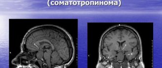
The MRI method is recognized as the most informative. With its help you can detect:
- liquor fluid in the sella turcica;
- a reduced and flattened pituitary gland, similar to a crescent;
- displacement of the pituitary tissue to the bottom, posterior wall;
- signs of intracranial hypertension - dilated sinuses, ventricles of the brain;
- asymmetrical sagging of the tank above the saddle;
- displacement of the pituitary funnel to the side, forward or backward, its thinning and lengthening.
MRI of the pituitary gland
Complications
Serious complications and consequences include hypopituitarism, optic chiasm prolapse, loss of vision, and infertility.
When pituitary gland function is insufficient, there is a disruption in the production of hormones in the adrenal glands, ovaries, seminal glands and thyroid gland. All of the above complications are extremely rare and, mainly, in the secondary form of the syndrome.
In the vast majority of cases, SPTS does not lead to any consequences. If symptoms are present, patients require medical supervision and health monitoring. Transformation of the pituitary gland and changes in its secretory function can provoke secondary pathologies of the thyroid gland, insufficiency of the adrenal cortex, changes in intracranial pressure and micro-strokes.