Structure
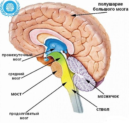
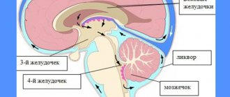
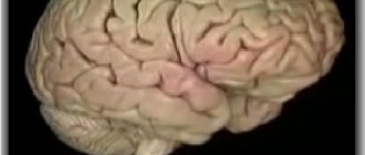
The division is part of the hindbrain. The structure and function of a bridge are very closely related, as in any other structure. It is located in front of the cerebellum, being a section between the midbrain and medulla oblongata.
It is separated from the first by the beginning of the 4th pair of cranial nerves, and from the second by a transverse groove. Outwardly, it resembles a roller with a groove, with nerves passing through it; they are responsible for the sensory abilities of the facial skin. In the groove there was also a place for the basilar arteries; their features include the fact that they supply blood to the back of the brain.
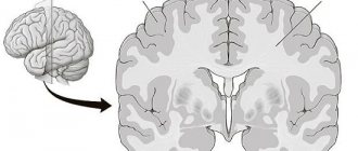
This section has a special diamond-shaped fossa located in the posterior part of the pons. The fossa is bordered at the top by the brain stripes, and above them are the facial colliculi.
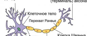
Above them there is a median eminence, and next to it is the locus coeruleus, which is responsible for the feeling of anxiety; it includes many nerve endings of the norepinephrine type. The pathways look like thick fibers of nervous tissue that run from the pons to the cerebellum. Thus, they form the arms of the pons and the peduncles of the cerebellum.
Among other things, the structure of the bridge has a “tire”, which is an accumulation of gray matter. This gray matter is the centers of the cranial nerves and the parts that contain the pathways. That is, the upper part of the brain is reserved for the centers that have a connection with the cranial nerves (fifth, sixth, seventh and eighth pairs).
Useful to know: Brain stem: features and functions
Speaking of pathways, the medial lemniscus and lateral lemniscus pass through this part. The same tegmentum contains the reticular formation; it is part of six nuclei and contains structures that are responsible for auditory analyzers.
At the base there are paths that pass from the cerebral cortex to different parts:
- pons brain;
- medulla;
- spinal cord;
- cerebellum.
And the blood supply occurs through arteries that belong to the vertebrobasilar region.
to contents ^
What functions does the bridge perform?
The pons is responsible for several important forms of activity.
Among them:
- Reflexive automatic and voluntary movements of the eyes and eardrum in response to loud sounds, as well as tissues of the oral cavity (palate). Any violation ends in problems.
- The ability for purposeful motor activity. Since the pons in the brain ensures the functioning of the cerebellum, any damage causes problems with the ability to control the body.
- Perception of vestibular stimuli. In this case, we are talking about the ability to perceive one’s body as a whole, its orientation and location in space, to respond to any changes in environmental conditions, and also to dampen unnecessary movements (for example, during sudden braking in public transport, stumbling, etc.). When affected, there is a lack of coordination. Ability to navigate in space.
- Providing olfactory function. The bridge has this ability partially. Other subcortical clusters are also responsible for it.
- Normal innervation of the skin and mucous membranes of the face.
- The pons is involved in the formation of sleep. This is a complex and coordinated work of several cerebral formations at once. Any violations immediately lead to problems with night rest. The patient becomes lethargic, asthenic processes appear.
- The functions of the pons include the acts of chewing and swallowing. Vital for nutrition and breathing.
- In fact, the body’s ability to perform normal gas exchange depends on the functioning of this structure. In the absence of adequate conduction of impulses, problems begin, even lethal disorders.
Basic actions are performed by nervous tissues constantly. Even minor changes are noticeable immediately.
The pons is part of the brain stem, therefore deviations in its activity become an indirect cause of dysfunction of this entire formation.
Complications, including rapid and fatal ones, are likely. High-quality medical care is not always possible due to the complex localization of structures and complex structure.
Motor and sensory functions
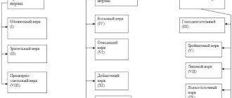
Speaking in more detail about motor and sensory function, let's talk about cranial nerves. When mentioning cranial nerves, it should be noted the ternary or mixed nerve (V pair). This pair of nerves is responsible for the movement of the masticatory muscles, as well as the muscles that are responsible for the tension of the eardrum and the palatine curtain.
To the sensory part of the trigeminal nerve there are afferent connections of nerve cells from receptors that are located in the skin of the human face, nasal mucosa, 60% of the tongue, eyeball and teeth. The sixth pair, or the so-called abducens nerve, is responsible for the movement of the eyeballs, namely for its rotation outward.
The 7th pair has one of the most important functions for human interaction; it is responsible for the innervation of muscles that allow the production of facial expressions. In addition, the facial nerve controls three glands: salivary, sublingual and submandibular. These glands provide reflexes such as salivation and swallowing.
The bridge also has a connection with the vestibulocochlear nerve. It is clear from the name that the cochlear part reaches the cochlear nuclei, but the vestibular part ends in the triangular nucleus. The eighth pair of nerves is responsible for analyzing vestibular stimuli; it determines the degree of their severity and where they are directed.
Useful to know: Gray matter of the brain, its structure, functions and properties
to contents ^
Possible pathologies and their diagnosis
The significance of the bridge can be assessed based on the influence of pathologies (syndromes) that damage individual functions of the body.
Common causes that lead to disruption of its functioning include: mechanical brain injuries, multiple sclerosis, stroke, cyst, tumor. In diagnosing pathologies, specialists primarily rely on the manifestation of symptoms, from which syndromes are formed.
The most common of them include:
- Bonnier syndrome is accompanied by damage to the nuclei of the auditory and vestibular nerves. In this case, the patient becomes dizzy, hearing loss occurs, and trigeminal neuralgia may occur. Common symptoms include weakness, depression, and sleep disturbances.
- Locked-in syndrome (ventral pontine syndrome) is a condition in which consciousness and full sensitivity are retained, but the ability to speak is completely lost. The function of the extraocular muscles is preserved. Communication with others is possible using non-verbal gestures. The condition is preceded by signs confirming insufficiency of the blood supply artery: double vision, dizziness, unsteadiness of gait.
- Raymond-Sestan syndrome (another name is oral tegmentum syndrome) is a combination of paralysis of the muscles responsible for the movement of the eyeball on the side opposite to the lesion. Etiological factors: atherosclerotic changes in cerebral vessels, tumors, ischemic strokes.
- Millard-Hubler syndrome is manifested by paralysis of the facial muscles on the affected side, along with partial paralysis on the opposite side. This disease manifests itself in pathologies at the base of the bridge. Constriction of blood vessels or micro-stroke predisposes to pathology, for example, if there is a cavernous angioma in a given structure with subsequent damage to the structures of the vascular system. Less commonly, the cause may be neurosyphilis or diffuse glioma.
- Foville syndrome is a combined lesion of individual elements of the facial and abducens nerves. The pathology is expressed in complete paralysis of the facial muscles in combination with strabismus. Often the cause of its development is ischemic stroke, less often tumor-like formations, inflammation.
- Gasperini syndrome is caused by the occurrence of pathology in the area of the bridge tire. It affects the nuclei of several nerves at once (facial, trigeminal, vestibulocochlear abducens). From the location of the pathological focus on the opposite side, a person feels a sensitivity disorder. The clinical picture includes strabismus, dizziness, and ataxia. This condition occurs due to ischemia, tumors, and inflammation.
- Grenet's syndrome is a sensory disorder with simultaneous damage to the muscles responsible for chewing on the affected side. On the opposite side, hemihypesthesia is noted. Often, pathology can occur due to ischemic changes in the branches of the posterior cerebral artery.
- Brissot-Sicart syndrome is a set of signs of damage to the nucleus of the facial nerve with partial paralysis of the limbs. Clinically manifested by a spasm of the facial muscles, which is accompanied by peripheral paralysis of the facial nerve and hemiparesis. Its occurrence is associated with ischemia and previous infectious diseases.
Modern methods of magnetic resonance imaging help to clarify the location, duration of the lesion, volume and other parameters of the pathological process.
Author of the article:
Dmitrieva Yulia (Sych)
– In 2014, she graduated with honors from Saratov State Medical University named after V.I. Razumovsky. Currently working as a cardiologist at the 8th City Clinical Hospital in the 1st clinic.
Like any organ in the human body, the VM can also stop functioning and this can be caused by the following diseases:
- stroke of the cerebral arteries;
- multiple sclerosis;
- head injuries. Can be obtained at any age, including during childbirth;
- tumors (malignant or benign) of parts of the brain.
In addition to the main reasons that can provoke brain pathologies, you need to know the symptoms of such a lesion:
- the process of swallowing and chewing is impaired;
- loss of skin sensitivity;
- nausea and vomiting;
- nystagmus is eye movements in one specific direction, as a result of such movements one can often feel dizzy, even to the point of loss of consciousness;
- may see double when turning the head sharply;
- disturbances in the functioning of the motor system, paralysis of certain parts of the body, muscles, or tremors in the hands;
- in case of disturbances in the functioning of the facial nerves, the patient may experience complete or partial anemia, lack of strength in the facial nerve;
- speech disorders;
- asthenia – decreased strength of muscle contraction, rapid muscle fatigue;
- dysmetria - incompatibility between the task of the movement being performed and muscle contraction, for example, when walking, a person may raise his legs much higher than necessary or, on the contrary, may stumble over small bumps;
- snoring, in cases where it has never been observed before.
Integrating function
These pons functions connect parts of the brain called the cerebral hemispheres. Also, all other paths, both ascending and descending, pass along the bridge, connecting it with many departments of the central nervous system. Among them are the spinal cord, cerebellum and cerebral cortex.
Impulses passing through the pontocerebellar pathways of the cerebral cortex have an impact on the functioning of the cerebellum. The cortex cannot exert influence directly, so it uses the bridge as an intermediary for these purposes. The pons regulates the medulla oblongata, influencing the centers that are responsible for the respiratory process and its intensity.
to contents ^
Formation in the prenatal period
Varolievye formation begins to form in the embryonic period from the rhomboid bladder. The bladder, in the process of its maturation and formation, is also divided into oblong and posterior. During the formation process, the hindbrain gives rise to the cerebellum, and the floor and its walls become components of the pons.
When a baby is born, the bridge is located just above the back of the sella turcica. Only after 2-3 years does it begin to rise and thereby become fixed in its permanent place - the upper part of the skull.
At the age of 8, the child’s spinal fibers begin to become overgrown with a myelin sheath.
CM pathologies
Like any organ in the human body, the VM can also stop functioning and this can be caused by the following diseases:
- stroke of the cerebral arteries;
- multiple sclerosis;
- head injuries. Can be obtained at any age, including during childbirth;
- tumors (malignant or benign) of parts of the brain.
In addition to the main reasons that can provoke brain pathologies, you need to know the symptoms of such a lesion:
- the process of swallowing and chewing is impaired;
- loss of skin sensitivity;
- nausea and vomiting;
- nystagmus is eye movements in one specific direction, as a result of such movements one can often feel dizzy, even to the point of loss of consciousness;
- may see double when turning the head sharply;
- disturbances in the functioning of the motor system, paralysis of certain parts of the body, muscles, or tremors in the hands;
- in case of disturbances in the functioning of the facial nerves, the patient may experience complete or partial anemia, lack of strength in the facial nerve;
- speech disorders;
- asthenia – decreased strength of muscle contraction, rapid muscle fatigue;
- dysmetria - incompatibility between the task of the movement being performed and muscle contraction, for example, when walking, a person may raise his legs much higher than necessary or, on the contrary, may stumble over small bumps;
- snoring, in cases where it has never been observed before.
Diseases leading to the development of syndromes
The structure of the Varoliev bridge suggests many possible lesions and an equally large number of manifestations. However, there is a group of diseases that become the foundation for the above syndromes.
This may include:
- Stroke. Acute disruption of cerebral blood flow in one area or another with death of nerve tissue and loss of some functions of cerebral structures. If the brain stem itself suffers, in the most favorable case it will end in a violation of higher activity.
- . Incorrectly called microstrokes. The same thing is observed, but there is no significant tissue death.
- . Impaired arterial patency as a result of blockage of the arteries by cholesterol plaques or spontaneous narrowing due to, for example, long-term smoking, hypertension (increased pressure).
- Infectious processes. Especially those that affect cerebral tissue. Encephalitis, meningitis.
- Demyelination. Multiple sclerosis.
The Varoliev bridge is responsible for a lot of important functions and has a systematic structure. Treatment of pathological conditions when the activity of this structure is already impaired is an extremely complex and sometimes impossible process.
Therefore, it makes sense to have a preventive effect on all diseases that may become a source of problems in the future. This is an important preventative measure.
The bridge (pons cerebri) is also called the Varoliev bridge (pons Varolii) in honor of Costanzo Varolii, an Italian anatomist of the mid-16th century, personal physician of Pope Gregory XIII.
Conducting function of the medulla oblongata. Participation of the pons in the sleep mechanism.
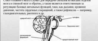
Conductor functions. All ascending and descending tracts of the spinal cord pass through the medulla oblongata: spinothalamic, corticospinal, rubrospinal. It originates the vestibulospinal, olivospinal and reticulospinal tracts, which provide tone and coordination of muscle reactions. In the medulla oblongata, the tracts from the cerebral cortex end - the corticoreticular tracts. Here the ascending pathways of proprioceptive sensitivity from the spinal cord end: the thin and wedge-shaped. Brain formations such as the pons, midbrain, cerebellum, thalamus, hypothalamus and cerebral cortex have bilateral connections with the medulla oblongata. The presence of these connections indicates the participation of the medulla oblongata in the regulation of skeletal muscle tone, autonomic and higher integrative functions, and analysis of sensory stimulation.
The reticular formation of the pons affects the cerebral cortex, causing it to awaken or sleep. In the reticular formation of the bridge there are two groups of nuclei that belong to the common respiratory center. One center activates the inhalation center of the medulla oblongata, the other activates the exhalation center. Neurons of the respiratory center located in the pons adapt the work of the respiratory cells of the medulla oblongata in accordance with the changing state of the body.
Midbrain. Functions of the superior and inferior colliculi. Functions of the red nuclei, their influence on alpha and gamma motor neurons of the spinal cord. Decerebrate rigidity. The meaning of the “substantia nigra”, its connection with the basal ganglia.
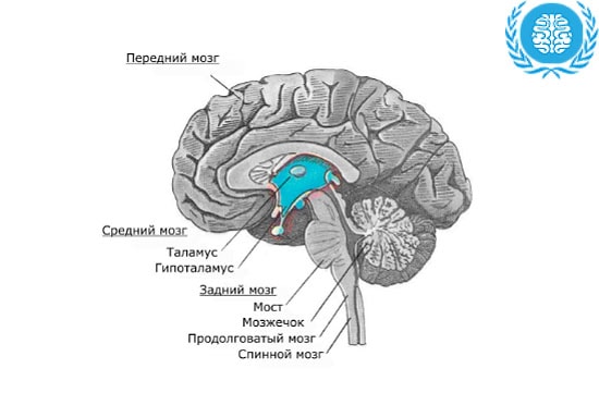
Morphofunctional organization. The midbrain (mesencephalon) is represented by the quadrigeminal and cerebral peduncles. The largest nuclei of the midbrain are the red nucleus, the substantia nigra and the nuclei of the cranial (oculomotor and trochlear) nerves, as well as the nuclei of the reticular formation.
Sensory functions. They are realized due to the receipt of visual and auditory information.
Conductor function. It consists in the fact that all ascending pathways to the overlying thalamus (medial lemniscus, spinothalamic tract), cerebrum and cerebellum pass through it. Descending tracts pass through the midbrain to the medulla oblongata and spinal cord. These are the pyramidal tract, corticopontine fibers, and rubroreticulospinal tract.
Motor function. It is realized through the nucleus of the trochlear nerve (n. trochlearis), the nuclei of the oculomotor nerve (n. oculomotorius), the red nucleus (nucleus ruber), and the black substance (substantia nigra).
The red nuclei are located in the upper part of the cerebral peduncles. They are connected to the cerebral cortex (pathways descending from the cortex), subcortical nuclei, cerebellum, and spinal cord (red nuclear-spinal tract). The basal ganglia of the brain and the cerebellum have their endings in the red nuclei. Disruption of connections between the red nuclei and the reticular formation of the medulla oblongata leads to decerebrate rigidity.
This condition is characterized by severe tension in the extensor muscles of the limbs, neck, and back. The main cause of decerebrate rigidity is the pronounced activating influence of the lateral vestibular nucleus (Deiters nucleus) on extensor motor neurons. This influence is maximum in the absence of inhibitory influences of the red nucleus and overlying structures, as well as the cerebellum. When the brain is transected below the nucleus of the lateral vestibular nerve, decerebrate rigidity disappears.
The red nuclei, receiving information from the motor zone of the cerebral cortex, subcortical nuclei and cerebellum about the impending movement and the state of the musculoskeletal system, send corrective impulses to the motor neurons of the spinal cord along the rubrospinal tract and thereby regulate muscle tone, preparing its level for the upcoming voluntary movement .
Another functionally important nucleus of the midbrain - the substantia nigra - is located in the cerebral peduncles, regulates the acts of chewing, swallowing (their sequence), and ensures precise movements of the fingers of the hand, for example when writing. The neurons of this nucleus are capable of synthesizing the neurotransmitter dopamine, which is supplied by axonal transport to the basal ganglia of the brain. Damage to the substantia nigra leads to disruption of plastic muscle tone. Fine regulation of plastic tone when playing the violin, writing, and doing graphic work is ensured by the substantia nigra. At the same time, when holding a certain position for a long time, plastic changes occur in the muscles due to changes in their colloidal properties, which ensures the least energy expenditure. Regulation of this process is carried out by the cells of the substantia nigra.
Neurons of the oculomotor and trochlear nerve nuclei regulate the movement of the eye up, down, out, toward the nose, and down toward the corner of the nose. Neurons of the accessory nucleus of the oculomotor nerve (Yakubovich's nucleus) regulate the lumen of the pupil and the curvature of the lens.
Reflex functions. Functionally independent structures of the midbrain are the quadrigeminal tuberosities. The upper ones are the primary subcortical centers of the visual analyzer (together with the lateral geniculate bodies of the diencephalon), the lower ones are the auditory centers (together with the medial geniculate bodies of the diencephalon). They are where the primary switching of visual and auditory information occurs. From the quadrigeminal tuberosities, the axons of their neurons go to the reticular formation of the trunk, the motor neurons of the spinal cord. Quadrigeminal neurons can be multimodal and detector. In the latter case, they react only to one sign of irritation, for example, a change in light and darkness, the direction of movement of the light source, etc. The main function of the quadrigeminal tuberosities is the organization of an alert reaction and the so-called start reflexes to sudden, not yet recognized, visual or sound signals. Activation of the midbrain in these cases through the hypothalamus leads to increased muscle tone and increased heart contractions; preparation for avoidance and a defensive reaction occurs.
The quadrigeminal region organizes indicative visual and auditory reflexes.
In humans, the quadrigeminal reflex is a sentinel reflex. In cases of increased excitability of the quadrigeminals, with sudden sound or light stimulation, a person begins to flinch, sometimes jump to his feet, scream, move away from the stimulus as quickly as possible, and sometimes run away uncontrollably.
If the quadrigeminal reflex is impaired, a person cannot quickly switch from one type of movement to another. Consequently, the quadrigeminal muscles take part in the organization of voluntary movements.
The cerebellum, its main functions. The meaning of the ancient, old, new cerebellar cortex. Characteristics of neurons in the cortex and cerebellar nuclei. Descending and ascending connections of the cerebellum with other parts of the central nervous system. The main symptoms that occur with damage to the cerebellum, their causes.
The cerebellum (cerebellum, small brain) is one of the integrative structures of the brain, taking part in the coordination and regulation of voluntary and involuntary movements, in the regulation of autonomic and behavioral functions.
Features of the morphofunctional organization and connections of the cerebellum. The implementation of these functions is ensured by the following morphological features of the cerebellum:
1) the cerebellar cortex is built in a fairly uniform manner, has stereotypical connections, which creates conditions for rapid information processing;
2) the main neural element of the cortex - the Purkinje cell, has a large number of inputs and forms the only axonal output from the cerebellum, the collaterals of which end on its nuclear structures;
3) almost all types of sensory stimulation are projected onto Purkinje cells: proprioceptive, cutaneous, visual, auditory, vestibular, etc.;
4) outputs from the cerebellum provide its connections with the cerebral cortex, with stem formations and the spinal cord.
The cerebellum is anatomically and functionally divided into old, ancient and new parts.
To the old part of the cerebellum
(archicerebellum) - vestibular cerebellum - belongs to the floccular lobe. This part has the most pronounced connections with the vestibular analyzer, which explains the importance of the cerebellum in the regulation of balance.
Ancient part of the cerebellum
(paleocerebellum) - spinal cerebellum - consists of sections of the vermis and the pyramid of the cerebellum, the uvula, the perioglocular region and receives information mainly from the proprioceptive systems of muscles, tendons, periosteum, and joint membranes.
New cerebellum
(neocerebellum) includes the cerebellar cortex and parts of the vermis; it receives information from the cortex, mainly along the fronto-pontocerebellar pathway, from the visual and auditory receptive systems, which indicates its participation in the analysis of visual, auditory signals and the organization of reactions to them.
The cerebellar cortex has a specific structure that is not repeated anywhere in the central nervous system. The upper (I) layer of the cerebellar cortex is a molecular layer, consisting of parallel fibers, dendritic branches and axons of layers II and III. In the lower part of the molecular layer there are basket and stellate cells, which provide interaction between Purkinje cells.
The middle (II) layer of the cortex is formed by Purkinje cells, arranged in one row and having the most powerful dendritic system in the central nervous system. There can be up to 60,000 synapses on the dendritic field of one Purkinje cell. Therefore, these cells perform the task of collecting, processing and transmitting information. The axons of Purkinje cells are the only way through which the cerebellar cortex transmits information to its nuclei and the nuclei of the cerebellar structure.
Under layer II of the cortex (under the Purkinje cells) lies a granular (III) layer, consisting of granule cells, the number of which reaches 10 billion. The axons of these cells rise upward, divide in a T-shape on the surface of the cortex, forming paths of contact with Purkinje cells. This is where the Golgi cells lie.
Information leaves the cerebellum through the superior and inferior peduncles. Through the superior legs, signals go to the thalamus, the pons, the red nucleus, the nuclei of the brain stem, and the reticular formation of the midbrain. Through the lower cerebellar peduncles, signals go to the medulla oblongata to its vestibular nuclei, olives, and reticular formation. The middle cerebellar peduncles connect the neocerebellum to the frontal lobe of the brain.
The impulse activity of neurons is recorded in the layer of Purkinje cells and the granular layer, and the frequency of impulse generation of these cells ranges from 20 to 200 per second. Cells of the cerebellar nuclei generate impulses much less frequently - 1-3 impulses per second.
Stimulation of the upper layer of the cerebellar cortex leads to long-term (up to 200 ms) inhibition of the activity of Purkinje cells. The same inhibition occurs with light and sound signals. The total changes in the electrical activity of the cerebellar cortex in response to stimulation of the sensory nerve of any muscle appear in the form of a positive oscillation (inhibition of cortical activity, hyperpolarization of Purkinje cells), which occurs after 15-20 ms and lasts 20-30 ms, after which a wave of excitation occurs, lasting up to 500 ms (depolarization of Purkinje cells).
In the cerebellar cortex, signals arrive from skin receptors, muscles, articular membranes, and periosteum along the so-called spinocerebellar tracts: posterior (dorsal) and anterior (ventral). These pathways to the cerebellum pass through the inferior olive of the medulla oblongata. From the olive cells come the so-called climbing fibers, which branch on the dendrites of the Purkinje cells.
The pontine nuclei send afferent pathways to the cerebellum, forming mossy fibers that end on the granule cells of layer III of the cerebellar cortex. There is an afferent connection between the cerebellum and the locus coeruleus of the midbrain using adrenergic fibers. These fibers are capable of diffusely releasing norepinephrine into the intercellular space of the cerebellar cortex, thereby humorally changing the state of excitability of its cells.
Axons of cells in layer III of the cerebellar cortex cause inhibition of Purkinje cells and granule cells of their own layer.
Purkinje cells, in turn, inhibit the activity of neurons in the cerebellar nuclei. The cerebellar nuclei have high tonic activity and regulate the tone of a number of motor centers of the intermediate, middle, medulla oblongata, and spinal cord.
The subcortical system of the cerebellum consists of three functionally different nuclear formations: the tent nucleus, the cortical nucleus, the spherical nucleus and the dentate nucleus.
The tent nucleus receives information from the medial zone of the cerebellar cortex and is connected with the Deiters nucleus and the RF of the medulla oblongata and midbrain. From here, signals travel along the reticulospinal tract to the motor neurons of the spinal cord.
The intermediate cerebellar cortex projects to the cortical and globular nuclei. From them, connections go to the midbrain to the red nucleus, then to the spinal cord along the rubrospinal tract. The second path from the intermediate nucleus goes to the thalamus and further to the motor zone of the cerebral cortex.
The dentate nucleus, receiving information from the lateral zone of the cerebellar cortex, is connected with the thalamus, and through it with the motor zone of the cerebral cortex.
Reticular formation of the bridge
The reticular formation is a branched network located in the brain and consisting of nerve cells and nuclei. It is present in almost all formations of the central nervous system and smoothly passes from one department to another. The reticular formation of the pons is located between the medulla oblongata and midbrain. Its long processes, called axons, form white matter and pass into the cerebellum.
In addition, signals can be transferred from the head to the back along the fibers of the nerve cells of the bridge. In addition, the reticular formation transmits signals to the cerebral cortex, due to which a person awakens or sleeps. The nuclei located in this part of the pons belong to the respiratory center located in the medulla oblongata.