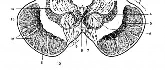
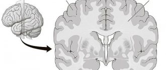
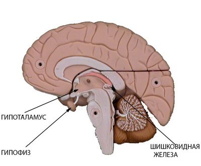
The intermediate cerebrum is located directly below the corpus callosum, just above the midbrain. Its structure includes the thalamic, subthalamic, suprathalamic regions, as well as the metathalamus and pituitary gland, consisting of the neurohypophysis and adenohypophysis. The cavity of the diencephalon is the 3rd ventricle, formed by six walls.
The boundaries of the diencephalon at the base of the brain are posteriorly the anterior edge of the posterior perforated substance and the optic tracts, and anteriorly the anterior surface of the optic chiasm. On the dorsal surface, the posterior border is the groove separating the superior colliculi of the midbrain from the posterior edge of the thalamus. The anterolateral border separates the diencephalon and telencephalon on the dorsal side. It is formed by the terminal strip (stria terminalis), corresponding to the border between the thalamus and the internal capsule.
You will learn in detail about the functional features and structure of the diencephalon by reading this material.
What areas belong to the diencephalon and their functions
The diencephalon develops from the caudal part of the forebrain, prosencephalon. During the process of ontogenesis, it undergoes significant changes. The ventral and dorsal walls become thinner and the lateral walls become significantly thicker. The cavity of this segment of the neural tube expands significantly and takes the form of a slit located in the median plane. It is called the third ventricle.
It should be noted that the dorsal (upper) wall of the third ventricle is represented only by ependymal epithelium. Above the ependymal epithelium is a process of the choroid, which delimits the diencephalon and the structures of the telencephalon (fornix and corpus callosum). The lateral parts of the diencephalon on the lateral side are directly fused with the structures of the telencephalon. The dorsal part of the lateral wall of the diencephalon develops from the pterygoid plate and is called the thalamic brain, thalamencephalon. The ventral part of the lateral wall of the human diencephalon, located below the subthalamic sulcus, develops from the main plate and is called the subthalamic region, or hypothalamus, hypothalamus.
Thus, the diencephalon includes the thalamic region, which is located in the dorsal areas, and the subthalamic (hypothalamic) region. The thalamic region includes the thalamus, metathalamus and epithalamus. Its cavity is the third ventricle.
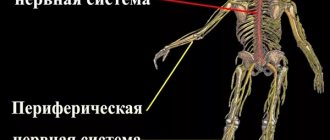
The diencephalon is the link between the telencephalon and the brain stem, and all its parts are grouped around the thalamus.
Table “Functions of the diencephalon”:
| Departments | Functions of the diencephalon | |
| Thalamus | Processing and integration of almost all signals going to the cerebral cortex from the spinal cord, midbrain, cerebellum, basal ganglia of the brain | |
| Suprathalamic region of the diencephalon, epithalamus | (corpuspineale, epiphysis, habenulae, comis - surahabenularumettrigonumhabenulae) is an endocrine gland | |
| Zatapama region, metathalamus (corpora geniculata mediates et laterales) | The medial and lateral geniculate bodies are the subcortical centers of hearing and subcortical centers of vision, respectively. | |
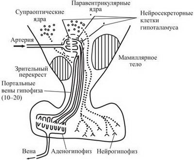
| Subthalamic region, or hypothalamus, hypothalamus | anterior group of nuclei | Neurons of the neurosecretory nuclei: (supraoptic, preoptic and paraventricular) produce neurosecretion for the posterior lobe of the pituitary gland - antidiuretic hormone (ADH) and oxytocin |
| intermediate group of nuclei | The nuclei of the subthalamic region proper, the nuclei of the gray tuberosity and the infundibulum: ventromedial hypothalamic, dorsomedial hypothalamic, arcuate, dorsal hypothalamic and posterior periventricular nuclei secrete releasing factors, under the influence of which the anterior lobe of the pituitary gland produces triple hormones (TSH, STH, GTG, ACTH, PTH and etc.) | |
| posterior group of nuclei | As part of the papillary bodies, which are the subcortical centers of smell. The function of this center of the diencephalon is to receive information from the parahippocampal gyrus. The axons of the cells of the papillary bodies are directed to the superior colliculus, forming the papillary-tegmental bundle, fasciculus mamillotegmentalis, and to the anterior nucleus of the thalamus, forming the papillary-thalamic bundle, fasciculus mamillotegmentalis. | |
| dorsolateral group of nuclei | For example, the posterior hypothalamic nucleus, nucleus hypothalamicus posterior (Luisy's nucleus), which serves as the integration center of the subthalamic region of the diencephalon | |
The next section of the article discusses the structure of such parts of the diencephalon as the thalamus and hypothalamus.
Divisions of the diencephalon: thalamus and hypothalamus
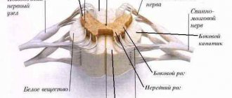
Thalamus. The thalamus, or posterior thalamus of the brain, or thalamus, consists mainly of gray matter, divided by layers of white matter into separate nuclei. The fibers arising from them form the so-called corona radiata, corona radiata, connecting the thalamus with other parts of the brain.
According to functional characteristics, the nuclei of the thalamus of the diencephalon are divided into three groups (according to Fulton):
- Nuclei that have no connection with the cerebral cortex. They are connected with the nuclei of the hypothalamus and the nuclei of the striopallidal system. This group of nuclei is located in the dorsolateral part of the thalamus.
- The nuclei in which the fibers of the general and special sensitivity pathways end. The axons of the cells of these nuclei are directed to the cerebral cortex. These nuclei are located in the ventral part of the thalamus and are somatosensitive.
- Associative nuclei that connect various centers of the diencephalon. These also include the nuclei of the dorsolateral part of the thalamus and the cushion nuclei.
Taking into account the different functional purposes of the thalamic nuclei, the following main groups can be distinguished.
- Anterior nuclei of the thalamus, nuclei anteriores thalami (anterior superior, anterior inferior, anteromedial). They are the subcortical center of smell. The anterior nuclei of the thalamus have connections with the papillary bodies of the corresponding side, which are also subcortical centers of smell. The bundle of nerve fibers originating from the neurons of the papillary body nuclei and ending in the anterior nuclei of the thalamus is called the papillary-thalamic bundle, fasciculus mamillothalamicus (Vic d'Azir bundle). It should be noted that some of the axons from the nuclei of the papillary bodies are directed to the superior colliculi of the midbrain, forming the papillary-tegmental bundle, fasciculus mamillotegmentalis. Nerve impulses are carried along this bundle, providing an unconditioned reflex increase in muscle tone and unconditioned reflex movements in response to strong olfactory stimulation. The axons of the cells of the anterior nuclei of the thalamus are sent to the limbic region of the cerebral cortex (mainly to the cortex of the medial surface of the frontal lobe). A small part of the axons ends on neurons of the medial nuclei of the thalamus.
- Ventrolateral nuclei of the thalamus , nuclei ventrolaterales thalami (posterior lateral, superior lateral, anterior inferior, intermediate inferior, medial inferior, posterolateral inferior, posteromedial inferior). They are the subcortical center of general sensitivity. Consequently, they end in fibers that are part of the spinal loop, lemniscus spinalis, medial loop, lemniscus medialis, and trigeminal loop, lemniscus trigeminalis. Viscerosensory fibers running as part of the trigeminal lemniscus are directed to the medial part of the ventrolateral nuclei of the thalamus, which are the subcortical center of interoceptive sensitivity. Most of the axons from the cells of the ventrolateral nuclei (80%) are sent as part of the internal capsule to the postcentral gyrus, forming the thalamocortical tract, tractus thalamocorticalis. A minority of axons (20%) end in the medial nuclei of the thalamus.
- Posterior nuclei of the thalamus , nuclei posteriores thalami, (pillow nuclei, lateral nucleus (geniculate body), medial nucleus (geniculate body). Along with the nuclei of the superior colliculi of the midbrain and the nuclei of the lateral geniculate bodies, they are subcortical centers of vision. In the posterior nuclei of the thalamus it ends part of the fibers passing through the optic tract. The axons of the cells of the posterior nuclei of the thalamus are directed to the medial nuclei of the thalamus, to the subthalamic and limbic regions of the brain.
- The median nuclei of the thalamus, nucleimediani thalami, (anterior and posterior paraventricular, rhomboid, connecting). These nuclei are subcortical centers of the diencephalon, responsible for vestibular and auditory functions. The fibers of the neurons of the auditory and vestibular nuclei of the pons partially end in them. In addition, the median nuclei have direct connections with the dentate and red nuclei. The axons of the cells of the median nuclei are sent to the medial nuclei of the thalamus and to the cortex of the temporal and frontal lobes of the cerebral hemispheres.
- Medial nuclei of the thalamus , nuclei mediates thalami , (dorsal medial). The main nucleus of this group is considered to be the dorsal medial nucleus, nucleus medialis dorsalis. It is a subcortical sensitive center of the extrapyramidal system, playing the role of an integration center of the diencephalon. The neurons of this nucleus end with part of the axons originating from the neurocytes of all the main nuclei of the visual thalamus. Thus, all types of information from the subcortical centers of general and special sensitivity come here. In turn, there is a two-way connection between the dorsal medial nucleus of the thalamus, the basal ganglia of the telencephalon (nuclei of the striopallidal system) and the areas of the cerebral cortex belonging to the limbic system. Some of the axons of the cells of the medial nuclei of the thalamus acquire a descending direction and end in the nuclei of the subthalamic region (Luizi's nucleus) and in the red nucleus.
- Reticular nuclei of the thalamus, nuclei reticulares thalami . Numerous small nuclei scattered in all parts of the visual thalamus are subcortical sensory centers of the reticular formation. These nuclei have bilateral connections with the nuclei of the reticular formation of the spinal cord, medulla oblongata, pons and midbrain.
- Paratenial nucleus.
- Subthalamic nucleus.
- Intraplate (intralaminar) nuclei located along the medullary plates of the thalamus (central median, paracentral, parafascicular, lateral central, medial central).
Hypothalamus. The nuclei of the subthalamic region are also very numerous (about 40), located mainly in the subthalamic region itself.
The hypothalamus of the diencephalon coordinates the nervous and humoral regulation of the activity of all internal organs, therefore it is considered the highest center of the autonomic functions of the body. The nuclei of the hypothalamus of the brain regulate cardiovascular activity, body temperature, salivation, gastric and intestinal juices, urine, sweat, etc.
In the light of modern ideas about the structure of the central nervous system, these higher centers of autonomic functions are under the control of the cerebral cortex. The subthalamic region forms the inferior wall of the third ventricle.
Bark
The cerebral cortex is a 3 mm thick superficial layer covering the hemispheres. It consists of vertically oriented nerve cells with processes. It also contains afferent and efferent nerve fibers, neuroglia. What is the cerebral cortex? This is a complex structure with horizontal layering.
The structure of the cerebral cortex: it has 6 layers (outer granular, molecular, outer pyramidal, inner granular, inner pyramidal, spindle cells), which have different density, width, size and shape of neurons. Due to the vertical bundles of nerve fibers, neurons and their processes present in the cortex, it has vertical striations. The human cerebral cortex, which contains more than 10 billion neurons, has an area of about 2200 sq.cm.
temporal lobe – hearing and smell;
occipital – vision;
parietal – touch and taste;
frontal – speech, movement, complex thinking.
association (connecting different cortical areas in one hemisphere);
commissural (connecting the hemispheres);
located in the convolutions between the furrows;
present in the outer parts of the hemispheres;
part of the internal capsule;
located in the corpus callosum.
The white matter of the brain is formed by nerve fibers that connect the gyral cortex of both hemispheres and the underlying formations. The subcortex of the brain consists of subcortical nuclei. The telencephalon controls all processes important for human life and our intellectual abilities.
Separate formations of the hypothalamus of the diencephalon
Considering that the subthalamic region includes a large number of individual formations, it is advisable to group them according to the topographic principle as follows:
Anterior hypothalamic region, regio hypothalamica anterior, or visual part, pars optica:
- Optic chiasm, chiasma opticum;
- Optic tract, tractus opticus.
The optic chiasm relates only in position to the hypothalamus of the brain, and in development to the telencephalon.
Intermediate hypothalamic region, regiahypothalamica intermedia:
- The subthalamic region proper, regio subthalamica propria;
- Gray tubercle, tuber cinereum;
- Funnel, infundibulum;
- Pituitary gland, hypophysis.
Posterior hypothalamic region, regio hypothalamica posterior, or papillary part, pars mamillaris.
- Mastoid bodies, corpora mamillaria.
Dorsolateral hypothalamic region, regio hypothalamica dorsolateralis.
- Posterior hypothalamic nucleus (Leuisy nucleus), nucleus hypothalamicus posterior.
The nuclei of the brain region of the hypothalamus are connected to the pituitary gland through the portal vessels (with the anterior lobe of the pituitary gland) and the hypothalamic-pituitary fascicle (with its posterior lobe).
Thanks to these connections, the hypothalamus and pituitary gland form a special hypothalamic-pituitary system.
Epithalamus and metathalamus of the diencephalon
Epithalamus. The suprathalamic region (epithalamus) includes:
- Pineal body, corpuspineale (epiphysis), - endocrine gland;
- Leashes, habenulae;
- Adhesion of leashes, comissura habenularum;
- Triangle of leashes, trigonum habenulae.
Under the epiphysis is the posterior commissure of the brain, comissura cerebri posterior; at the base of the epiphysis there is a pineal-shaped depression, recessus pinealis, which is a cavity that is a continuation of the third ventricle.
The pineal body (epiphysis) , develops in the form of an unpaired protrusion of the roof of the future third ventricle of the brain, belongs to the epithalamus of the diencephalon and is located in a shallow groove between the upper colliculi of the roof of the midbrain. The outside is covered with a connective tissue capsule containing a large number of blood vessels anastomosing with each other. The cellular elements of the parenchyma are specialized glandular cells - pinealocytes and glial cells - gliocytes.
The endocrine role of the pineal gland is the secretion by its cells of a substance that inhibits the activity of the pituitary gland until the onset of puberty, as well as participation in the fine regulation of almost all types of metabolism. During various periods of adulthood, and especially often in old age, cysts and deposits of brain sand may appear in the pineal gland.
Metathalamus . Behind the thalamus there are two small elevations - the geniculate bodies, corpus geniculatum laterale et mediate.
The medial geniculate body, smaller in size but more pronounced, lies in front of the handle of the inferior colliculus under the cushion, pulvinar, of the thalamus, separated from it by a clear groove. The fibers of the auditory loop, lemniscus lateralis, end there, and the medial geniculate body projects them onto the auditory area of the cerebral cortex. As a result, it, together with the lower colliculi of the roof of the midbrain, is a subcortical center of hearing.
The lateral geniculate body, larger, in the form of a flat tubercle, is placed on the lower lateral side of the pillow. The lateral part of the optic tract ends for the most part in it (the other part of the tract ends in the thalamic cushion). From here, visual stimuli are transmitted to the visual cortex. Therefore, together with the pillow and the superior colliculi of the roof of the midbrain, the lateral geniculate body is the subcortical center of vision.
The following describes the structure and functions of the pituitary gland.
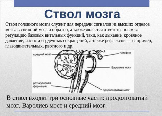
Midbrain
This department is located above the bridge. It is here that the signals received by the retina are transmitted to the brain, where they are processed by the nuclei of the superior colliculus, allowing us to see. The lower nuclei are responsible for the functioning of the human auditory system. They receive impulses produced in the outside world, implementing the human guard reflex, that is, the body can instantly engage in an action that requires a quick reaction.
This section extends from the anterior edge of the pons to the papillary bodies and optic tracts. It contains a cluster of nuclei, which are called quadrigeminal tubercles. The midbrain is responsible for hidden vision. It also contains the center of the orienting reflex, which ensures the body turns in the direction of a sharp noise.
The structure of the pituitary gland of the human brain and what it is responsible for
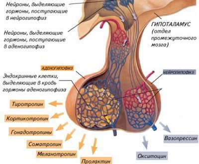
The pituitary gland (hypophysis) of the brain is located on the ventral surface of the brain at the base of the skull, in the fossa of the sella turcica. The pituitary gland is not homogeneous in its structure and embryogenesis. The pituitary gland of the brain has two main parts: the neurohypophysis and the adenohypophysis, which have different embryonic origins and structures.
The neurohypophysis is a derivative of the bottom of the infundibulum of the diencephalon. It is in close morphological and functional connection with the hypothalamus; the fibers of the hypothalamic-pituitary tract, coming from the supraoptic and paraventricular nuclei of the hypothalamus, end there.
The adenohypophysis (anterior lobe) develops from the epithelial protrusion (Rathke's pouch) of the roof of the intestinal tube. The anterior lobe of the pituitary gland has a close vascular connection with the hypothalamus. Here the arteries branch into capillaries, forming a dense, mantle-shaped plexus on the surface of the median eminence. The capillary branches of this plexus form veins that reach the anterior lobe of the pituitary gland of the human brain, here the veins again break up into capillaries that penetrate the entire lobe. This entire complex system of blood vessels is called portal. Through it, peptide hormones (liberins and statins) enter the adenohypophysis from the hypothalamus, regulating the synthesis and secretion of adenohypophysis hormones. The neurohypophysis has its own blood supply system, independent of the portal system.
What is the pituitary gland responsible for in the human brain? The adenohypophysis secretes two types of hormones - effector, i.e. realizing their properties directly in the body, and triple ones - having a regulatory effect on the peripheral endocrine glands. In total, six hormones are synthesized in the adenopituitary gland - growth hormone, prolactin, thyrotropin, adrenocorticotropic hormone (ACTH), follicle-stimulating hormone, luteinizing hormone. Follicle-stimulating and luteinizing hormones are combined into the group of gonadotropic hormones.
In recent years, it has been established that almost all biologically active substances secreted by neurons of the hypothalamic-pituitary system are of peptide nature.
In the nervous system there are special nerve cells - neurosecretory cells. They have a typical structural and functional (i.e., they have the ability to conduct a nerve impulse) neuronal organization, and their specific feature is the neurosecretory function associated with the secretion of biologically active substances. The functional significance of this mechanism is to ensure regulatory chemical communication between the central nervous and endocrine systems, carried out with the help of neurosecreted products.
During the process of evolution, the cells that made up the primitive nervous system specialized in two directions: ensuring fast processes, i.e. interneuronal interaction, and ensuring slow processes associated with the production of neurohormones acting on target cells at a distance. In the process of evolution, specialized neurons, including neurosecretory ones, were formed from cells combining sensory, conductive and secretory functions. Consequently, neurosecretory cells did not originate from the neuron as such, but from their common predecessor, the proneurocyte of invertebrate animals. The evolution of neurosecretory cells led to the formation in them, like classical neurons, of the ability to process synaptic excitation and inhibition, and generate action potentials.
All vertebrates have this type of cells, and they mainly constitute neurosecretory centers. Electrotonic gap junctions were found between neighboring neurosecretory cells, which probably ensure synchronization of the work of identical groups of cells within the center.
The axons of neurosecretory cells are characterized by numerous extensions that arise due to the temporary accumulation of neurosecretion. Large and giant expansions are called “Hering bodies”. Within the brain, the axons of neurosecretory cells, as a rule, lack a myelin sheath. Axons of neurosecretory cells provide contacts within neurosecretory areas and are connected to various parts of the brain and spinal cord.
One of the main functions of neurosecretory cells is the synthesis of proteins and polypeptides and their further secretion. In this regard, in cells of this type, the protein synthesizing apparatus is extremely developed - the granular endoplasmic reticulum and the Golgi apparatus. The lysosomal apparatus is also highly developed in neurosecretory cells, especially during periods of intense activity. But the most significant sign of the active activity of a neurosecretory cell is the number of elementary neurosecretory granules visible in an electron microscope.
In the hypothalamus, three main groups of neurosecretory cells should be distinguished:
- Peptidergic;
- Liberin- and statinergic;
- Monoaminergic.
However, this division is very arbitrary, since the same cells can synthesize two types of neurohormones. The paraventricular and supraoptic nuclei are connected to the neurohypophysis by the growth of axons of nerve cells into it, forming these nuclei and forming the hypothalamic-neurohypophyseal system. In the supraoptic and paraventricular nuclei, two peptide hormones are synthesized, secreted from the neurohypophysis. These are vasopressin and oxytocin.
The hypothalamus is the highest subcortical center for the integration of nervous and endocrine influences, autonomic and emotional components of behavioral reactions and thereby ensures the regulation of the constancy of the internal environment.
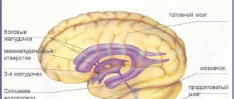
Next, you will learn about the structure and size of the 3rd ventricle of the brain.
Structure
Protection from mechanical damage and negative phenomena of the human brain is its location in the cranial cavity. It is protected on all sides by cranial bones. The shape of the brain and its parts in the process of growth become similar to the structure of the skull. Brain tissue is based on lipids, which determine its structure and color. It is jelly-like in shape and light yellow in color.
The functions of the brain are protected by soft, hard and arachnoid (intertwined with blood capillaries) tissues. The provided link between them was cerebrospinal fluid. Thanks to the diagram presented below, you can clearly see how the human brain works.
Referring to the diagram reflecting the structure of the brain, let's look at the departments and what they are responsible for. Using the example of the interaction of neurons with each other within a system unit, it will not be difficult to determine the functions of the brain.
How does the human brain work from a neurobiological point of view? “First of all, it is distinguished not so much by its complexity as by the lack of knowledge of the functional activity of neurons” (A. R. Luria). From the point of view of visual perception, the brain and its structure can be considered using the example of the main component, two parts of the cerebral hemispheres.
They are covered with a relief substance - bark, which is so dominant in volume that it occupies the majority of the area in percentage terms. It is accepted that the mass of a brain lobe is determined by the presence of the number of convolutions. On average, the bark has up to seven layers. Neurons are the main component of these layers. They ensure the flow of information from a central point to a peripheral point and vice versa.
Below the two cerebral hemispheres is the brain stem. This “trunk” name is justified by the arrangement of the hemispheres according to the principle of branches on the trunk on both sides.
Below the two hemispheres in the back is the cerebellum. The structure of its tissue differs from the main grooved surface. The cerebellum and pons (one of the components of the structural and functional blocks of the brain) belong to the posterior section. It is customary to mark five compartments:
- main, occupying 82% of the total mass, or final;
- the posterior section includes the pons and cerebellum;
- the next part is the middle one;
- oblong, or stem.
Also, according to a recognized definition, the main organ is divided into: two hemispheres, the cerebellum and the medulla oblongata.
The structure and functions of the brain underlie all vital processes of the body. Using an example, let’s look at the parts of the brain and what they are responsible for in the human body:
- The two hemispheres control speech, motor skills, and sensory capabilities.
- In the gyrus of the parietal lobe there is a region of the cortex responsible for motor activity.
- The posterior gyrus, located in the center, is part of the parts of the brain that are responsible for sensitivity; here is also the center for correction of proprioceptive perception.
- The structure of the human brain in the region of the transition of the frontal part to the temporal part contains a center that triggers taste buds and the sense of smell.
- In the temporal lobes, the function of the brain is designed to provide a person’s auditory capabilities.
- The visual center is located in the occipital region.
- Considering the functions of the brain regions, it can be noted that particularly important receptors are located in the medulla oblongata. All centers important for life are collected here: heartbeat, taste/food reflexes, breathing, regulation of smooth muscles of internal organs.
- The functions of the hindbrain include control of the vestibular apparatus. Here are the main passages of information from high points to lower centers and vice versa.
The thalamus is an (intermediate) department - its function is to regulate the sensitivity of all organs, it is responsible for memory. The hypothalamus controls the endocrine hormonal system and the central nervous system (CNS). For a better understanding of the operation of the entire system, you can refer to the table.
The cerebellum is formed from white and gray matter. The first is located under the bark, and the second is located outside, forming the cortex of the department. The cerebellum is responsible for such important parameters as coordination of movements and maintaining body balance. This department is also responsible for muscle contraction. People whose cerebellum is affected suffer from problems with spatial orientation, speech disorders and smooth movement. The growth of the department ends by the age of 15.
This department plays an important role in fine motor skills and the acts of chewing and swallowing, ensuring their correct sequence. Like the parts of the brain described above, the midbrain is directly related to muscle function. Thus, it controls work during prolonged stress, for example, when some part of the body must remain in one position for a long time, then it maintains muscle tone so that it can suddenly move to another position. The development of the midbrain directly depends on the formation of other parts.
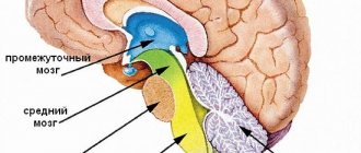
The structure of the brain, pictures of which are presented below, can be considered in several aspects.
final (80% of the total mass);
intermediate;
posterior (cerebellum and pons);
average;
oblong.
cerebral hemispheres;
brain stem;
cerebellum.
Structure of the brain: drawing with the names of the departments.
In addition to the physiological division of the brain into lobes, the need arose to divide it into areas that are responsible for certain functions.
Frontal lobes
This is the so-called command center. What is the frontal lobe responsible for? It is a point of independence, self-awareness, and initiative. The defeat of these areas or the presence of pathologies in their functioning will affect a person’s attitude towards the world - almost everything will become indifferent to him, motivation will disappear, interest in current events will disappear, and laziness will appear.
The main functions of the frontal lobes are to control human behavior. It generates responses to social phenomena. When zones are violated, the limiter is deactivated, establishing a ban on certain actions, called uncultural.
The frontal lobes also allow you to analyze, plan, and learn new skills. Repeated repetition of the same sequences of movements over time becomes automatic and does not require effort or thought to perform them. Damage will force you to repeat monotonous movements every time as if for the first time, putting a lot of effort into it.
Perseveration is another consequence of deviations in the functioning of the frontal lobes. This is looping or repetition, such as repeating one phrase or word during a conversation. On the left side (for right-handers) there are centers that are responsible for speech and attention.
These areas of the brain are also involved in coordinating and maintaining the body in an upright position when sitting and walking.
Temporal
They are located on the sides in the upper part of the brain, in the temple area. Thanks to them, the sound perceived by sound receptors turns into images, a person understands what he heard, certain sound vibrations are associated with images and are assigned to them. With the help of this part of the brain, people understand each other, their sound vibrations are filled with meaning, they choose the necessary words to describe certain phenomena.
Usually the left, non-dominant lobe is involved in determining the intonation of speech and reads emotions from facial expressions alone. Thanks to a small formation, the hippocampus, access to long-term memory is provided. Where it is located, on what medium and in what way our memories are recorded, this should be found out. The non-dominant part is used in visual memory, but the dominant part is used in verbal memory.
With problems with the temporal lobes, deviations in the functioning of the speech apparatus appear, in particular aphasia.
Parietal
MRI has shown that for left-handers and right-handers these lobes perform different functions, in fact directly opposite.
The left one gives us the ability to create a whole from fragments, that is, it helps to form a holistic picture of the world from small, at first glance, unrelated pieces.
They allow you not only to put together a mosaic from fragments, but also from letters into words, from a sequence of actions into a dance or technique, etc.
The non-dominant part allows you to perceive the world in three dimensions by processing information coming from the occipital lobes. Due to impairments, a person loses the ability to recognize faces, outline objects, and determine the distance to them and between them. These areas are also involved in the perception of pain and cold.
Occipital
Visual information processing center. They interpret the photons reflected from obstacles arriving at the biological light sensor - the retina - and form the resulting image, rotating it 180 degrees. Data about the size, color, shape, and material of an object are processed in separate centers and then recombined to form a single three-dimensional picture.