Brain compression is observed in 5% of patients with traumatic brain injury and intracranial hematoma. In addition, this condition can be caused by cerebral edema, hydrocephalus, or the presence of a tumor. The main clinical signs of brain compression are severe cerebral symptoms that develop against the background of increased intracranial pressure. In this article we will introduce you to the causes, main manifestations, rules for providing emergency care to the victim and methods of treating compression of brain tissue.
Compression of brain tissue is a dangerous condition and threatens the life of the victim, since their compression, caused by progressive intracranial hypertension, leads to the death of brain cells and the development of, in most cases, irreversible neurological symptoms. The proportion of deaths in this condition is about 30-50%, and disability of victims after traumatic brain injuries reaches 30%.
With injuries, increasing hydrocephalus, edema or strokes, compression of the brain occurs acutely. Such rapid development of this dangerous condition leads to massive death of cells in various parts of the brain and irreversible changes in their functions. With the development of tumor processes, tissue compression occurs more slowly, and the process proceeds chronically. Such long-term and slowly developing cerebral compression allows the tissues to adapt to some extent to the created pathological conditions.
Symptoms of brain compression are caused by many factors and are not immediately noticed at the initial stages, because they manifest themselves weakly. In most cases, after injuries, patients experience a “clear gap”, which can last several minutes or about 36-48 hours (and in the case of subarachnoid hematomas, up to 6-7 days). During this period, the victim has no signs of severe brain tissue injury and is conscious. After this period is completed, the patient’s general condition sharply deteriorates, and consciousness may be impaired from stupor to coma.
Pathogenesis
In the pathogenesis of S. g. m., not only the volume of intracranial formation is important, but also the speed of its development and the multiplicity of lesions. The faster the volume of the formation increases and the more foci of brain damage, the more violently the S. hemorrhage occurs. Compression of the brain with a hematoma caused by arterial bleeding develops more acutely than with the formation of the same hematoma due to venous bleeding. Wedge, manifestations of S. hemorrhage occur when the volume of the epidural hematoma reaches 50-70 ml, and the subdural hematoma - approx. 100 ml. This is explained by the presence between the brain and the skull of the so-called. reserve spaces due to the difference in their volumes, edges individually range from 8 to 15%. The difference in the volumes of epidural and subdural hematomas, in which they cause a wedge, manifestations of S. g. m., is explained by the fact that the epidural hematoma is usually located relatively locally and does not extend beyond the bone sutures, to which the dura mater is tightly attached . A subdural hematoma usually spreads over the surface of the brain, therefore the mass of blood shed subdurally, leading to S. hematoma, should be significantly greater than with epidural bleeding. In the pathogenesis of S. g.m., other factors are also of great importance, in particular the development of venous stagnation, which causes an increase in brain volume and its hypoxia (see). This, in turn, contributes to increased brain swelling. Violation of the outflow of blood from the choroid plexuses of the lateral ventricles into the great cerebral vein (vein of Galen) leads to an increase in the production of cerebrospinal fluid (see) and a decrease in its absorption. This causes an increase in hydrocephalus (see), increasing compression of the brain. That. a vicious circle arises: increasing internal hydrocephalus in combination with intracranial patol. focus leads to dislocation of the brain (see) with occlusion of the cerebrospinal fluid pathways and severe disturbances of the vital functions of the brain stem, up to disruption of the breathing rhythm of the Cheyne-Stokes type (see Cheyne-Stokes breathing), Biot (see Biot breathing), development terminal types of breathing or its complete stop, cardiovascular disorders in the form of tachycardia or bradycardia, arrhythmias, a sharp rise or fall in blood pressure, cardiac arrest. The development of the described pathology also depends on the localization of the primary patol. focus: with a hematoma in the posterior cranial fossa, vital disturbances occur in a shorter time and with a significantly smaller volume of the hematoma than when it is localized on the surface of the brain. An epidural hematoma at the base of the skull (which is very rare), even with a volume of 10-20 ml, compresses the brain stem and leads to irreversible changes. Local or general hypoxia developing as a result of compression of the brain leads to disruption of all types of brain metabolism and redox processes in it. At the same time, the intensity of anaerobic and aerobic glycolysis increases, which, however, does not ensure the restoration of high-energy phosphorus compounds and leads to rapid depletion of energy resources, the accumulation of lactic acid and an increase in acidosis in the brain, its edema, and, consequently, to an even greater increase in intracranial pressure and S. g. m. An important role in the development of vasospasm and cerebral ischemia is played by reflex mechanisms and active chemicals. substances coming from the blood or damaged brain tissue (oxyhemoglobin, serotonin, acetylcholine, etc.).
Clinical picture
The clinical picture of acute and chronic compression of the brain is different.
Acute
S. g. m. is characterized by a stormy beginning. In case of traumatic brain injury, S. g. m. in most cases develops against the background of its bruise. Therefore, the initial manifestation may be loss of consciousness lasting from a few seconds to several hours (see Brain Contusion). This initial post-traumatic loss of consciousness is caused by direct trauma to the brain. Despite the fact that traumatic intracranial hematomas, as a rule, develop in the first minutes after the injury, the compression itself due to compensatory mechanisms caused by the presence of reserve space, the elasticity of the vascular wall and brain tissue can be revealed later - several minutes, hours or even days after hematoma formation. Therefore, in a number of patients the so-called a bright period* when the patient’s consciousness is fully restored and the victim can perform purposeful actions - walk, talk, answer simple questions, and sometimes even perform official duties. This lucid interval can often be reduced (the degree of impairment of consciousness decreases, but it is not completely restored). After a different period of time, individually for each patient (from several minutes to several hours or 1 - 2 days), the patient again loses consciousness due to the direct influence of the factor compressing the brain.
Chronic
S. g. m. is characterized by persistent, increasing headaches, accompanied by nausea, vomiting, congestion in the fundus (see) with decreased visual acuity (see), slowly increasing focal symptoms (paresis of cranial nerves, pyramidal insufficiency, disorders of speech, sensitivity, etc.)* At a certain stage of development, chronic S. g.m. can acquire an acute character with signs of an intracranial catastrophe. This is observed with hemorrhages in brain tumors, hypertensive-hydrocephalic crises during chronic. space-occupying formations of the posterior cranial fossa, etc.
Clinically, S. g. m. has a phase course. A phase of clinical compensation is distinguished, when consciousness is fully preserved, there are no general cerebral symptoms, and secondary focal symptoms are not detected. This phase is observed mainly in chronic S. g. m. The phase of clinical subcompensation corresponds to the light interval. Against the background of satisfactory or moderate severity of the condition, the patient complains of headaches of varying intensity, sleep disturbances, and mild psychomotor agitation are noted. At the location of the patol. focus near the cerebral cortex, epileptic seizures may occur (see Jacksonian epilepsy). Focal symptoms are either absent or mildly expressed: tendon reflexes increase and abdominal reflexes decrease on the side opposite to the source of compression, and sometimes mild contralateral hemiparesis appears (see Hemiplegia). In some cases, moderate bradycardia and unstable anisocoria are detected (see). In the phase of moderate clinical decompensation, the patient’s condition is moderate or severe. Consciousness is impaired from stupor to stupor (see Stun), psychomotor agitation, severe headache, and repeated vomiting are noted. Motor, speech, sensory disorders (hemiparesis, hypoesthesia), mental disorders of the “frontal type” (see Psychoorganic syndrome), and sometimes anisocoria increase or appear. Muscle tone changes, abdominal reflexes disappear, unilateral, rarely bilateral, pathological foot signs appear - Babinski's symptom (see Babinski reflex), symptoms of Oppenheim, Schaefer, etc. (see Pathological reflexes). In some cases, meningeal symptoms are detected (see Meningitis), bradycardia, increased blood pressure, moderate shortness of breath, and increased body temperature. The phase of severe clinical decompensation is characterized by disturbance of consciousness from stupor to severe coma (see), gross disturbances of stem functions against the background of pronounced focal brain lesions: gaze paresis, anisocoria, convergence disorders, decreased pupillary response to light, bilateral patol. signs, a sharp violation of muscle tone, disturbances in vital functions - breathing, cardiovascular activity. Some patients develop severe bradycardia, and in deep coma - tachycardia. Blood pressure rises sharply to 200/120 mmHg. Art., but in deep coma it can drop to subnormal numbers. Body temperature rises to 40-41°. Focal symptoms are revealed in the form of pronounced hemiparesis or hemiplegia on the side opposite to the compression of the brain, and with pronounced dislocation phenomena - homolaterally. In deep coma and severe dislocation disorders, tetraparesis may develop. Anisocoria is sometimes absent; pupils are pinpoint or evenly wide (with deep coma), without reaction to light. The terminal state is characterized by deep coma, muscle atony, absence of all reflexes, and a catastrophic state of vital functions (respiration, blood circulation).
S. g.m. does not necessarily go through all of the listed phases. Depending on the intensity and speed of compression, as well as the pathogenetic treatment undertaken, individual phases may or may not occur. Thus, with an epidural hematoma, all phases of the development of the process or their regression can be observed with timely removal of the hematoma. Acute hemorrhages in hypertension after the phase of clinical subcompensation can immediately go into the phase of severe clinical decompensation or a terminal state (see).
First aid
If there are signs of traumatic brain injury or compression of the brain, the following measures are necessary:
- Carefully move the victim to a safe place.
- Call an ambulance.
- Lay on your back. If there is no consciousness, then the head should be turned to the side (to prevent aspiration of vomit).
- Apply cold to your head.
- If there are no contractions of the heart or breathing, perform resuscitation measures (artificial respiration and chest compressions). Until doctors arrive, constantly monitor your breathing.
- If there is a wound, apply an aseptic bandage.
- Use a rigid stretcher to carry the victim.
Transportation of the patient to a medical facility should be as gentle as possible. The surface on which it will lie must be completely motionless and not too hard. To ensure head immobility, the cervical spine is fixed.
Diagnosis
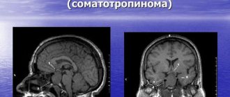
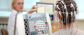
The diagnosis is established on the basis of anamnesis, wedge, pictures, as well as results obtained using instrumental studies. During an ultrasound examination of the brain (see Echoencephalography), a shift of the median echo signal to the healthy side and the absence of pulsation of echo signals are noted. On the EEG you can often see a local decrease in bioelectrical activity, respectively patol. focus and its disorganization up to a flat curve with severe decompensation. The angiographic picture indicates both the localization and type of patol. process, and for the presence of vasospasm, signs of hydrocephalus and brain dislocation (displacement of the main trunks of the cerebral arteries). With chronic process, using skull radiography, one can identify signs of intracranial hypertension, pronounced finger-like impressions, and an expanded sella turcica. Computed tomography (see Computed tomography) in most cases provides accurate information about both the nature and localization of the patol. focus, and about the state of the ventricles of the brain and the phenomena of its dislocation.
Examination of cerebrospinal fluid does not provide accurate data on the presence of intracranial hematoma; the liquid can be either colorless or blood-stained; the cerebrospinal fluid pressure can be increased, less often normal or decreased. It should be noted the danger of a spinal puncture (see) for diagnosing S. g.m. caused by an intracranial hematoma, especially in the phase of occlusion of the cerebrospinal fluid tract, since a decrease in the cerebrospinal fluid pressure in the subarachnoid space of the spinal cord after the puncture can lead to the emergence or increase symptoms of brain herniation.
Pathomorphology
Pathomorphologically, SHM is characterized by a volumetric accumulation of:
- liquid and/or coagulated blood (according to localization, the accumulation can be epidural, subdural, intracerebral, intraventricular);
- cerebrospinal fluid (subdural localization);
- detritus mixed with clotted blood (intracerebral localization);
- air (subdural and/or intraventricular localization).
The presence of a foreign accumulation causes local and general compression of the brain substance, displacement of the midline structures, deformation and compression of the cerebrospinal fluid reservoirs, dislocation and infringement of the brain stem.
The presence of a foreign accumulation causes local and general compression of the brain substance, displacement of the midline structures, deformation and compression of the cerebrospinal fluid reservoirs, dislocation and infringement of the brain stem.
Treatment
Radical treatment for S. g. m. is surgical intervention, which should not be limited only to the removal of patol. focus (hematoma, tumor, etc.) or decompressive craniotomy (see). The operation should, in addition, provide prevention or treatment of existing dislocation and herniation: dissection of the cerebellar tentorium - tentoriotomy, drainage of the cerebral ventricles (see Hydrocephalus), drainage of the residual cavity at the site of the patol. hearth. Surgical treatment must be carried out against the background of intensive drug therapy (dehydration and cardiovascular drugs). To compensate for respiratory failure, artificial ventilation is used (see Artificial respiration) in the mode of moderate hyperventilation.
Etiology
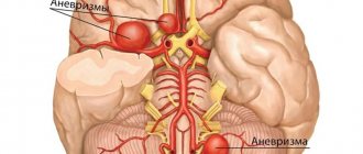
Among the causes of compression, intracranial hematomas (epidural, subdural, intracerebral, intraventricular) come first. This is followed by depressed fractures of the skull bones, focal crush injuries of the brain, subdural hygromas, and pneumocephalus. Cerebrovascular diseases (CVD).
Among the causes of compression, intracranial hematomas (epidural, subdural, intracerebral, intraventricular) come first. This is followed by depressed fractures of the skull bones, focal crush injuries of the brain, subdural hygromas, and pneumocephalus. Cerebrovascular diseases (CVD).
Among the causes of compression, intracranial hematomas (epidural, subdural, intracerebral, intraventricular) come first. This is followed by depressed fractures of the skull bones, focal crush injuries of the brain, subdural hygromas, and pneumocephalus. Cerebrovascular diseases (CVD).
Forecast
The prognosis depends on the duration of acute S. g. m., i.e., on the timeliness of hospitalization, diagnosis and surgery. Complex surgical and drug treatment of acute S. g. m., carried out in the early (up to 6 hours) terms after its occurrence, can reduce mortality from 50-60 to 25%. With surgical intervention in the phase of deep clinical decompensation, mortality is 60-75%, and in a terminal state - 100%. The prognosis for chronic S. g. m. in cases of timely recognition and elimination of the compression factor (tumor, hematoma, abscess, etc.) is favorable.
Bibliography:
Arseni K. and Nonetantinescu A. Intracranial hypertension, trans. from Romanians, Bucharest, 1978; Isakov Yu. V. Acute traumatic intracranial hematomas, M., 1977; Klumb and L.A. Neurophysiology of acute traumatic brain injury, Vilnius, 1976; Lebedev V.V. and Gorenshtein D.Ya. Treatment and its organization for traumatic brain injury, M., 1977; Likhterman L. B. and Khitrin L. X. Traumatic intracranial hematomas, M., 1973, bibliogr.; Guide to neurotraumatology, ed. A. I. Arutyunova, part 1, M., 1978; Samotok N. A. Principles of classification of acute closed craniocerebral injury, Vopr. neurosurgeon, c. 4, p. 3, 1978; Singur N. A. Brain contusions, M., 1970; Phasing of the clinical course of traumatic brain injury, ed. M. G. Grigoriev and L. B. Likhterman, Gorky, 1979; Surgical treatment of bruises and brain dislocations, ed. B. D. Komarova, M., 1974; Surgery of the central nervous system, ed. V. M. Ugryumov, part 1, L., 1969; HaseU. A. O. The influence of the decompressive operation on the intracranial pressure and the pressure-volume relation in patients with severe head injuries, Acta neurochir., v. 45, p. 1, 1978; Johnston IH a. Rowan JO, Raised intracranial pressure and cerebral blood flow, J. Neurol. Neurosurg. Psychiat., v. 37, p. 585, 1974; Miller JD ao Significance of intracranial hypertension in severe head injury, J. Neurosurg., v. 47, p. 503, 1977.
V.V. Lebedev.