Human anatomy is undoubtedly a major core subject for study in medical schools. Despite the fact that normal human anatomy is a discipline that stood at the origins of the development of medicine, a large number of scientific works still appear that make adjustments to modern anatomical atlases.
It would seem that human anatomy cannot change so quickly with the course of evolution, but our understanding of it is constantly improving as new research methods appear - evidence of this is provided by ever new versions of the anatomy atlas.
Atlas of anatomy Sinelnikova R.D. in 4 volumes, this is perhaps the most authoritative and time-tested source of knowledge on this topic. It is constantly republished, delighting us with its visual illustrations and text accessible to everyone. Many students tried to download Sinelnikov’s atlas for study, but the links either did not work, or there was a virus in the folder... We solved this problem by making a website dedicated to this source.
The main goal of studying human anatomy is to create a fundamental knowledge base for students for further study of other medical disciplines. It is difficult to imagine mastering a curriculum in physiology, pathological physiology, pathological and topographic anatomy, operative surgery, and a whole range of clinical disciplines without a thorough study of normal human anatomy.
It is very important for a student to have a visual image of the material studied; for this it is necessary to study human anatomy in pictures. The main feature of this science. of course, is the structuring of its sections and subsections, as well as a clear systematization of the entire nomenclature.
Thus, we can distinguish the following directions that correspond to each system:
- osteology (section on the bones of the human skeleton). Studies the skeleton, both as a whole mechanism and as individual bones. There is also a study of age-related changes in bones.
- syndesmology (joints, ligaments). An extremely important section for future orthopedists and traumatologists.
- myology (muscular system). He studies not only the structure, but also the development and physiology.
- splanchnology (internal organs). Includes the anatomy of the endocrine, digestive, respiratory, excretory and genitourinary systems.
- angiology (vessels and their derivatives). Information is presented on the structure of blood and lymphatic vessels.
- neurology (central and peripheral nervous system). An extremely important section for successful diagnosis of diseases and perhaps the most difficult.
- aesthesiology (the science of the sense organs). Everything about vision and hearing. And also about taste, olfactory and tactile sensitivity. Closely related to neurology.
Read online “Human Anatomy” by Prives Mikhail Grigorievich – RuLit – Page 344
The upper two mounds, colliculi superióres
, are subcortical centers of vision, both lower,
collículi inferióres
, are subcortical centers of hearing.
The pineal body lies in a flat groove between the superior tubercles. Each colliculus passes into the so-called handle of the colliculus, bráchium colliculi, which goes laterally, anteriorly and upward, to the diencephalon. The handle of the superior colliculus, bráchium colliculi superióris
, goes under the pillow, púlvinar, of the thalamus to the lateral geniculate body, corpus geniculatum laterale.
The handle of the inferior colliculus brachium colliculi inferióris
, passing along the upper edge of the trigonum lemnisci to the sulcus lateralis mesencephali, disappears under the medial geniculate body, corpus geniculatum mediale. The named geniculate bodies already belong to the diencephalon.
2. The ventral part, the cerebral peduncles, pedunculi cérebri , contains all the pathways to the forebrain.
The cerebral peduncles look like two thick semi-cylindrical white cords that diverge from the edge of the pons at an angle and plunge into the thickness of the cerebral hemispheres.
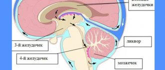
3. The midbrain cavity, which is a remnant of the primary cavity of the midbrain bladder, has the appearance of a narrow canal and is called the cerebral aqueduct, aqueductus cérebri . It is a narrow, ependymal-lined canal 1.5–2.0 cm long, connecting the IV ventricle with the III. Dorsally, the aqueduct is limited by the roof of the midbrain, ventrally by the tegmentum of the cerebral peduncles.
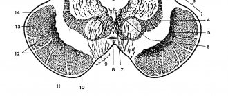
Internal structure of the midbrain . In a cross section of the midbrain, three main parts are distinguished: I) roof plate, lamina técti ; 2) tire, tegmentum , representing the upper section of the pedunculi cérebri; 3) the ventral section of the pedunculi cerebri, or the base of the cerebral peduncle, basis pedunculi cerebralis . According to the development of the midbrain under the influence of the visual receptor, it contains various nuclei related to the innervation of the eye.
In lower vertebrates, the superior colliculus serves as the main termination of the optic nerve and is the main visual center. In mammals and in humans, with the transfer of visual centers to the forebrain, the remaining connection of the optic nerve with the superior colliculus is important only for reflexes. In the nucleus of the inferior colliculus, as well as in the medial geniculate body, the fibers of the auditory loop (lemniscus lateralis) end. The roof of the midbrain has a bilateral connection with the spinal cord - tractus spinotectalis
and
tráctus tectobulbáris et tectospinális
. The latter, after crossing in the tegmentum, go to the muscle nuclei in the medulla oblongata and spinal cord. This is the so-called visual-sound reflex pathway, which was mentioned when describing the spinal cord. Thus, the plate of the roof of the midbrain can be considered as a reflex center for various types of movements that arise mainly under the influence of visual and auditory stimuli.
The cerebral aqueduct is surrounded by central gray matter, which in its function is related to the autonomic system. In it, under the ventral wall of the aqueduct, in the tegmentum of the cerebral peduncle, the nuclei of two motor cranial nerves are located - n. oculomotórius (III pair) at the level of the superior colliculus and n. trochlearis (IV pair) at the level of the inferior colliculus. The nucleus of the oculomotor nerve consists of several sections, corresponding to the innervation of several muscles of the eyeball. Medial and posterior to it there is also a small, also paired, vegetative accessory nucleus, nuccleus accessorius , and an unpaired median nucleus. The accessory nucleus and the unpaired median nucleus innervate the involuntary muscles of the eye, m. ciliaris and m. sphincter pupillae. This part of the oculomotor nerve belongs to the parasympathetic system. Above (rostral) the nucleus of the oculomotor nerve in the tegmentum of the cerebral peduncle is the nucleus of the medial longitudinal fasciculus.
Lateral to the cerebral aqueduct is the nucleus of the midbrain tract of the trigeminal nerve, nucleus mesencephálicus n. trigémini.
The cerebral peduncles are divided, as already noted, into the ventral part, or the base of the cerebral peduncle , basis pedunculi cerebralis
, and
tire
, tegmentum . The border between them is the black substance, substántia nigra, which owes its color to the black pigment contained in its constituent nerve cells - melanin (Fig. 284).
Rice. 284. Transverse section through the cerebral peduncles.
Age characteristics and prevention
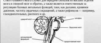
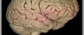
The brain is a complex structure. It operates with close interaction between all segments. The center that controls the middle section is the cerebral cortex. With age, connections become weaker, and reflex activity weakens. Since the area is responsible for motor function, even minor disruptions in this tiny segment lead to the loss of this important ability.
First of all, you should avoid hitting your head. If this happens, it is necessary to begin treatment immediately after the injury. It is possible to preserve the functions of the midbrain and the entire organ until old age if you train it with regular exercises:
- The lifestyle a person leads is important for physical and mental health. Drinking alcohol and smoking destroy neurons, which gradually leads to a decrease in mental and reflex activity. Therefore, you should give up bad habits, and the sooner you do this, the better.
- Moderate physical activity and walks in nature supply the brain with oxygen, which has a beneficial effect on its activity.
- You shouldn’t give up reading, solving charades and puzzles: intellectual activity keeps the brain active.
- An important aspect of the functioning of brain structures is nutrition: fiber, protein, and greens must be present in the diet. The midbrain responds positively to intake of antioxidants and vitamin C.
- It is necessary to control blood pressure: the health of the vascular system affects the general condition of a person.
The brain is a flexible system that can be successfully developed. Therefore, by constantly improving your mind and body, you can maintain clarity of thoughts and motor activity until old age.
The midbrain, its structure and functions are determined by the location of the structure, providing movement, auditory and visual reactions. If you have difficulty maintaining balance or lethargy, you should consult a doctor and undergo an examination to find the cause of the disturbances and eliminate the problem.
Anatomical structure and functions
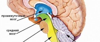
The midbrain is a very small formation, it contains several structures at once and is responsible for many functions, including reproduction. It would be a mistake to assume that the midbrain consists of the cerebral peduncles and the tegmentum. In addition to the base of the NM and the substantia nigra, it contains the quadrigeminal tract and the tegmentum. It is formed from a complex mesencephalic, ventral and dorsal part.
The main difficulty in considering the anatomical structure is the decision whether to include only the ridges from which the cerebral peduncles are formed, or whether to include in their composition the lid of the midbrain, the bottom and the specific substantia nigra:
- A traditional examination will not show anything other than the body of the legs itself, with a fossa located between them (tarina or interpeduncular). It is part of the interpeduncular cistern, located at the base of the human skull, from where the midbrain receives the cerebrospinal fluid necessary for life.
- In the interpeduncular area there is the exit of the nerve that moves the eyeball, eyelid and ensures the reaction of the pupil to a light stimulus, and on the outer surface there is the trochlear nerve, without which the movement of the oblique muscles and some characteristic turns of the eyes in the orbit are indispensable.
- The black mass, in one interpretation – a demarcating structure, and in another – an integral part of the operating system, develops from the mesencephalon and regulates the coordination of movements. Its remarkable feature is that this is the only area of the NCC in which the cells are stained with melanin.
- The base of the NM (base of the brain) is nothing more than the lowest part located in front. There are discrepancies in determining the identity of this part not only in old anatomy textbooks, where these terms were considered equivalent or were simply not distinguished as a separate entity. Now the ONM is precisely the lower part, in which there are axons stained with myelin.
- In contrast to the base, the tegmentum is located between the substantia nigra and the aqueduct of the brain, it forms both one and the other segments located nearby. Its structures include the red nucleus, its ventral and dorsal parts.
- It also contains gray (periaqueductal) matter, which is anatomically classified as the tegmentum, although it is located next to the cerebral aqueduct. OSV is considered the main center of regulation of the pain symptom, going along the descending line. It is capable of projecting to the nuclei of the thalamus and spinal cord, as well as to the raphe nuclei and locus coeruleus.
The importance of anatomical formation, regardless of its composition in the first or second case, is undeniable. After all, it is precisely this that provides orientation based on the visual image and the received stimulus, ensures the maintenance of a certain body posture and the regulation of muscle tone at the reflex level.
The connection of an anatomical formation with other areas of the brain is carried out in the most variable ways - from the fibers of the corticospinal tract going to the most important analyzers (ascending and descending), to information transmitted by neurons of all adjacent formations of gray, white and black.
Functions
All areas of the brain work interconnectedly, together creating a unique system for supporting human life. The main functions of the midbrain are designed to perform the following role:
- Sensory functions. The load for sensory sensations is carried by the neurons of the quadrigeminal nuclei. Signals from the organs of vision and hearing, the cerebral cortex, the thalamus and other brain structures arrive to them along pathways. They provide accommodation of vision to the degree of illumination by changing the size of the pupil; his movement and turns of his head towards the irritating factor.
- Conductor. The midbrain plays the role of a conductor. The base of the legs, nuclei and substantia nigra are mainly responsible for this function. Their nerve fibers are connected to the cortex and underlying brain regions.
- Integrative and motor. Receiving commands from sensory systems, the nuclei convert the signals into active actions. Motor commands are given by the stem generator. They enter the spinal cord, making possible not only muscle contraction, but also the formation of body posture. A person is able to maintain balance in various positions. Reflexive movements are also made when moving the body in space, helping to adapt so as not to lose orientation.
Consequences of damage to the peduncles of the human brain
Any damage in this area leads to the development of numerous syndromes described in the specialized literature and even named after the scientists who first recorded the phenomenon for science.
The nature of the manifestations depends on what exactly has undergone destructive changes:
- damage to the oculomotor nerve, depending on the dislocation, results in vertical nystagmus, incoordination or ophthalmoplegia of various kinds (internal total, external), Nothnagel syndrome;
- destruction of the base of the pedicle leads to paralysis of the oculomotor nerve and destruction of the pyramidal fasciculus;
- injury in this area results in cerebellar disorders, loss or impairment of the functionality of the organs of vision and hearing;
- atrophic changes in the cerebral peduncles can lead to loss of the ability to respond to external stimuli and can lead to loss of the ability to independently support life.
Being relatively small in size, the midbrain and the control and management structures located in it play a vital role in ensuring the normal functioning of the organs of hearing and vision. They are the control center for sleep and even cerebellar activity. A lesion of any type - injury, the appearance of a neuroinfection, a failure of cerebral circulation, malignant and benign neoplasms will inevitably cause disturbances in the functioning of the systems for which they are responsible.