Definition
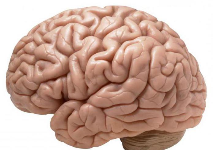
The striatum is an anatomical structure of the telencephalon, which belongs to the basal ganglia of the human hemispheres.
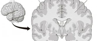
The body got its name because, in frontal and horizontal sections of the brain, it looks like alternating stripes of white and gray matter.
The earliest studies showed that the peak activity of the striatum occurred when a person turned 15 years old. But recent work shows that the body's real activity begins closer to the age of 25, and hyperactivity occurs at the age of 30.
In addition, after conducting a rather interesting study, scientists found that the brain reacts when pay does not cover the effort that a person puts into work. Thus, if an employee understands that his colleague receives more for the same amount of work, then the motivation for long-term work capacity decreases. Conversely, when work is revalued, the desire to work increases.
Neuroscience for everyone. Details: striatum
Whenever scientific news talks about human behavior, motivation for activity, or how to learn better, the reward system is almost always mentioned. We will talk about the anatomical structure of the brain, which is precisely responsible for this.
This structure is called the striatum ( corpus striatum
) or striatum. It is located in the very front part of the brain. The striatum is part of the basal ganglia (and is considered the largest of the 5 cellular components of the basal ganglia), the main function of which is the conscious control and management of movements, as well as learning to do so.
Historically, it was believed that the main function of the entire basal ganglia complex was the embodiment of movement. For example, it was noticed that in patients with Parkinson's disease, along with impaired motor activity, there were also changes in the ganglia. It is quite easy to connect these two observations into one whole.
Then, as scientific knowledge accumulated, it became known that the basal ganglia can control eye movements and even influence behavior.
Their violations can lead to diseases in which either hypokinesis (decreased motor activity) or hyperkinesis (excessive, sometimes “extra” movements) is manifested.
Anatomical structure
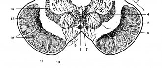
The striatum received its second name in Russian (“striated body”) due to the fact that alternating light and dark stripes are visible on its sections. In Latin, this word (“striatum”) also means “striped.”
Usually the striatum is divided into two parts - the dorsal (located on top), which is represented by the caudate nucleus
), the lentiform nucleus (
lentiform nucleus
or
lenticular nucleus
) and the putamen, and the ventral one, which contains the
nucleus accumbens
.
Caudate and lentiform nucleus. Illustration: Wikimedia Commons
Structures called nuclei are specific areas in the brain that can be separated or, conversely, combined with other nearby neurons by function or connectivity.
Inside them there are also “subnuclei” ( subnuclei
) due to the fact that even in such a small formation as the nucleus, a more accurate and subtle division of neuronal functions can occur.
The dorsal part of the striatum is important for the decision-making process and for choosing how to respond to an event, for choosing actions.
The nucleus accumbens is associated with reward and reinforcement systems, and depending on the correctness of its operation, a transition can occur from simply performing actions that bring pleasure to constant targeted desires to perform these same actions (addiction).
The striatum constantly receives nerve impulses from many parts of the cerebral cortex: motor, sensory (with some exceptions), associative, limbic and paralimbic. And each of these areas is projected onto a strictly defined zone in it.
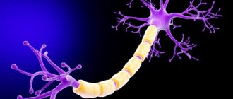
The striatum contains a variety of cells, but the majority (approximately 90%) are medium-sized styloid GABAergic projection neurons.
Such neurons collect and analyze information that the striatum receives and transmit it to neurons in the various basal ganglia.
The striatum is divided into two parts in humans and macaques by a thin strip of white matter, compared to rats and mice, in which the caudate nucleus and putamen are not separated. This white matter forms the internal capsule, however, many connections remain between the caudate nucleus and the putamen.
The size of the human brain is 13-18 times larger than that of a rhesus monkey, but the size of the striatum is only 6 times larger. By the way, in one of the studies, scientists found that the volume of the striatum in people with Parkinson's disease, oddly enough, is larger (7.5 cm3) than in healthy people (6.3 cm3).
Functions
The striatum has many functions. This is the planning and control of movements, various cognitive processes that are needed to perform various actions. The striatum is involved in learning, and its interactions with dopamine-containing neurons in the midbrain are very important for this.
Striatum on MRI. Illustration: Wikimedia Commons
The striatum shares its role in decision making with the prefrontal cortex. They are very closely interrelated with each other and can often even be mutually activated. Therefore, some scientists even suggest deep stimulation of one thing in order to indirectly influence another.
The striatum is important in the functioning of the reward system, and, moreover, scientists have shown that it is possible to derive pleasure not only from past and ongoing events or actions, but even from simply anticipating the events themselves.
Pathology
But how does the striatum behave in pathology? What could even happen in it?
The most dangerous thing is the cessation of the production of dopamine, which is exactly what is needed for correct movements. During old age, the number of dopamine receptors decreases, and in general the concentration of this neurotransmitter decreases.
Interestingly, not only physical changes in the striatum can lead to deviations in its functioning, but genetic factors also play a role. For example, overexpression of dopamine D3 receptor genes in the striatum disrupts motivation and motivational behavior in mice, but does not affect other behaviors.
It is with problems with the nucleus accumbens that addiction begins - behavior when people have an obsessive need for a specific activity. This may be expressed in the fact that a person gets used to medications and feels a constant need for them, or it may be, for example, a desire to perform monotonous actions.
In Huntington's chorea, the HTT gene, which encodes the hangtintin protein, contains repeats of three CAG nucleotides, and if the number of such repeats is more than 36, then the spatial structure of the protein changes. And a person develops chorea.
In this case, the hangtintin protein begins to form aggregates with other proteins, and therefore interferes with normal cellular transport in neurons and can lead to their death.
Nadezhda Potapova
Previous materials on neuroanatomy (Details series): thalamus, hypothalamus, Purkinje cells.
Read the materials on our website on VKontakte and the Telegram channel , and also follow the new pictures of the day on Instagram .
Source: https://zen.yandex.ru/media/neuronovosti.ru/neironauki-dlia-vseh-detali-striatum-5a56e34a3c50f77e4eecb7a9
Structure
The striatum consists of:
- Caudate nucleus.
- Lenticular nucleus.
- Fences.
If we examine the body under a microscope, it consists of large neurons with long tails extending beyond the boundaries of the striopallidal system.
The parts of the caudate are the head, body and tail. The head forms the lateral wall of the anterior horn of the lateral ventricle; the body of the nucleus is extended along the central part of the ventricle; the tail is located on the upper wall of the inferior horn of the ventricle and ends at the level of the lateral geniculate body.
The posterior wall of the nuclear head is located on the border with the thalamus, separated by a strip of white matter.
The lenticular kernel, as the name implies, is shaped like a lentil.
It is located sideways to the caudate nucleus and thalamus. If you cut the kernel in half, it has a wedge-shaped shape, with the top facing the middle and the base facing the side.
And small layers of white matter divide the nucleus into several parts:
- The shell.
- Lateral globus pallidus.
- Medial globus pallidus.
The globus pallidus is a specific ancient formation (ancient body), which differs from other parts of the striatum both in macroscopic and histological appearance.
The fence is located outside the lenticular core. Externally, it is a thin, up to two millimeters, plate of a gray substance. The middle of the plate is smooth, and on the lateral edge there are small bulges of gray matter.
Main functions
The striatum of the brain is considered one of the main subcortical regulatory and coordinating centers of the motor system.
Thanks to experiments, it has been proven that the body contains vegetative coordinating centers that regulate heat generation, heat release, metabolism and vascular reactions.
The main functions of the striatum include:
- Regulation of muscle tone.
- Decreased muscle tone.
- Participation in the regulation of the work of internal organs.
- Participation in behavioral reactions.
- Participation in the formation of conditioned reflexes.
function
The ventral striatum, and the nucleus accumbens in particular, primarily mediates reward, cognition, reinforcement, and motivational convexity, while the dorsal striatum is a major mediator of cognitive function involving motor function, certain executive functions (such as inhibitory control and impulsivity), and also stimulus-response learning;
There is a small degree of overlap, as the dorsal striatum is also an integral part of the reward system that, along with the nucleus accumbens, mediates the encoding of new motor programs associated with future reward acquisition (for example, a conditioned motor response to a cue reward).
Metabotropic dopamine receptors are present on both spiny neurons and axonal cortical terminals. Second messenger cascades caused by activation of these dopamine receptors can modulate pre- and postsynaptic function, both in the short term and in the long term. In humans, the striatum is activated by stimuli associated with reward, but also by aversive, novel, unexpected, or intense stimuli, and by cues associated with such events.
fMRI evidence suggests that a common binding property of these stimuli to which the striatum responds is salience under presentation conditions. A number of other brain regions and circuits are also associated with reward, such as the frontal. Functional maps of the striatum reveal interactions with widespread cortical regions important for a variety of functions.
The interaction between the striatum and the prefrontal cortex is relevant to behavior, especially adolescent development, as proposed by the dual system model.
Damage to the striatum and consequences
When the striatum stops functioning, a person experiences the following disorders:
- Athetosis. Banal alternating movements of the limbs.
- Chorea. Incorrect movements that are performed without any sequence or order, involving the entire musculature of the body.
- Strengthening unconditioned reflexes (defensive, indicative, etc.).
- Hyperkinesis. Significant strengthening of auxiliary movements that accompany each main movement.
- Hypotonicity. Upset muscle tone, its decrease.
- The appearance of Tourette's syndrome.
- The onset of Parkinson's disease contributes to the death of neurons in the body, which is why domafin, which is responsible for the motor system of the human body, is not produced.
- The appearance of Huntington's disease.
In addition, damage to the striatum and caudal nucleus in particular:
- Completely or partially prevents the perception of painful, visual, auditory and other types of stimulation.
- Reduces or increases salivation.
- Makes it difficult to orient in space.
- Impairs memory.
- Slows down the growth of the body.
- Promotes the disappearance of conditioned reflexes for a long time. Human behavior can be inert and stagnant.
How the brain works: the striatum
As promised, today we are talking about the second basal nucleus of the brain - the striatum (corpus striatum).
It is not surprising that it is “striped”, because it consists of layers of white and gray matter and in cross-section the color is like that of a zebra. This area of the brain consists of the caudate nucleus (nucleus caudatus), the lentiform nucleus (nucl. lentiformis) and the claustrum.
The lentil-shaped kernel, as is already clear, is similar to a lentil grain. Small layers of white matter divide it into three parts (nuclei):
– shell of the lenticular kernel (putamen);
– lateral globus pallidus (globus pallidus lateralis);
– medial globus pallidus (globus pallidus medialis).
The fence is located outward from the lenticular core. It is a plate of gray matter up to 2 mm thick.
And the next layer envelops the caudate nucleus, which is similar in appearance to the hippocampus.
The striatum receives afferent impulses (from the peripheral nervous system to the center) mainly from the thalamus, partly from the cortex, and sends efferent impulses (from the central nervous system to the peripheral) mainly to the globus pallidus.
The striatum is currently considered the highest subcortical regulatory and coordination center of the motor apparatus.
It was found that low-frequency electrical stimulation of the caudate nucleus changes the behavior of the animal - drowsiness and a sleepy state occurs and the reaction time of neurons in the cerebral cortex is prolonged.
The striatum regulates muscle tone, reducing it; participates in the regulation of the functioning of internal organs, plays a role in various behavioral reactions (for example, food-procuring behavior); participates in the formation of conditioned reflexes.
With lesions of the striatum, athetosis (stereotypical rhythmic movements of the limbs) and chorea (strong irregular movements that occur without any order or sequence and involve almost all the muscles, the second name is “St. Vitus’s dance”) are observed in humans. Both athetosis and chorea are considered to be the result of a loss of the inhibitory influence that the striatum has on the pallidum.
Chorea or “Dance of St. Vitus”
Also, with damage to the striatum, there is a significant increase in unconditioned reflexes - defensive, orientation, etc. The auxiliary movements accompanying each main movement are also significantly increased (this increase in auxiliary movements is called hyperkinesis). At the same time, muscle tone is upset, usually in the direction of lowering it - hypotonicity.
Damage to the striatum can result in Tourette's syndrome. Neurons in the striatum die in Parkinson's disease. When neurons are damaged in the striatum, dopamine, which is responsible for motor functions in the human body, ceases to be produced.
The striatum (as well as other brain structures in the future) is also affected in Huntington's disease.
Previously, researchers believed that the striatum reached its peak activity at the age of 15, but work in 2020 showed that real maturation begins 10 years later - by the 25th year of life. At a time when young people begin a completely independent life. Scientists suggest that the “hyperactivity” of the striatum continues until the age of 30.
Another interesting work was carried out in 2014, where a group of researchers from the Norwegian School of Economics found out how the brain reacts to the situation when the payment for our work does not “cover” the effort.
The subjects always worked in pairs, completing the task for 30, 60 or 90 minutes, but so that the sum of the working time in the pair was 120 minutes. After the end of the experiment, participants were paid the money they earned.
The problem was that the amount due to partners for two hours of work was not always distributed fairly.
During the study, scientists recorded the neural activity of the striatum in response to a similar situation. The results showed that if a person received an honestly earned amount, the activity of the striatum did not change in any way. If there was less or more money, the device recorded a surge in neural activity.
Interestingly, if a person understands that his colleague receives more compensation for similar work, his motivation decreases, and vice versa - the feeling that the work is overvalued gives an even greater desire to work.
Anastasia Sheshukova
Especially for the portal “Neurotechnologies.rf”
Source: https://neurotechnologies.ru/articles?id=224