American neurosurgeons Joseph Bogen and Philip Vogel , as well as neuropsychologist Roger Sperry , established in the mid-20th century that the right and left hemispheres of the brain perform different cognitive functions. However, the results of their research were misunderstood by many, which led to the belief that all people have a dominant one of the brain hemispheres: the right is responsible for logic and prudence, and the left for imaginative thinking and creativity.
In fact, all people use both the right and left hemispheres of the brain to almost the same extent. However, each of them provides different principles for the perception of reality, the organization of speech and color recognition.
Article on the topic Living supercomputer. Why does the brain need water and why is it more active at night?
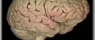
General information about the structure of the brain
They have been trying to study it for a long time, but for all this time scientists have not been able to accurately and unambiguously answer the question 100% of what it is and how this organ works.
Many functions have been studied, for some there are only guesses. Visually, it can be divided into three main parts: the brain stem, the cerebellum and the cerebral hemispheres. However, this division does not reflect the full versatility of the functioning of this organ. In more detail, these parts are divided into departments responsible for certain functions of the body.
Oblong section
The human central nervous system is an inextricable mechanism. A smooth transitional element from the spinal segment of the central nervous system is the medulla oblongata. Visually, it can be imagined as a truncated cone with the base at the top or a small onion head with thickenings diverging from it - nerve tissues connecting to the intermediate section.
There are three different functions of the department - sensory, reflex and conductive. Its tasks include controlling the basic protective (gag reflex, sneezing, coughing) and unconscious reflexes (heartbeat, breathing, blinking, salivation, secretion of gastric juice, swallowing, metabolism). In addition, the medulla oblongata is responsible for such senses as balance and coordination of movements.
Midbrain
The human midbrain is responsible for such an important ability of the body as sleep.
The middle section has a complex structure. There are 4 clusters of nerve cells - tubercles, two of which are responsible for visual perception, the other two for hearing. Nerve clusters are connected to each other and to other parts of the brain and spinal cord by the same nerve-conducting tissue, visually similar to legs. The total size of the segment does not exceed 2 cm in an adult.
Diencephalon
The department is even more complex in structure and functions. Anatomically, the diencephalon is divided into several parts: Pituitary gland. This is a small appendage of the brain that is responsible for the secretion of necessary hormones and regulation of the body's endocrine system.
The pituitary gland is conventionally divided into several parts, each of which performs its own function:
- The adenohypophysis is a regulator of peripheral endocrine glands.
- The neurohypophysis is connected to the hypothalamus and accumulates the hormones it produces.
Everyone knows that if the brain stops functioning, then a person does not react to any external factors, does not show any activity, and turns into a “vegetable.” The structure of the brain is symmetrical and consists of the right and left hemispheres.
Disputes between scientists do not subside, but some facts have been proven and approved.
Important facts:
- The human brain consists of 25 billion neurons.
- The adult brain makes up about 2% of body weight.
- The organ consists of three membranes: hard, soft, arachnoid. The shells perform the main – protective function.
It is generally accepted that the left hemisphere is responsible for all mental processes, and the right hemisphere for the perception of the outside world. Roughly speaking, the left is the logical hemisphere, and the right is the creative hemisphere.
From an anatomical point of view, the brain consists of the following parts:
- Medulla. Responsible for vegetative functions.
- Midbrain. Controls reflexes to external stimuli.
- Hindbrain. Responsible for coordination of movements.
- Diencephalon. Includes sensory centers (hunger, thirst, satiety, sleep regulation).
- Forebrain. The largest part, which is covered with grooves (convolutions). Provides better brain function.
Frontal lobe
Returning to the question of which part of the brain is responsible for speech, it is necessary to dwell on the study of the frontal lobe. First of all, there is a statement that the left hemisphere of the brain is responsible for the ability to speak. Speech centers are located here.
The frontal part of the cerebral hemispheres is of great importance in human daily life. She is responsible for:
- The nature of thinking.
- The process of urination.
- Maintaining the body in an upright position.
- Motivation and behavior control.
- Speech and handwriting.
The frontal lobe takes responsibility for the semantic construction of human speech.
The role of this part of the brain is not so extensive, but much more narrowly focused. The temporal lobes are located in both the left and right hemispheres of the brain, which affects their basic functions.
The left temporal lobe is responsible for:
- Perception of sound information.
- Short-term memory.
- Selection of words during a conversation (role in speech formation).
- Synthesis of visual and auditory information.
- Interaction of music and emotions.
The right temporal lobe is responsible for:
- Facial expression recognition.
- Perception of rhythm and musical tone.
- Perception of speech intonation.
- Recording visual facts.
This part of the brain allows a person to understand by the intonation of the interlocutor’s speech about his emotions and attitude to the issue under discussion.
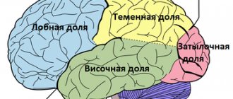
The cerebral cortex has four lobes:
- occipital;
- parietal;
- temporal;
- frontal
Each share has a pair. All of them are responsible for maintaining the vital functions of the body and contact with the outside world. If injury, inflammation, or disease of the brain occurs, the function of the affected area may be completely or partially lost.
Functions of the right hemisphere
The highest brain function is thinking. The brain is responsible for planning, decision-making, emotions and feelings. To determine which hemisphere is dominant, just do a few simple tests. After clapping your hands or crossing your arms over your chest, you need to pay attention to the palm or shoulder, which in these situations is on top. The functions of the right hemisphere are assigned to separate areas of the brain:
- Occipital lobe. Processing and storing information coming from the surrounding space. Thanks to the work of the occipital brain structures, a person distinguishes the shape and color of objects, recognizes facial expressions, manner of gestures, and individual facial features of other people.
- Temporal lobe. Processing and storing information about the nature and characteristics of non-speech sounds, which include the noise of the sea and wind, rustling leaves, birdsong, technical knocking and rumble, musical chords. The temporal region of the brain perceives the timbre and pitch of the voice, sound intonation and features of speech pronunciation.
- Parietal lobe. Responsible for the accumulation of spatial experience. The database is formed from early childhood and includes motor skills (the ability to independently put on clothes, use cutlery, wash, walk, hold a needle and sew, ride a bicycle). Controls the tactile function, allowing you to identify an object by touch, with your eyes closed, thanks to tactile sensations upon contact with it.
- Frontal lobe. Controls the implementation of non-verbal actions.
If a person has developed parts of the brain on the right, he exhibits creative abilities. The right hemisphere is responsible for all types of creativity: poetic, dance, visual, acting, artistic inclinations and skills. The main functions of the right side of the brain structures:
- Processing of non-speech information. Perception, analysis, interpretation of information that is expressed not in verbal form, but in the form of symbols and images.
- Imagination. Thanks to the brain structures located in the cranium on the right, a person has the ability to dream, imagine and fantasize. The right-sided parts of the brain give us the ability to play music, draw, and create objects of fine art.
- Orientation in space. A person objectively assesses his location. Thanks to the work of the right side of the brain, it is possible to find a way in a forest or metropolis, to put together whole images from small, scattered fragments (puzzles, mosaics).
- Control of motor activity in the left half of the body (for right-handers) - movements of the left hand, left leg.
- Creative thinking. Simultaneous processing of a large volume of heterogeneous information and the formation of a spatial understanding of the surrounding world. Any task or object is considered as a whole and from different points of view.

The right hemisphere of the brain is responsible for intuition, which in women manifests itself in the form of the ability to subconsciously identify the essence of a situation, personality traits, and the reasons for the actions of other people. In women, the right side of the brain is usually dominant, which is expressed in increased sensitivity and the predominance of an intuitive assessment of events.
The right hemisphere of the brain is responsible for understanding allegorical phrases - metaphors, which in men is manifested by the ability to adequately interpret allegorical, figurative statements. In men, the left hemisphere is often more developed; a logical, balanced approach to solving problems and perceiving environmental factors predominates.
Brain functions
It is almost impossible to list all the functions. Areas of the brain are responsible for all human actions in everyday life.
Main functions:
- Reasonable function, or human thinking.
- Processing of external signals that coordinates taste, vision, hearing, and smell.
- Managing psychological state and emotions.
- Regulation of basic movements, reflex function.
In ordinary life, a person does not think about why he acts one way or another. The brain is responsible for all actions.
The motor area is located in the front part of the frontal lobe of the left hemisphere, next to the motor center, which is responsible for muscle activity. Main function of the motor area (Broca's area):
- Responsible for the motor ability of the tongue. In case of any violations in this department, the person continues to understand speech, but is not able to respond.
The sensory area is located in the posterior part of the temporal lobe of the brain. The main task of this center (Wernicke Center) is:
- Perception and storage of oral speech, both one’s own and those of others. If disturbances occur in this area, then the person ceases to perceive the speech of others, although he himself retains the ability to speak, albeit with defects.
If for some reason the sensory speech zone has to be removed, then the person completely loses the ability to perceive and produce speech.
Connection between hearing and the left hemisphere of the brain
The first thing that attracts attention when testing speech hearing after switching off or damaging the left hemisphere is a clear lack of interest in the sounds of human speech. The subject does not notice that he is being asked a question. In order for him to hear the speech addressed to him and pay attention to these sounds, they must be somewhat amplified compared to what is required in a normal state. And yet the subject from time to time ceases to perceive them. The experimenter constantly has to repeat the question or task, ensuring that the subject finally hears them and does what is required. The patient does not recognize many words, and answers questions slowly, not immediately, as if he needs a second or two every time to collect his thoughts.
Let's try to figure out what happens to hearing, why the test subject hears simple sounds and distinguishes them well from each other, but does not recognize or understand words. Let's start with the most elementary units - phonemes. With extensive damage to some areas in the temporal region of the left hemisphere or temporary deep shutdown of the entire left hemisphere, difficulties arise in distinguishing them. Confusion occurs when listening to phonemes that differ in vowels and consonants. Subjects cannot clearly distinguish and repeat
,
o
,
e
or
i
.
They are usually confused with
e
,
o
s
y
, but
a
does not cause any particular difficulties and is usually perceived correctly.
It is even more difficult to distinguish be
from
ne
,
do
from
to
,
ka
from
ha
.
It is very difficult to notice the difference between s
and
z
,
w
or
g
.
No less difficulties are caused by meaningless combinations of speech sounds such as: leb
,
mut
,
pur
,
bir
.
They are often perceived incorrectly, and subjects repeating them introduce additional distortions. Fep
,
put
,
vur
,
ber
appear .
The ear is not a flawless device. Every healthy person, after listening to several meaningless sound combinations, can make a mistake. We are more accustomed to dealing with ordinary words that have a very specific meaning. It is not surprising that we often perceive a familiar word behind poorly heard sounds. Among the few mistakes a healthy person makes, the most common are cases of comprehending the sounds he hears. Instead of meaningless sound combinations of the word bread
or
lion
,
mule
or
mat
,
puk
or
tur
,
drill
,
bar
or
bor
.
When the functioning of the sound-perceiving centers of the left hemisphere is disrupted, such errors do not occur; more often, ordinary, well-known words are perceived as a random set of sounds. Trying to repeat them, the subject generates a chain of sounds that is consonant with the word he heard, of the same length, with the same rhythmic and sound pattern, but which has nothing in common with any of the words known to the subject. And strangely enough, the world of meaningless sounds in which a person is now immersed does not cause him any particular surprise, irritation, or protest.
When the function of the left hemisphere is suppressed or due to its diseases, memory for sounds, including speech, suffers sharply. Therefore, even if individual tones are easily identified, distinguishing complexes of three to five sounds is impossible. Recognizing speech sounds is even more difficult. Most often, the subject is able to accurately repeat them separately, but usually cannot cope with several at once. He is able to repeat a simple combination of three sounds “a-o-u” only immediately after listening, and after a minute he begins to get confused. Already forgot!
For the same reason, even the rhythms cease to differ. To check, tap a simple rhythm with your finger: knock-knock, knock-knock, knock-knock or knock-knock-knock, knock-knock-knock. If it is played at a fast pace and the subject did not have time to count the number of beats in “packs” between longer pauses, then he does not notice any rhythm and cannot repeat it.
The memory capacity for sounds in such people is narrowed, and its duration is significantly shortened. With a fairly well-preserved ability to recognize individual speech sounds and repeat them, a person will become confused if there are three to five of them. Although he recognized each individual sound, the process of analyzing the next sound prevents him from retaining the previous one in memory. When he reached the third sound, the first was already forgotten. Analysis of a whole word is very difficult for him, especially if it contains poorly differentiated sounds, such as p
and
b
:
beech
and
bunch
,
base
and
groove
,
bike
-
soldering
,
gang
-
panda
,
reality
-
dust
,
board
-
port
.
And when a word contains two or three difficult sounds ( fornication
-
plut
), the barrier cannot be overcome, despite repeated listening.
Due to the difficulties of sound analysis, synthesis also suffers. A person loses the ability to select the necessary sounds and arrange them in long chains so that words or sentences arise from them. That is why a painful process that affects the auditory center will certainly disrupt speech.
In severe cases, patients do not speak at all. Although articulation is intact, the stream of sounds they emit can become completely unintelligible. Experts call this symptom word salad. It seems that ordinary speech is chopped into small pieces, everything is thoroughly mixed and presented to the listeners in this form. The patient actually mixes speech sounds almost in random order.
In mild forms of the disease, the patient is able to recognize and reproduce simple words, such as table
,
chair
,
glasses
. But try to pronounce the same words not together, but with a tiny interval between individual sounds - s-t-o-l, s-t-u-l. The patient recognizes them and even remembers the sequence, but will not be able to compose or synthesize a word from them.
A very characteristic symptom of switching off Wernicke’s area or the entire left hemisphere is loss of perception of the meaning of words. How could it be otherwise if a person is not able to notice the difference between the words hammer
and
poradok
or
malovok
.
Sometimes the patient has a vague idea that the word he heard belongs to a certain category. For example: a hammer
is a tool and is somehow connected with repair, with a workshop. It is especially difficult to remember the name of the desired item if it is not in front of your eyes. Prompting the first syllable or even the first two syllables usually does not help recall it. When communicating with others, patients try to replace lost words with similar ones.
A very interesting feature of the loss of functions of the Wernicke center: the immediate meanings of words suffer more than the more general ones. When replacing a lost word, these general meanings and associations are used, often very distant and unexpected. Therefore the hammer
can be replaced by the word
blacksmith
or more often
forge
. Guessing that they are not understood, patients are forced to utter several sentences instead of one word: “Well, that’s... what they’re hammering with,” “Well, that’s... they’re fixing...”, “Well, there... in the workshop.”
In the speech of such a person there are few names of objects and their qualities, and if they exist, they are often distorted beyond recognition or replaced with words denoting the action or relationship of objects to each other. Thanks to this, even with gross defects, it is still possible to understand what the person wanted to say. Moreover, the preservation of abstract, abstract concepts like direction
,
cost
,
value
,
circumstances
may be satisfactory. In general, speech consists mainly of connectives, prepositions, adverbs and interjections, and the more serious the brain damage, the greater the place they occupy in the patient’s statements.
Speech impairment cannot but affect thought processes. In the past, such profound brain pathology was explained as a serious intellectual disorder. In reality, only thought processes based on retaining systems of speech cords in memory suffer. An attempt to force one to think out loud by repeating a chain of necessary logical operations several times not only does not help the thinking process, but, on the contrary, worsens it, causing memory overload.
How great memory exhaustion is can be seen from a simple example. With mild forms of damage to Wernicke's area, knowledge of many simple and common words can be preserved. The patient can correctly repeat and show the eye, nose, ear. However, if he is asked to show the same parts of his face again, then confusion soon begins. The patient is unable to repeat the already mentioned detail again, distorting the familiar word more and more: eye
...
gas
...
class
. And now he shows completely wrong parts of his face. It is quite natural that patients are completely unable to understand such a phrase as “Masha is lighter than Tanya, but darker than Tamara,” no matter how many times it is repeated. The patient cannot say which of the named girls is the lightest or the darkest.
The preservation of intelligence is evidenced by the ability to operate with abstract concepts, the ability to grasp and understand the meaning of a metaphor, and to correctly use linking words, prepositions, and conjunctions in speech. Turning off Wernicke's center does not lead to disruption of spatial relationships and does not interfere with the classification of objects based on more general meanings of words.
The less participation of speech and speech memory an intellectual task requires, the easier it is to perform. Freely operating with plot pictures, laying out and combining them in front of him on the table, the patient is able to build them into a coherent, sequential story. He is completely unable to number the pictures in the required sequence without moving them.
The ability to perform arithmetic operations, if they are performed in writing, can be preserved even with severe speech impairment. There is a known case of such a patient who continued to perform the functions of chief accountant. He submitted annual reports efficiently and on time, but could not remember the names of his closest employees, and constantly confused and distorted them. However, the operations of mental counting are completely destroyed. After all, in this case it is necessary to keep in mind the results of intermediate operations, and the memory is lame on both legs.
Memory impairment is the most noticeable symptom. Whatever the Wernicke center does in a healthy person, it acts like a sieve, sifting out and retaining the necessary linguistic material. When he becomes ill, holes appear in the sieve and it ceases to perform its function.
Wernicke's zone
In the Greek province of Phocis, on the southern slope of Parnassus, the rocky peaks of which are covered with snow almost all year round, in ancient times there was Delphi - a sacred city, a pan-Greek religious center. Here, almost thirty centuries ago, the Temple of Apollo was erected, one of the most famous and richest temples in the world, closed in 390, during the rise of Christianity, by Emperor Theodosius I.
The temple stood on a high terrace in the center of the site, enclosed by an arc of a high stone wall. The glory of the temple was due not only to its wealth. It was here, inside, that the navel of the earth was located, its center, marked by the sacred stone omphalos and sculptures of golden eagles. Tradition claimed that Zeus once wanted to find out where the center of the earth was. Not being able to measure it, he simultaneously released two eagles from both ends of the earth, east and west. In the place where the birds met, the omphalus was installed.
The oracle located here gave the temple even greater grandeur. No one dared to enter the inner part of the temple except the Pythia, the priestess-broadcaster of the will of Apollo. Prophecies were made nine months a year, except for three winter months.
Pythia had been preparing for them for a long time. For the last three days before the scheduled day, she did not eat anything, bathed in the sacred waters of the Kastalsky spring, and did not communicate with anyone. When the long-awaited hour finally arrived, she took a sip of sacred water and, having chewed the leaves of the sacred laurel, took a place on a golden tripod at the crevice of the rock, from which poisonous fumes rose.
Perhaps from the laurel juice swallowed on an empty stomach, from hunger and poisonous fumes, or perhaps simply under the influence of suggestion, the priestess fell into ecstasy and began to shout out some words. Her task was not difficult, she could shout out anything she wanted without worrying about anything, since between Apollo and the Greek people there was one more authority - the priests - interpreters of the will of God, conveyed through the lips of the Pythia. They were the most responsible link in the cult of divination. They needed not to make a mistake, not to miss their benefits, and in case of an always possible mistake, to protect themselves from reproaches and the threat of disqualification. It is no coincidence that Apollo’s will was conveyed in a deliberately unclear and ambiguous form.
The organization of brain functions is in many ways reminiscent of the distribution of responsibilities of the Delphic oracle. Some centers, such as the Wernicke Center, receive information from specific analyzers. Other centers deal with already processed information and interpret it.
When we look at a photograph, visual information is sent for analysis to various parts of the cortex, where the slope of straight lines, the magnitude of angles, the radii of arcs, or the color of the image are assessed. Other centers systematize it, and still others interpret what they saw, concluding that this is a female head. Similarly, one center of the dolphin's echolocator analyzes the acoustic parameters of the echo - the reflection of its location messages from oncoming objects, and the others give a conclusion about the distance to these objects, their size, shape, the material from which they are made, the direction and speed of their movement.
Wernicke's area is located in the upper part of the temporal lobe of the left hemisphere in right-handed people. The middle part of the temporal lobe does not receive acoustic information, but nevertheless it is also associated with speech analysis, and in addition, it maintains close contacts with the cortical areas of the visual analyzer, although it does not receive information directly from the eyes. This is why damage to these areas of the brain also leads to speech defects.
The main difference between this part of the brain and Wernicke's area is that when it is turned off, phonemic hearing does not suffer. Distinguishing even close phonemes like be
or
not
violated. When repeating words, they are not distorted; when writing, no gross errors occur, unless an unfamiliar and difficult word is used or the patient is given too much stress.
In the medial temporal region of the left hemisphere there is a “sieve” for ready-made words. When it is damaged, words are not retained here and verbal memory is inactive. The patient, confidently repeating individual words, even difficult ones, cannot reproduce a series of three to five words. This is an enormous burden on his memory. The inertia of nervous processes prevents you from remembering the right word. Because of it, perseveration arises, a monotonous repetition of the same word. Instead of the words set for repetition: “nose, eye, mouth, ear, chin,” the patient says: “nose... eye... eye... eye...” It is even more difficult to repeat several short phrases: “The dog is barking. The car is rushing. The water is pouring." Even if the phrases can be remembered, their order is lost.
The slightest pause (5...10 seconds) between listening to the task and the subject’s answer, especially if it is filled with something, is enough to make it impossible to reproduce what was heard. It is verbal memory that suffers. If you show the patient three to five objects or drawings, he will remember them perfectly.
Switching off the middle parts of the temporal region does not impair the understanding of words. True, if the task is given to show the eye, nose and ear, the subject will most likely get confused and point to the eye, nose and forehead or chin. There is no doubt that he understood the task correctly, but while he was doing it, he forgot some part. However, he cannot name most objects on his own. Where can I remember this in the absence of verbal memory? In mild cases, presenting them separately, you can get a satisfactory result, but the patient is no longer able to name two or three objects in a row. He cannot withstand such a load on his memory and begins to get confused.
The loss of names of objects appears even more clearly in the patient’s active speech. He stumbles all the time, is unable to remember the right word and often makes mistakes, giving the wrong names. The librarian woman forgot the word book
, but tries to explain what he wants to talk about, listing how this item is used, where it can be encountered, or giving it other names. “Well, this one... well, they read... well, schoolchildren carry... in their briefcases... to school... school textbooks... no, not textbooks... ugh, I forgot... thick with pictures... well, on our shelves... there are a lot of them..."
Speech disorders also occur when the functions of the parieto-temporo-occipital region of the left hemisphere are lost. In this case, it is also difficult for a person to remember the names of many objects. However, for such patients, the slightest hint helps them immediately remember the forgotten word. It is enough to pronounce the first sound, or at least the first syllable, and the patient joyfully responds: “I remember, pencil.” Such patients do not experience difficulties in using abstract concepts such as emancipation
,
progressive
,
combative
and in conversation on abstract topics.
The main symptom of damage to this part of the brain is the loss of the ability to grasp the meaning in the set of words that make up a phrase, to understand its logical and grammatical structures. This is explained by the loss of the meaning of prepositions: above
and
under
,
before
and
after
and adverbs
on the right
or
left
. Therefore, the patient cannot cope with the simplest task: draw a triangle to the right of the circle or put a dot under it; he draws the required elements in the sequence in which they were listed in the task, and helplessly gives up, admitting that he does not understand what he is doing. are achieving. He will never say or understand such a phrase as: “Put the notebook under the book.” Despite years of persistent efforts to train patients to use these excuses, recovery is slow and rarely produces tangible results.
Along with verbal expressions that convey the spatial relationship between objects, the ability to understand other comparative constructions is also lost: “The trunk is heavier than a backpack, but lighter than a suitcase.” Patients are unable to understand the difference between expressions such as mother's daughter
, and
my daughter's mother
,
father's brother
and
brother's father
,
lent to Ivan
and
borrowed from Ivan
,
the owner of the dog
and
the owner's dog
. If the subject understands a sentence with a simple construction: “The dog bit the cat,” then he will not be able to analyze a phrase where the word order is opposite to the order of the designated action: “The cat was bitten by the dog.” He is not able to decide which of two phrases is true: “The Volga flows into the Caspian Sea” or “The Caspian Sea flows into the Volga.”
When, after a temporary shutdown of the left hemisphere, gradual restoration of functions occurs, speech disorders remain the most noticeable symptom. At first, the subject is able to pronounce only individual words, then the simplest sentences consisting of only two words. The subject is unable to construct complex sentences. Proficiency in a foreign language is even more impaired.
The organization of speech functions in polyglots, people who speak several languages, is interesting. If such a person has damaged speech centers, then speech restoration usually goes through the following four stages: understanding the speech of the main language, then the ability to use it. Then the foreign language is restored in the same sequence. The primary does not have to be native. Often it becomes foreign to them due to the fact that this language is constantly spoken. Here are some examples from the history of illnesses of polyglots.
The patient, Russian by nationality, was fluent in English, which she taught at a language university. In addition, she knew German, French, Latin and Old Town. Damage to the left hemisphere caused her serious disorders of Russian speech and even more profound impairments of the English language. To facilitate the recovery process, she was strictly forbidden to use any language other than Russian. Systematic training in my native speech for three months led to significant success. At the same time, it turned out that my English language skills also improved. Typically, restoration of one language stimulates restoration of the other.
Sometimes restoring the speech of polyglots goes against the general rules. A case of speech disorders in a Georgian who spoke both his native and Russian languages since childhood is described. His written speech was especially impaired in a particularly complex way. The patient began to write any word in Georgian, and ended in Russian. During the period of recovery, he was surrounded by Russians, which, obviously, was associated with successes in restoring Russian speech. However, unlike the previous case, the restoration of Russian literacy led to the complete loss of Georgian.
There are cases when a foreign, seemingly long-forgotten language was the first to be restored. During the First World War, a young German soldier was shell-shocked and captured by the British. In England, he quickly mastered the English language, which he studied at school, and worked as a translator for two years. Then, for almost 20 years, he practically did not have to actively use English. He later learned other languages and could speak, read and write French and Spanish.
In 1936, he fought in Spain as a volunteer with the International Brigade and suffered a cranial wound that left him speechless. It was only on the fifth day that he was able to say his first sentence, but only in English. On the seventh day, he regained the ability to speak Spanish with his ward mates and German with friends from the International Brigade who visited him in the hospital. From that moment on, English speech began to deteriorate noticeably. The patient was at a loss for words.
Later, already in a Soviet hospital, the patient’s German speech was completely restored, French and Spanish were slightly worse, and English came in last place. During treatment, he began to learn to speak and write Russian and made great progress.
It is difficult to say what explains the cases of atypical course of recovery processes. A detailed analysis of the features of speech impairment and recovery in polyglots will undoubtedly help to understand the structure of the multilingual brain.
Frontal lobe
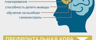
These lobes have a frontal location, they occupy the forehead area. Let's figure out what the frontal lobe is responsible for. The frontal lobes of the brain are responsible for sending commands to all organs and systems. They can be figuratively called a “command post.” It would take a long time to list all their functions. These centers are responsible for all actions and provide the most important human qualities (initiative, independence, critical self-esteem, etc.).
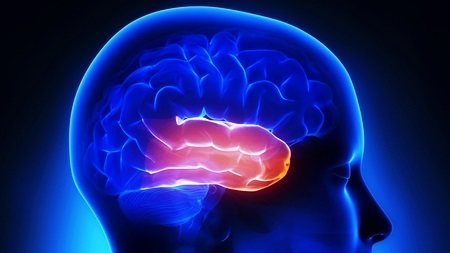
It is the frontal lobes that control human behavior. This part of the brain sends commands that prevent a specific antisocial action from being performed. It is easy to notice how this area is affected in dementia patients. The internal limiter is turned off, and the person can tirelessly use obscene language, indulge in obscenities, etc.
The frontal lobes of the brain are also responsible for planning, organizing voluntary actions, and mastering the necessary skills. Thanks to them, those actions that seem very difficult at first become automatic over time. But when these areas are damaged, the person performs the actions as if anew each time, and automaticity is not developed. Such patients forget how to go to the store, how to cook, etc.
When the frontal lobes are damaged, perseveration can occur, in which patients literally become fixated on performing the same action. A person may repeat the same word, phrase, or constantly move objects around aimlessly.
The frontal lobes have a main, dominant, most often left, lobe. Thanks to her work, speech, attention, and abstract thinking are organized.
It is the frontal lobes that are responsible for maintaining the human body in an upright position. Patients with their lesions are distinguished by a hunched posture and a mincing gait.
Forebrain
The functions of the forebrain are the most complex. It is responsible for mental activity, learning ability, emotional reactions and socialization. Thanks to this, it is possible to predetermine the characteristics of a person’s character and temperament. The anterior part is formed at 3-4 weeks of pregnancy.
To the question of which parts of the brain are responsible for memory, scientists found the answer - the forebrain. His cortex is formed during the first two to three years of life, for this reason a person does not remember anything before this time. After three years, this part of the brain is able to retain any information.
A person's emotional state has a great influence on the front part of the brain. Negative emotions have been found to destroy it. Based on experiments, scientists answered the question of which part of the brain is responsible for emotions. They turned out to be the forebrain and cerebellum.
The front part is also responsible for the development of abstract thinking, computational abilities and speech. Regular mental training can reduce the risk of developing Alzheimer's disease.
Interaction of brain parts
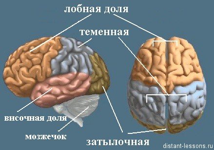
If you delve into the topic, in order to determine which part of the brain is responsible for speech, you need to know what main sections this human organ consists of. They are usually called shares. The structure and functions of the cerebral hemispheres play a vital role in the life of each of us.
The human brain has the following lobes:
- Frontal.
- Temporal.
- Parietal.
- Occipital.
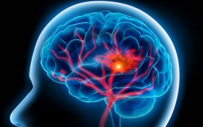
Separate from the structure and functions of the cerebral hemispheres, the cerebellum, which is responsible for coordinating the body in space, and the pituitary gland, which regulates the production of hormones, are distinguished.
Not in all cases, scientists agree on which part is responsible for what. This speaks first of all about the great lack of knowledge about the areas of the brain and the imperfections of modern medicine.
In addition to the fact that each part of the brain has its own tasks, the holistic structure determines consciousness, character, temperament and other psychological characteristics of behavior. The formation of certain types is determined by varying degrees of influence and activity of one or another segment of the brain.
The first psychotype or choleric. The formation of this type of temperament occurs under the dominant influence of the frontal lobes of the cortex and one of the subsections of the diencephalon - the hypothalamus. The first generates determination and desire, the second section reinforces these emotions with the necessary hormones.
The characteristic interaction of the departments that determines the second type of temperament - sanguine - is the joint work of the hypothalamus and hippocampus (the lower part of the temporal lobes). The main function of the hippocampus is to maintain short-term memory and convert acquired knowledge into long-term memory. The result of such interaction is an open, inquisitive and interested type of human behavior.
Melancholic people are the third type of temperamental behavior. This variant is formed due to increased interaction between the hippocampus and another formation of the cerebral hemispheres - the amygdala. At the same time, the activity of the cortex and hypothalamus is reduced. The amygdala takes on the entire “blow” of exciting signals. But since the perception of the main areas of the brain is inhibited, the reaction to excitement is low, which in turn affects behavior.
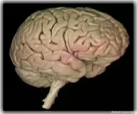
In turn, by forming strong connections, the frontal lobe is able to set an active pattern of behavior. When the cortex of this area interacts with the tonsils, the central nervous system generates only highly significant impulses, while ignoring unimportant events. All this leads to the formation of a Phlegmatic model of behavior - a strong, purposeful person with an awareness of priority goals.
Brain cells or neurons transmit and process signals that perform related work. The brain is divided into cavities consisting of sections. Each department is responsible for different functions. The activity and functioning of the body depends on their work. The brain is divided into 5 sections, each of which is responsible for separate functions:
- Rear. This section is divided into the pons and the cerebellum. Responsible for coordination of movements.
- Average. Responsible for innate reflexes to environmental stimuli.
- The intermediate is divided into the thalamus and hypothalamus. Responsible for emotions, processing signals coming from receptors, regulates vegetative work.
- Oblong. Responsible for managing vegetative functions: breathing, metabolism, cardiovascular system, digestive reflexes.
- Forebrain. This section is divided into the right and left hemispheres, covered with convolutions, which increases the volume of the surface. Makes up 80% of the mass of all departments.
What are shares?
They are responsible for hearing, turning sounds into images. They provide speech perception and communication in general. The dominant temporal lobe of the brain allows you to fill the words you hear with meaning and select the necessary lexemes in order to express your thoughts. The non-dominant helps to recognize intonation and determine the expression of a human face.
The anterior and middle temporal regions are responsible for the sense of smell. If it is lost in old age, it may signal incipient Alzheimer's disease.
If both temporal lobes are affected, a person cannot assimilate visual images, becomes serene, and his sexuality goes through the roof.
Parietal
In order to understand the functions of the parietal lobes, it is important to understand that the dominant and non-dominant side will do different jobs.
The dominant parietal lobe of the brain helps to understand the structure of the whole through its parts, their structure, order. Thanks to her, we know how to put individual parts into a whole. The ability to read is very indicative of this. To read a word, you need to put the letters together, and you need to create a phrase from the words. Manipulations with numbers are also carried out.
The parietal lobe helps to link individual movements into a complete action. When this function is disrupted, apraxia is observed. Patients cannot perform basic actions, for example, they are not able to get dressed. This happens with Alzheimer's disease. A person simply forgets how to make the necessary movements.
The dominant area helps you feel your body, distinguish between the right and left sides, and relate parts and the whole. This regulation is involved in spatial orientation.
The non-dominant side (in right-handed people it is the right side) combines information that comes from the occipital lobes and allows you to perceive the world around you in three-dimensional mode. If the non-dominant parietal lobe is disrupted, visual agnosia may occur, in which a person is unable to recognize objects, landscapes, or even faces.
The parietal lobes are involved in the perception of pain, cold, and heat. Their functioning also ensures orientation in space.
Associative speech center
Various human speech disorders have motivated scientists to study how this fact is affected by the functioning of the brain. It has been determined that there are several speech centers that are located predominantly in the left hemisphere. In joint interaction, they maintain a person’s speech at the proper level. If any part is injured, this will certainly affect the quality and ability to speak.
There are two main speech areas of the brain:
- Motor zone.
- Sensory area.
- Association Center.
Each of them is responsible for clearly defined functions.
This part of the brain does not develop in a person from birth, but only by the age of 2, when the child begins to try to pronounce conscious phrases. This zone is located in the parietal part of the cerebral cortex and also plays one of the most important roles in the formation of human speech.
An integrated approach to the development of both halves of the brain

As I already said, it is important to coordinate the work of both halves in order to expand their capabilities and functions for which they are responsible. Then you will be provided with a creative approach to solving even the most complex problems, and the speed and efficiency of information processing will also increase.
- Sit comfortably with a straight back, choose a point in front of you, you will have to concentrate on it. After about a minute, try using your peripheral vision, without taking your eyes off the selected point, to see what is to your left, and then to your right.
- With one hand, stroke your stomach, and with the other, perform tapping movements on your head. Slowly at first to adjust, building up the pace over time.
- Also, the development of both hemispheres will provide you with the following task: place the finger of one hand on the tip of your nose, and with the other hand grab the ear opposite to it. For example, the right hand should take the left ear. As soon as you take it, clap your hands and do the same, changing the position of your hands. That is, the fingers of a completely different hand touch the nose, exactly the same pattern with the ears.
- Stretch your arms in front of you, draw a square in the air with one of them, for example, and a circle with the other. When you feel that you have made progress, come up with new figures to master.
Cerebral hemispheres
The main feature of the large brain is that it is divided into the right and left hemispheres. Each of them is responsible for different functions: for controlling one of the sides of the body, receiving signals from a certain side.
The right hemisphere is responsible for the following:
- the ability to perceive the situation as a whole;
- development of intuition;
- making decisions;
- recognition abilities: pictures, faces, images, melodies.
The left hemisphere is responsible for the functioning of the right side of the body, and also processes information coming from the right side. The left hemisphere is responsible for the following:
- speech development;
- analysis of the situation and related actions;
- ability to generalize;
- logical thinking.
The brain is a very complex organ with many sections. Even a minor injury or inflammation of one part of the brain can cause hearing, vision or memory loss.
The hemispheres are connected by the corpus callosum. This is a large plexus of nerve fibers through which the signal is transmitted between the hemispheres. Adhesions are also involved in the joining process. There is a posterior, anterior, and superior commissure (fornix commissure). This organization helps to divide the functions of the brain between its individual lobes. This feature has been developed over millions of years of continuous evolution.
Which half is dominant?
Which of the two hemispheres is dominant? Previously, scientists believed that the left. However, it is now known that the left and right hemispheres of our brain work together, and the dominance of one of them is associated with the nature of a particular person. You're probably wondering which hemisphere is dominant for you. To determine this, you can take special tests. You can also analyze what types of activities you are better at and what you are capable of. In order for the hemispheres of the brain to work harmoniously, it is useful to perform special exercises that increase the potential of the weaker one.
During childhood, the right side of the brain is more active. We perceive the world through images. However, our entire education system and our lifestyle develop the functions of the left. Thus, the right hemisphere is often inactive, its functions do not receive proper development and it gradually loses its potential. This imbalance has a negative impact on our lives in the future.
The ability to achieve great success thanks to the harmonious work of the hemispheres is shown to us by examples of brilliant people. For example, Leonardo da Vinci was excellent with both hands. It is known that he was not only an excellent artist and sculptor, but also a scientist. The work of the hemispheres of his brain was harmonious. Their development was uniform, thanks to which he was able to create discoveries and inventions that change the life of not only a particular person, but also society as a whole.
Conclusion
So, each department has its own functional load. If a separate lobe suffers due to injury or disease, another zone may take over some of its functions. Psychiatry has accumulated a lot of evidence of such redistribution.
It is important to remember that the brain cannot function fully without nutrients. The diet should have a variety of foods from which the nerve cells will receive the necessary substances. It is also important to improve blood supply to the brain. It is promoted by sports, walks in the fresh air, and a moderate amount of spices in the diet.
If you want to maintain full brain function until old age, you should develop your intellectual abilities. Scientists note an interesting pattern - people with intellectual work are less susceptible to Alzheimer's and Parkinson's diseases. The secret, in their opinion, lies in the fact that with increased brain activity in the hemispheres, new connections are constantly created between neurons. This ensures constant tissue development. If a disease affects one part of the brain, its functions are easily taken over by the neighboring zone.

