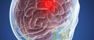
Brain neuroma is a neoplasm that grows from Schwann cells, which form the myelin sheath of nerve fibers. Neuroma has a benign course: it is not prone to rapid growth, metastasis and aggressive effects on neighboring tissues.
The disease is more often recorded in women and has no age limit. The tumor can grow anywhere where there is a myelin sheath, but is most often found on the auditory nerve.
Externally, a neuroma looks like a dense round node with a bumpy surface. The tumor is covered with a thin capsule of connective tissue. If you cut it open, you can see alternating pale and gray areas inside. If there is fat in the neuroma, spotted yellow lesions are observed. Sometimes brown dots are found on the section of the tumor - these are small hemorrhages.
What's happened
The structure of the neuroma contains auxiliary cells that make up the myelin sheath. They are necessary for the rapid transmission of impulses from the brain to the organs and back. They are called Schwann cells. Neuroma is formed from them. Hence its second name – schwannoma.
Schwann cells are present in all nerves, so a neuroma can form in any area. These formations are found in the brain and spinal cord. Neuroma is usually a benign tumor, but there are also malignant types.
The development of neuroma is caused by a failure in the process of cell division, as a result of which their number increases. If the tumor is benign, their functions are preserved, so the organs continue to function. The structure of these cells is similar to that of healthy tissue.
On this topic
- central nervous system
How does a headache with a brain tumor hurt?
- Natalya Gennadievna Butsyk
- December 3, 2020
Benign neoplasia develops slowly. If the nature of the neoplasia is malignant, then the cells multiply quickly and penetrate into neighboring tissues, interfering with their normal activity.
When a schwannoma grows rapidly, pressure occurs on the nerve, which impairs the functioning of the organ. With a cerebral neuroma, problems with sensory organs and thought processes are observed (depending on the location of the tumor).
There are several types of brain schwannoma:
- Epileptoid. Such formations look like dense bodies consisting of fibers.
- Xanthomasic. The tumor cells contain pigment, so it may be green, yellow or yellow-gray.
- Angiomatous. These schwannomas lead to vasodilation and the appearance of cavities.
Since these neoplasms form in different areas, there is no classification based on location. But more often, adverse changes affect the eighth pair of cranial nerves.
Main types
The classification of neuroma is not numerous; it reflects the multiplicity, prevalence, and nature of the course. The structural features of the neoplasm are of key importance:
- epithelioid schwannoma - consists of densely packed connective tissue cells with a small volume of nerve fibers;
- angiomatous - a tumor with an abundance of blood vessels, which, when expanded, lead to the formation of cavernous cavities;
- xanthomatous - the neoplasm consists of more than 85% xanthochrome (or pigment) cells, visualized under the skin in the form of dense yellowish nodules.
ICD-10 disease code G95 - benign neoplasm and other diseases of the spinal cord. Tumor malignancy occurs extremely rarely, but with complicated cancer heredity and precancer, the risk of cancer increases significantly.
Causes
What causes the pathology has not yet been established. There is an assumption that the main factor is genetic predisposition. If parents had such a disease, there is a high probability of its occurrence in children.
On this topic
- central nervous system
What are the dangers of a porencephalic cyst of the brain?
- Olga Vladimirovna Khazova
- May 27, 2020
The gene responsible for uncontrolled cell division is initially in a quiet state. Under the influence of unfavorable external factors, it is activated, as a result of which a neuroma begins to form. Among the provocateurs are:
- stress;
- weakness of the immune system;
- severe illness;
- abuse of harmful substances;
- sedentary lifestyle;
- unfavorable environmental conditions;
- poor nutrition.
These circumstances, individually or in combination, negatively affect a person’s condition. Because of this, the body’s protective functions cease to control the process of cell division, which leads to the formation of neoplasia.
When a brain neuroma is detected in people whose family did not have this disease, it is assumed that its appearance is caused by a negative external effect on the body.
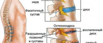
What is spinal neuroma
This disease refers to a pathological process in which the proliferation of Schwann cells occurs. The latter form the myelin sheath of every nerve in the body.
Schwannoma can form from nerves of both the central and peripheral systems. The formation always has a round shape, grows gradually, usually up to 2 mm annually, and is covered with a capsule.
More often the process is benign, but it can become malignant under the influence of certain predisposing factors. In such situations, tumor growth becomes more intense, it can reach significant sizes with the appearance of characteristic clinical symptoms.
Can it develop into cancer?
Schwannoma of the brain in most cases is a benign neoplasm. It slowly increases in size, a process that can take years. But in the absence of treatment and active harmful effects on the body, its cells can degenerate into malignant ones. Therefore, the possibility of cancer exists.
Sometimes, due to the presence of a predisposition, cell degeneration occurs immediately. Then malignant neoplasia forms, requiring urgent medical intervention.
Development of neoplasm
In medicine, 2 classifications of this disease are used. According to KOOS there are 4 stages:
- the tumor is located in the internal auditory canal;
- extends into the cerebellopontine angle, increases to 20 mm in diameter;
- extends to the brain stem, but does not put pressure on it, reaches 21 - 30 mm;
- puts pressure on the brain stem, the size of the formation is more than 30 mm.
The famous neurosurgeon Majid Samii divided stages 3 and 4 into subsections:
- T3a – tumor fills the cerebellopontine cistern;
- T3b – spreads to the brain stem;
- T4a – causes its compression;
- T4b - grossly deforms the trunk and IV ventricle.
Symptoms
To avoid complications and death, it is necessary to start treatment in a timely manner. And for this you need to contact a specialist in time for diagnosis. This can be done by knowing the symptoms of cerebral neuroma. They may vary depending on which part of the nerve is under pressure.
Observed:
- unsteady gait;
- difficulty maintaining balance;
- dizziness;
- headache ;
- numbness of the face;
- tingling of the skin on the face;
- deterioration in the functioning of facial muscles;
- change in taste sensations;
- decreased appetite;
- fast fatiguability;
- impairment ;
- noise in one of the ears.
On this topic
- central nervous system
Everything you need to know about pituitary adenoma
- Olga Vladimirovna Khazova
- February 28, 2020
As the tumor grows, symptoms intensify. Patients complain of hearing loss, visual impairment, difficulty swallowing, significant deterioration in coordination, problems with memory, concentration, and mental activity.
If these signs are detected, you should undergo examination at a medical facility.
Symptoms of spinal tumors
In the clinical organization of spinal cord tumors, 3 syndromes are distinguished:
- radicular;
- Bronsekar - damage to half the diameter of the spinal cord;
- complete transverse damage.
Regardless of the location of the tumor, damage occurs to the entire diameter of the spinal cord. Intracerebral tumors and tumors that do not grow into the substance of the spinal cord develop differently. In the first case, the onset of tumor growth is manifested by dislocated sensory disturbances of a segmental type. In the second case, radicular syndrome begins with further injury to the diameter of the spinal cord.
Radicular syndrome is manifested by severe headaches of the radicular type, which increases with sneezing, coughing, trying to bow the head, or physical stress. When lying down, the headache intensifies; when sitting, the pain decreases. Patients sleep in a forced position - half-sitting. With the subsequent course of the disease, sensory perception and reflexes in the area of innervation disappear. At the level of the tumor, upon percussion of the spinous roots, pain is formed, which radiates to the lower part of the body.
Neuroma has manifestations of a typical radicular syndrome. This manifestation may trigger an error in diagnosis. It is necessary to exclude radiculitis, infectious diseases and spinal deformities. Dislocated sensory disturbances and homolateral central paresis are signs of Brown-Séquard syndrome. There is a violation of vibration, muscle and articular sensitivity on the side of the tumor, on the opposite side - pain and temperature perception is reduced.
General damage to the diameter of the spinal cord provokes the appearance of bilateral violations of the deep and superficial sensory, bilateral paresis below the level of the tumor.
Secondary pathology in the form of urosepsis develops when pelvic activity is disrupted. Bedsores - as a result of vegetative-trophic disorders.
Diagnostics
Using diagnostic procedures, you can not only identify pathology, but also determine its stage and characteristics. To do this:
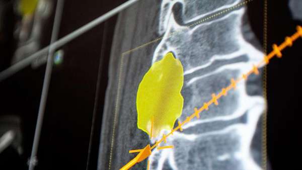
- CT or MRI. With their help, you can see small tumors and other disorders.
- X-ray. It allows you to detect changes in the bones caused by the presence of neoplasia.
- Audiometry. This method is necessary for acoustic neuroma - to determine how severe the hearing problems are.
- Ultrasound. Through it, abnormalities in tissues near the tumor are detected.
- Biopsy. It is designed to determine the cellular composition of the tumor. Based on its results, it is judged whether a benign or malignant formation has been formed.
The main method is considered to be MRI. It allows you to diagnose pathology at an early stage. The presence of a neuroma is indicated by the detected volumetric neoplasm of a round or oval shape.
Prognosis for treatment of malignant disease
Treatment of neuroma should be carried out regardless of stage and form. A benign form can develop into a malignant one, which will lead to the death of the patient. With a benign form, the five-year survival rate is 100%, with a malignant stage – from 37 to 65%. The tumor grows slowly; the location of the disease and the possibility of surgery are important. After removal, a neuroma can lead to loss of hearing, facial expressions, and some motor functions may fail. Surgery for neuroma in most cases does not have negative consequences, the patient is prescribed postoperative care and recovery.
Treatment
Features of therapy are determined depending on the stage of the disease and the patient’s well-being.
For conservative treatment, Mannitol solution (20%) is used, which is administered intravenously. This reduces intracranial pressure. In addition to it, glucocorticoids (Prednisolone or Dexamethasone) are used. They are taken orally or parenterally. When results from their use appear, stop using Mannitol.
If necessary, radiation therapy or surgery is used, after which the dose of glucocorticoids is gradually reduced. Special drugs (Nitsergoline, Cavinton) help improve blood circulation in the brain. Antipsychotics and sedatives are prohibited.
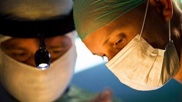
Surgery is the optimal way to treat pathology. It allows you to eliminate the tumor and avoid complications. But it is performed only on patients in serious condition with large, fast-growing tumors. The patient's age should not exceed 60 years.
On this topic
- central nervous system
What threatens a cyst in the head?
- Olga Vladimirovna Khazova
- February 28, 2020
During the operation, the neoplasia is eliminated completely or the pathological tissue is excised as much as possible. After this, the tumor is sent for a biopsy to identify the characteristics of the disease.
Radiation therapy is used for unadvanced malignancies. This treatment method also helps with inoperable neuromas. During the procedure, a gamma knife is used.
If the tumor is small, radiation treatment is used. For malignant schwannoma, chemotherapy is prescribed in addition to radiation methods.
Treatment of neuroma
The most effective treatment method in cases where a neuroma is detected is surgery. It is prescribed for progressive and active growth or the appearance of new unfavorable symptoms of schwannoma.
Contraindications to surgery are cardiovascular diseases, old age and serious condition of the patient.
Removal of a neuroma occurs microsurgically (acoustic schwannoma), partially when the tumor grows with nerve fibers, and stereotactically (local irradiation). Radiosurgical treatment is prescribed for contraindications to surgery. Reviews for this treatment method are only good, because it has virtually no side effects.
A conservative treatment method consists of taking medications and monitoring water-electrolyte balance and diuresis. In addition to basic medications, drugs that improve brain activity are also prescribed.
Timely ways to detect symptoms and diagnose neuroma will help avoid the unpleasant consequences of this disease. A well-performed operation will allow you to forget forever about the existence of such a pathology in the past.
Possible complications
Without treatment, neuroma leads to complications. They depend on the location of the tumor, its size and nature. With small neoplasias that are detected and treated in time, there are no significant consequences, but large neoplasms put pressure on the nerves and interfere with brain activity.
Deafness is a complication that develops when the auditory nerve is irreversibly damaged. Proper therapy contributes to the full or partial restoration of hearing, but in its absence the patient stops hearing.
Prolonged compression of the auditory nerve leads to its atrophy, which cannot be overcome even by removing the tumor. With unilateral schwannoma, problems are observed in one ear. But there is a 5% chance that a person will lose their hearing completely.
The second complication can be called paresis of the facial nerve. It also develops in advanced cases, when the facial nerve is exposed to pressure from the neuroma for a long time. This leads to its atrophy. The result is loss of taste, increased salivation on the injured side, and facial asymmetry.
Cerebellar disorders occur with advanced trigeminal neuroma. Prolonged pressure on the cerebellum causes poor coordination of movements. The patient has difficulty maintaining balance and his gait becomes uneven.
Another pathology caused by schwannoma is intracranial hypertension syndrome. It occurs in the presence of large inoperable neoplasia. Because of them, difficulties arise with the circulation and outflow of cerebral fluid. It accumulates in the brain structures, which causes increased pressure inside the skull. This disorder is indicated by attacks of nausea, headaches, dizziness, convulsions, fainting, and visual disturbances. When there is excessive accumulation of cerebral fluid, the patient develops hydrocephalus.
A dangerous complication of brain schwannoma is its degeneration into a cancerous tumor. If left untreated, the patient faces death.
Treatment tactics
The only treatment for spinal schwannoma is surgery, but surgery is performed for special indications: impressive size and neurological symptoms of varying severity, suspicion of a malignant process. If surgery is not possible, then radiation therapy is used.
In treatment centers in Moscow and the regions, neuroma treatment is carried out under the compulsory medical insurance policy. In the Stavropol Territory, removal operations are performed by doctors of the highest qualifications and training.
Conservative or drug therapy is intended to relieve symptoms before and after surgery and prevent further growth of a small tumor. Doctors often choose a wait-and-see approach, especially if there are no symptoms and the tumor is discovered by chance. This is required to determine the growth trend of the tumor.
If the tumor is growing slowly, the following drugs may be prescribed:
- muscle relaxants;
- water-salt balance stabilizers (glucose, sodium bicarbonate 3-5%);
- non-steroidal anti-inflammatory drugs for pain relief.
Warming topical medications may provide temporary relief due to their distracting effects. In case of intense radiating pain and the ineffectiveness of NSAIDs, narcotic analgesics can be prescribed.
For schwannoma of the lumbar spine, hormonal drugs and cerebral circulation stimulants are indicated. Therapeutic gymnastics is acceptable. It is mandatory to follow a diet with limited salt and liquid (no more than 1.5 liters per day). To enhance the therapeutic effect and relieve pain, massage, manual therapy, and physiotherapy (acupuncture, pulse therapy, electrophoresis) are indicated.
During surgery, the tumor is removed along with the capsule and tissues near the tumor are captured. For neuroma of the thoracic spine, as well as other parts of the spinal column, the degree of entrapment and compression of nerves, features of innervation, and irradiation to the limbs are assessed.
The operation is performed through small incisions in the area where the schwannoma is located. After removal, the wound is sutured and the patient is transferred to the ward. When the tumor is localized in the lower part of the brain, surgery is practically not performed, since schwannomas constantly recur. A complicated tumor with necrotic changes requires more surgical intervention.
The operation itself does not pose a direct threat, except in cases of damage to nerve endings, which is why the intervention is recommended to be carried out when the volume of the capsular membrane is insignificant and the involvement of nerve structures in the pathological process.
The operation to remove a spinal neuroma in the Stavropol diagnostic center is performed by the best surgeons in the field of oncology and neurology.
In most cases, clinicians reassure patients, since according to statistics, only 2-6 operations are performed per 100 cases of neuroma. In 85% of patients, a tumor that occurs accidentally does not provoke the development of serious neurological symptoms after 3-5 years.
Surgery on the back and spine is a serious burden for the entire musculoskeletal system and body. Recovery is slow; it is important to achieve high-quality and correct fusion of all membranes and tissues excised at the time of surgery.
In the early postoperative period, patients are prescribed strict bed rest, protective rest, and regular dressings. After the stitches are removed, adequate physical activity, physiotherapy, and massage are allowed.
The duration of recovery varies depending on the extent of the surgical intervention, the specifics of the operation, and the technique used. In 95-98% of cases, after total resection of spinal schwannoma there is no recurrence (except in cases of the lower part of the brain), but such risks still occur.
Forecast
The severity of the consequences of the pathology is determined by the degree of its severity, the endurance of the body, the timeliness and correctness of therapy. If a neuroma is detected at an early stage of its development, before the tumor has reached a large size, treatment can eliminate it. There will be no complications in this case.
On this topic
- central nervous system
Causes and consequences of cyst formation in the head in children
- Olga Vladimirovna Khazova
- February 28, 2020
If the disease is detected at an advanced stage, even complex therapy cannot guarantee a favorable outcome. Removing a schwannoma does not provide complete recovery. Compression of a nerve over a long period of time leads to its atrophy or disturbances in its activity. This can cause hydrocephalus, facial nerve paresis, hearing loss or deafness, and visual disturbances.
When malignant tumors form, there is a possibility of death. When a cancerous tumor is removed, there is still a possibility of recurrence.
Complications and consequences
Complications of spinal neuroma are caused by growth and constant compression of the nerve roots. Thus, with spinal localization, the risk of compression myelopathy, vertebral displacement, the formation of a hernia of the lumbar spine, and impaired innervation in the pelvic organs increases. Considering the special relationship between the nervous system and its structures throughout the body, the following possible complications can be identified:
- spastic paresis and paralysis;
- unsteadiness of gait, loss of coordination;
- impaired vision, speech, hearing;
- headache;
- neuropathic laryngeal paresis.
If the innervation of the lower abdomen is disrupted, disorders of urination, defecation, and sexual activity in men and women are likely. Acute pain syndrome develops even with a slight influence of spinal neuroma on the nerve endings.
Prevention
Due to the lack of information about the causes of cerebral neuroma, it is impossible to name rules that will eliminate the risk of its occurrence. Since it is caused by a genetic predisposition, prevention is ineffective.
You can reduce the likelihood by reducing the impact of negative factors on the body:
- To refuse from bad habits.
- rationally .
- Do not use medications without a doctor's prescription.
- Treat in a timely manner .
- Exercise .
- Avoid physical and emotional overload.
- Strengthen the immune system.
These measures will minimize traumatic effects on the body, which will reduce the likelihood of uncontrolled cell division.
It is also important to monitor changes in your health. If pathological symptoms are detected, you should consult a doctor and undergo an examination. Early detection of neuroma provides a better chance of eliminating it without consequences.
Brain neuroma is one of the types of neoplasms that arise in the human body. Its development is due to the characteristics of heredity in combination with the influence of unfavorable environmental factors. Early detection of pathology increases the patient's chances of recovery.
Causes and predisposing factors
To date, the only adequate theory for the occurrence of spinal neuroma is a genomic mutation of chromosome 22 or a hereditary predisposition. Predisposing factors to disorders at the genetic level may be:
- the impact of teratogenic factors on the fetus during pregnancy;
- long-term intoxication of the body of various nature;
- neurofibromatosis in close relatives;
- family history of cancer;
- benign tumors.
The probability of developing a neuroma in a child increases sharply to 50% if one of the parents is a carrier of an altered chromosome 22, to 70% if both parents suffer from this pathology.
Types of disease
Neuroma is divided into two types - benign and malignant. Benign is a slowly growing node that does not cause disruptions or disturbances in the functioning of the body. Location: head, spine, neck.
Malignant neuroma is the result of the transition of a benign tumor to a malignant sarcoma of the nerve. It is difficult to define and establish clear boundaries of the disease. The node grows quickly, and metastases may develop in other organs. The tumor can reach large sizes; the larger, the more difficult it is to treat. Location: forearms, feet, hands. Take into account! The malignant form is typical for people aged 20 to 40 years, and the benign stage is typical for people aged 50 to 60 years. The clinical symptoms of both forms are similar, which complicates the situation; the doctor needs to clearly understand what stage the disease belongs to.
The following types of neuroma are also distinguished:
- tumor of the thoracic and cervical regions. The disease affects the spine, when the tumor grows through the intervertebral spaces, a schwannoma of the neck begins to develop;
- Morton's neuroma. The tumor affects the foot, touching the nerve endings. In the affected area, both one and both limbs often grow between the fingers;
- a brain tumor. Slow development, the tumor is limited to neighboring tissues;
- acoustic schwannoma. Localization - right or left ear, slow development.
The disease can affect the trigeminal nerve and can develop in the lower leg area, between the right and left pleural cavities.
Treatment tactics
The choice of method is determined individually and depends on the size of the schwannoma and the general condition of the patient’s body. Usually the tumor is removed surgically, and radiation is used less frequently.
This method is quite new, with its help it is possible to treat without surgery. The idea is to irradiate the tumor area with photons using a special device.
Healthy tissues are not affected in any way; the formation itself quickly regresses. The caveat is that this technique is only available on a commercial basis.
Find out the difference between a protrusion and a hernia.
This technique is suitable for small neuromas (up to 5-6 mm). Only this condition allows the tumor to be removed along with its capsule. The procedure is characterized by effectiveness and safety, since it does not affect other nearby structures of the spinal cord and spinal column.
The method is used for large neuromas. The tumor itself is removed first, followed by the capsule. When implementing this method, there is a risk of affecting other tissues, which can lead to unpleasant complications. The latter, however, do not pose a danger to life.
Clinical picture
Many years may pass before the first symptoms of schwannoma appear. Therefore, the pathology is characterized by a long asymptomatic course. In the initial stages of the disease, dull pain in the affected area may periodically appear.
As the tumor progresses, it compresses the surrounding tissues, which causes the appearance of negative symptoms:
- increased pain in the back and limbs;
- muscle atrophy and weakness;
- deterioration of motor function and sensitivity below the tumor location (paresis, paralysis);
- numbness in the limbs;
- parasthesia (“goosebumps”, numbness in the limbs, burning);
- dysfunction of the pelvic organs;
- sexual dysfunction.
If the neoplasm is more than 5 cm in diameter, then it can be felt under the skin. It is dense, elastic, painful. Neuromas of the cervical spine are especially easy to visualize.
On a note! The appearance of symptoms of spinal neuroma should be an impetus for immediate diagnosis and treatment.