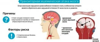
A brainstem stroke is distinguished by the fact that it damages the central parts of the brain, which play a major role in a person’s motor activity. Mental abilities do not deteriorate, that is, the patient continues to correctly perceive and analyze information entering the brain, but he cannot react to it.
The brainstem consists of the cerebellum, pons, medulla oblongata and midbrain. Their defeat due to acute disruption of blood flow provokes a disruption in the functioning of the corresponding part of the brain. A brainstem stroke can occur as a result of hemorrhage or blockage of a vessel - these are the leading causes of a brainstem stroke.
The main consequences of an attack are disruption of the respiratory process, swallowing, blood flow, thermoregulation and paralysis of the arms and legs. In addition, the brain stem contains nerve cells that are responsible for the functioning of facial muscles and eye movement. It turns out that the pathology poses a great threat to humans, and the success of subsequent treatment depends on timely first aid.
If patients survive a brainstem stroke, they almost always have neurological deficits. Sometimes pathology requires lifelong use of additional equipment - a ventilator, etc.
How does a stem lesion differ from other types of stroke?
The peculiarities of the course of an attack are that when blood flow stops in one of the parts of this brain structure, vital functions suffer. Let's see why damage to the medulla oblongata is life-threatening:
- Irregularities in heart rhythm. Any disturbances can occur: from bradycardia and extrasystole to arrhythmia and fibrillation. A distinctive feature is that when the brainstem is damaged, the rhythm is poorly restored with the help of traditional cardiac medications.
- Breathing problems. Respiratory function may become difficult, shortness of breath may appear, and in severe cases, a person loses the ability to breathe independently and only connection to a ventilator can save the person.
- Swallowing and speech. If speech dysfunction is relatively harmless, then problems with swallowing lead not only to drooling. In some body positions, saliva enters the respiratory tract and provokes aspiration pneumonia.
In addition, a stroke of the brain stem negatively affects the victim’s vision, sensitivity, motor activity and coordination. The attack is severe and without timely medical care can be fatal.
What is a columnar stroke?
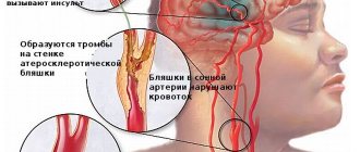
The reasons why a stroke occurs: thrombosis, embolism, hypertension, aneurysm, severe trauma, leading to the formation of a hematoma, blockage of blood vessels. If blood circulation in the brainstem is disrupted, a brainstem stroke is diagnosed. This important area is the link that transmits signals from the brain to organs throughout the body.
The brainstem is the oldest part of the brain; it affects the functioning of both hemispheres, providing a controlling and regulating effect. The nuclei of this department are the closing link of simple and complex reflex arcs; they regulate swallowing, breathing, are responsible for the functioning of the heart, and thermoregulation of the body.
In severe cases, it causes the cessation of nutrition of this part of the brain, its atrophy and the impossibility of functioning of all internal organs.
What is the brain stem
The human brain stem is a special organ that connects the brain and nervous system with the rest of the organs of our body. This is where the signals that make our heart beat, lungs breathe, and arms and legs move occur.
There is not a single system in our body that does not depend on the brain stem. The trunk is located deep in the skull under the hemispheres.
If the blood circulation is impaired, a tumor immediately occurs here, the trunk moves and is pinched, which leads to disability or death of the patient.
Source: //golovnoj-mozg.ru/zabolevaniya/krovoobraschenie/stolbovoj-insult-chto-eto-takoe
Causes of brainstem stroke
There are 2 main causes of the pathological process:
Provoking factors for the development of pathology are:
Having considered what it is - a pillar stroke, let's look at what signs can be used to recognize the pathological process.
Preventing Stroke
The main cause of stroke is usually atherosclerosis (hardening of the arteries or blood vessels clogged with deposits of fat and other substances). That is why preventive measures are aimed at eliminating the causes of atherosclerosis and proper rehabilitation after its occurrence.
The main preventive measures are to ensure dietary restrictions on animal fats, salt and cholesterol. It is important that a person's daily meals include more fruits, vegetables, whole grains and legumes.
Another goal is to prevent weight gain, which is why an active lifestyle is recommended.
Last but not least, it is recommended to quit smoking. Smoking, clearly and in any case, is harmful to human health, but with atherosclerosis it doubles the likelihood of developing a cerebrovascular stroke.
The next factor is high blood pressure, which must be carefully monitored as you age (at least after 50 years), after consulting with your doctor, try to follow his recommendations or take appropriate medications. High blood pressure is one of the most common causes of many diseases.
Symptoms of brain stem damage
With a brainstem stroke, the symptoms are varied. Signs of impaired blood flow in the medulla oblongata include:
- hyperemia of part of the face against the background of pallor of other areas of the skin;
- breathing problems (increases frequency, wheezing appears, and if a person develops a coma, the frequency of respiratory movements may decrease to 8-10 per minute):
- difficulty speaking (the victim speaks slurred or makes an inarticulate moo);
- increased sweating;
- changes in heart rate (tachycardia or bradycardia);
- hypertension;
- increase (less often - decrease) of body temperature;
- problems with coordination;
- visual impairment (loss of fields, double vision).
A brainstem infarction is characterized by the fact that a person has a small clear gap before the condition worsens, and intracerebral hemorrhage provokes a sharp increase in symptoms.
With a brainstem stroke, patients lose consciousness in 70-80% of cases. Sometimes there is complete paralysis and loss of speech while the patient is conscious. This condition in medicine is called “locked-in person” syndrome (isolation syndrome, deefferentation syndrome).
CT scan shows a brainstem stroke that damages the ventral striatum corresponding to the pyramidal fasciculi. This condition can lead to “locked-in person” syndrome.
Stem symptoms depend on which part of the trunk is affected. The patient may have predominant brainstem symptoms related to breathing, cardiac activity, or motor function.
Rehabilitation therapy
A stroke in the brain stem causes a condition of muscle weakness. To eliminate the consequences of circulatory disorders, the following types of therapy are used:
- treatment with nootropic drugs;
- special gymnastics;
- massage;
- physiotherapeutic procedures;
- occupational therapy
The consequences of an ischemic stroke are eliminated with the help of drugs that stimulate the activity of the nervous and muscular systems. Use B vitamins and glucose. Active gymnastics are used to restore motor functions. The patient is taught to sit, stand, and walk. To quickly achieve results, use the following devices:

After a stroke in the brain stem, the patient’s pelvic organ functions are often impaired. In this case, the doctor recommends exercises that help strengthen the muscles and external sphincters.
During the recovery period, physiotherapeutic procedures are carried out:
- electrophoresis;
- paraffin therapy;
- ozokerite treatment.
During a course of special gymnastics, breathing exercises are used to improve the functioning of the heart and blood vessels.
Prognosis for recovery from brainstem stroke
The most dangerous period of damage to the brain stem is the first hours after an attack. If medical assistance is not provided, the following may occur:
- lethal outcome (brain stem stroke without medical assistance leads to death in 2/3 of cases), and why damage to the medulla oblongata is fatal can be understood by remembering what functions this structure is responsible for;
- unfavorable prognosis (in most patients, complete recovery after a brainstem stroke does not occur; the person remains completely or partially incapacitated).
It doesn’t matter whether there is a brainstem infarction or a hemorrhage in the brainstem - it is equally dangerous. Only by calling emergency help when the first pathological signs appear, there is a chance to prevent serious consequences.
Features of brainstem hemorrhagic stroke
Rupture of an artery with hemorrhage causes an acute clinical picture. Hemorrhagic stroke of the brainstem contributes to the formation of extensive symptoms:
- Loss of balance;
- Painful sensations inside the eyes;
- Numbness of the skin of the face;
- Paralysis of limbs.
In comparison with the ischemic type, brainstem hemorrhages provoke severe hemiparesis and paralysis of the muscular sphere. The accumulation of blood (hematoma) provokes an increase in intracranial pressure at the beginning of development. Brain stem coma gradually develops. The person loses consciousness. Even if it is possible to get a person out of the condition, negative consequences remain:
- Muscle paralysis and paresis cause disturbances in gait and swallowing, and difficulties with eating persist. Parenteral nutrition provides the necessary nutritional components;
- Speech disorders make it difficult to communicate with other people. Preservation of consciousness and intelligence allows for rehabilitation after a stroke;
- The cause of death in most patients is swelling of the stem structures with strangulation of the structure in the area of the foramen magnum;
- Hematoma and cerebral infarction are treated on an inpatient basis under the supervision of specialists.
Recovery after a stroke takes a long time. Experts do not guarantee a complete return to health, but some of the lost functions return.
Signs of a brainstem stroke
The pathology is accompanied by destruction of the conductive tracts between the brain and spinal cord. Infringement of cranial nerves leads to disruption of the functioning of internal organs.
First signs:
- Occipital pain;
- Bradycardia or tachycardia;
- Loss of consciousness;
- Significant daily fluctuations in body temperature;
- Dizziness.
Manifestations of brainstem infarction resemble ischemic stroke with cerebral and focal symptoms. Alternating syndromes are formed due to the involvement of accompanying cranial nerves.
Signs of cerebral symptoms:
- Tongue shift to the side;
- Ocular nystagmus;
- drooping eyelid;
- Paralysis of the facial muscles on the side opposite to the lesion;
- Hemiplegia of body parts.
The symptoms of the pathology are more extensive, but the described signs are leading.
Diagnostic measures
Symptoms of damage to the brain stem allow the neurologist to predict in advance the location of the damage. Further research is aimed at clarifying:
- there was a brain stem infarction or hemorrhagic hemorrhage;
- what important centers are damaged and the size of the pathological focus;
- tissue became saturated with blood or a hematoma formed (if hemorrhage was detected in the brain stem).
To clarify these data, CT or MRI of the brain is used. Hardware examination provides a complete picture of the size of the lesion and the nature of damage to brain structures.
Diagnostics
Anamnesis is especially important for diagnosis. Whether a person has high blood pressure and diabetes, as well as a family history of atherosclerosis and other types of vascular diseases, plays an important role. This is followed by a simple neurological examination - for example, a “handshake” test. In the case of a stroke, as already mentioned, paresis and plegia occur, therefore, there is a noticeable difference in the strength of the hand grip.
A more detailed diagnosis is additionally performed in the hospital using CT (computed tomography) data, which can be supplemented by MRI (magnetic resonance imaging). Based on the results of these tests, the extent of brain damage is determined, whether ischemia or bleeding is present, and a prognosis for treatment is made for the patient.
Therapeutic measures
Treatment of brainstem stroke can be conservative or surgical. Therapeutic tactics depend on whether a stroke has occurred in the brain stem or hemorrhage, as well as on the nature of the hemorrhage (tissue impregnation or hematoma formation).
Conservative
Brainstem lesions are treated conservatively if brainstem stroke or hemorrhagic impregnation is detected. The patient is placed in intensive care, where he is prescribed:
- diuretics (Mannitol, Lasix) to prevent cerebral edema;
- intensive oxygen therapy (if the patient breathes on his own, then through a nasal catheter, and if respiratory function is impaired, mechanical ventilation is used);
- sedatives (Phenazepam, Relium, etc.);
- muscle relaxants to prevent possible seizures;
- antihypertensive drugs (drugs of this group in small doses to prevent recurrent stroke are prescribed to patients even with normal blood pressure);
- blood-thinning medications (for hemorrhagic impregnation of brain tissue).
At an early stage, damage to the brain stem can cause a recurrent stroke or provoke an increase in the ischemic focus, so therapy is aimed at preventing possible consequences. When the patient’s condition has stabilized, he is transferred to a ward, where rehabilitation begins, allowing the impaired functions to be fully or partially restored.
Operational
Hemorrhage into the brain stem may be accompanied by the formation of large hematomas. The blood clot compresses nearby tissues, aggravating the pathological process.
Hemorrhagic stroke of brain stem cells is treated according to the following scheme:
- craniotomy is performed over the area of the hematoma;
- Blood clots are removed without affecting brain tissue.
In the future, the pathology is treated in the same way as an ischemic stroke or hemorrhagic impregnation - with the help of drugs that prevent possible complications.
The doctor decides how to treat the pathology individually, taking into account the nature and degree of brain damage.
Treatment
Cerebral infarction is treated with drug therapy, which includes anticoagulants and drugs for high blood pressure.
The patient is also prescribed treatment for concomitant diseases.
Hemorrhagic stroke is most often treated with surgery. Open craniotomy can relieve the patient of a hematoma, which poses a mortal threat. After the operation, the patient is prescribed drug therapy.
Possible consequences
Is recovery possible after brain stem injury?
Unfortunately, the prognosis for recovery from stem lesions, especially for old patients, is unfavorable. Only a small proportion of patients return to a full life, and the majority receive disability. Let's see what consequences happen with stem lesions.
Treatment of brainstem strokes with speech impairment is almost always successful if the patient and his relatives follow medical recommendations:
- regularly attend classes with a speech therapist;
- repeat the material covered at home;
- the patient actively communicates with his family, trying to pronounce difficult sounds.
It is difficult to say how long it takes to restore speech. This process depends on the diligence of the patient and the help of loved ones.
Swallowing
If a person has difficulty swallowing food, then the chances of eliminating the disorder are minimal. To alleviate the patient’s condition, relatives are advised to:
- make liquid puree dishes;
- Give food warm and in small portions.
In this case, the prognosis is uncertain. In some patients, disorders persist for life.
Movement
Often, survivors of hemorrhagic or brainstem ischemic stroke experience:
- involuntary contraction of muscles and movement of limbs;
- attacks of sudden weakness in an arm or leg on one side.
If the condition does not improve within 2-3 months after the attack, then the hope for a favorable outcome of rehabilitation decreases.
Coordination
Coordination disorder with frequent dizziness due to damage to the cerebellum. Your doctor will tell you how to recover from a brainstem stroke in this area. But even following rehabilitation programs does not always ensure recovery.
Breath
Impaired respiratory function is not always restored. Some patients after stroke damage to the brainstem, even after restoration of consciousness and some motor functions, are forced to breathe with the help of a ventilator.
Hemodynamics
If we consider what is dangerous about the pathology of stroke damage to the brain stem, then it is a disorder of cardiac activity. Patients develop:
The danger is that these conditions are difficult to treat with drugs to normalize heart rhythm.
Thermoregulation
Typically, non-infectious fever occurs in the first hours of the pathological process and causes the death of brain cells. In the late stage, thermoregulatory disorders do not develop. In rare cases, patients experience chilliness or suffer from excessive sweating.
Vision
A stroke of the occipital part of the head with damage to the brain stem causes:
- loss of visual fields;
- difficulty fixing gaze.
The most dangerous disorder is the inability to recognize. The patient sees objects, but cannot determine what is in front of him.
Post-stroke consequences can be temporary and disappear during the rehabilitation process, or they can persist for a long time and cause disability.
Treatment of movement disorders
The consequences of ischemic stroke are eliminated using various methods of restoring impaired functions. In the early period of the disease, treatment measures are aimed at saving the patient’s life. Before starting rehabilitation therapy, the general condition of the patient and the presence of associated complications are studied. The doctor prescribes drugs that activate the functioning of nerve cells:
- Gammalon;
- Actovegin;
- Piracetam;
- 0.1% nasal drops Semax.
For decreased muscle tone, medications are used:
Special gymnastics helps prevent increased muscle tone. Position therapy is prescribed. The patient lies on his back and healthy side for several hours. The paralyzed arm is placed on a pillow, and the affected leg is bent at an angle of 15-20°. Position therapy avoids stress on the same muscle groups and reduces the likelihood of developing pain in the shoulder joint.

The patient is prescribed a course of massage for post-stroke movement disorders. Use acupuncture, gentle rubbing, shallow kneading. Active gymnastics is of great importance for stabilizing a person’s motor function.
How to prevent a stroke
There are no recommendations that are 100% guaranteed to prevent an acute ischemic attack in the brainstem or other parts of the brain. But if a stroke cannot be completely prevented, then you can reduce the risk of developing pathology by following simple tips:
- Blood pressure control. Hypertensive crises are a common cause of cerebral ischemia.
- Reducing cholesterol. Taking statins and following a diet will help cope with atherosclerotic plaques.
- Normalization of blood viscosity. If you are prone to blood clots, you must take blood thinners.
A brain stem stroke is difficult for patients to tolerate and the rehabilitation process is always lengthy. The success of post-stroke recovery depends on the area of the injury and how quickly the victim is taken to a medical facility.
Video: Anatomy and functions of the brain stem
Disorders of this kind are quite rare, since for them to occur it is not enough to damage the occipital lobe on only one side; the symmetrical lobe appears to be affected as well. More frequent are cases of partial visual agnosia, in which the patient does not distinguish familiar faces from unfamiliar ones and does not distinguish colors. Of course, in parallel with visual agnosia, alexia also develops. A disorder in which the correct recognition of the contours and outlines of familiar objects is impaired is called metamorphopsia. In the mind of a patient suffering from a similar disorder, objects have some kind of broken, irregular shape.
For the formation of disorders of this kind, the disruption of connections between the occipital region and the temporal region, which is directly related to the adequate perception of spatial relationships, is important.
In pathological processes that are localized in the occipital lobe, a number of irritation phenomena are observed.
If irritation of the projection visual field occurs, visual hallucinations appear in the form of shadows, sparks, etc. More complex hallucinations appear when the outer surface of the occipital lobe is irritated. These hallucinations may take the form of figures or objects that have a frightening appearance. In this case we are talking about metamorphopsia
, which indicates the localization of the lesion in an area close to the temporal lobe. Visual hallucinations, as a rule, are harbingers of an incipient epileptic seizure. The first spasm of such a seizure is a turning of the eyes and head in the opposite direction.
A very striking picture is distinguished by strokes, in which disorders of motor functions (up to paralysis) are observed on the side of the body opposite to the affected hemisphere. This is due to the fact that the pathways connecting the cerebral hemispheres with the spinal hemisphere cross, moving from one side to the other.
Brain stroke
– this is an acute disorder of cerebral circulation, usually resulting from atherosclerosis and arterial hypertension; Somewhat less often, strokes are caused by pathologies of the valvular apparatus of the heart, myocardial infarction, congenital anomalies of cerebral vessels, hemorrhagic syndromes, and post-traumatic changes.
There are five main groups of acute cerebral vascular lesions: 1) transient cerebrovascular accidents; 2) ischemic stroke (cerebral infarction); 3) cerebral embolism; 4) hemorrhagic stroke (bleeding in the brain); 5) subarachnoid hemorrhage, which may cause psychomotor agitation and seizures.
With strokes, disorders of higher mental functions are not uncommon, in particular, written and oral speech is difficult (or impossible), and mental impairment is possible.
Cerebral palsy
is a group of diseases of newborns characterized by non-progressive motor disorders. Factors such as birth asphyxia, birth trauma, and prenatal pathology are important in the development of this group of diseases. With congenital hemiplegia, intelligence is often reduced and approximately half of patients are susceptible to epileptic seizures.
Cerebral dysfunction (a rudimentary variant of cerebral palsy) is manifested by mild behavioral disturbances, some defects in praxis and gnosis, as well as epileptic episodes.
Of course, any form of cerebral palsy is characterized by motor impairments, more or less pronounced.
| State | patrolled |
| Ischemic stroke | |
| CT scan of the brain showing an infarct in the right hemisphere of the brain (the infarcted area is outlined in red.) | |
| ICD-10 | I 63 63. |
| ICD-10-CM | I63.9 and I63 |
| ICD-9 | 434.01 434.01 , 434.11 434.11 , 434.91 434.91 |
| ICD-9-CM | 433.01[1], 433.31[1], 434.91[1], 433.81[1] and 433.21[1] |
| OMIM | 601367 |
| MeSH | D002544 |
Ischemic stroke
- disturbance of cerebral circulation with damage to brain tissue, disruption of its functions due to difficulty or cessation of blood flow to one or another part. Accompanied by softening of a section of brain tissue - cerebral infarction. It may be caused by insufficient blood supply to a certain area of the brain due to decreased cerebral blood flow, thrombosis or embolism associated with diseases of the blood vessels, heart or blood [2]. It is one of the main causes of death among humans [3].
A cerebral stroke is an acute disruption of the blood circulation in the brain, leading to damage and death of nerve cells. A stroke occurs when a blood vessel in the brain is either blocked (by a plaque, a blood clot—an ischemic stroke) or ruptured (a hemorrhagic stroke). After some of the nerve cells die, the body loses one of the functions for which the dead cells were responsible: paralysis, loss of speech and other serious disorders occur. The larger the area of death in the brain, the more serious the consequences of a stroke.
Contents: 1. Symptoms 2. Causes 3. First signs 4. Consequences 5. Diagnosis 6. Prevention 7. Rehabilitation 8. Probability of death 9. Left side
Symptoms
Symptoms of a cerebral stroke or impending brain catastrophe are varied. However, there are three main symptoms indicating acute cerebrovascular accident.
- The victim cannot smile directly. As a rule, the smile is asymmetrical, the corner of the lips may be lowered.
- Can't speak normally. Speech is slow and slurred.
- Weakness in the arm and leg on the affected side. The arms cannot rise at the same time. The presence of these three symptoms requires an urgent call to the ambulance.
As a rule, an attack has a precursor, the so-called ischemic attack. Its clinical manifestations should be alarming:
- speech disorder;
- causeless intense headache;
- varying severity of motor dysfunction in the arm or leg;
- severe dizziness;
- fainting state.
Stroke symptoms can be divided into two categories: general cerebral and focal.
1. General cerebral symptoms
- severe headache accompanied by nausea or vomiting;
- the appearance of drowsiness, for some the opposite effect is characteristic - strong excitement;
- impairment and loss of consciousness;
- dizziness;
- loss of orientation in space or time;
- heartbeat;
- sweating and feeling hot.
2. Focal symptoms
Symptoms will depend on which part of the brain is being deprived of normal blood supply. Each area of the brain is responsible for a specific activity, therefore, a violation of the blood supply to different areas of the brain will manifest itself in different symptoms - impaired balance, vision, speech. Loss of coordination may be accompanied by dizziness and vomiting.
Ischemic stroke is considered the most dangerous, since it often does not have pronounced symptoms in the initial stages. It may appear, for example, in the form of headaches, dizziness and nausea, or disappear. Hemorrhagic stroke has pronounced symptoms and already in the initial stages makes itself felt with severe headaches, loss of consciousness, vomiting, and clouding of mind.
There is a so-called transient cerebrovascular accident, also called a mini-stroke. It is characterized by a sharp change in a person’s health - speech impairment, partial or complete blindness, and weakness in the limbs. A microstroke causes temporary impairment of a person’s health, which is restored after a certain time (several minutes or hours). However, a person’s normal state of health does not last long and in such cases hospitalization is required, since this pathological condition is a harbinger of a real stroke.
How to properly treat a cerebral aneurysm Nutrition after a cerebral stroke here
Find out what a recurrent stroke is
Causes
The causes of brain stroke vary depending on the type.
With a hemorrhagic stroke of the brain, a violation of the integrity of the artery occurs due to a number of factors:
- aneurysmal dilatation and thinning of the artery wall;
- congenital pathology of the bloodstream, called arteriovenous malformation;
- inflammatory and traumatic damage to the inner membrane.
In this case, increased pressure in the cerebral blood flow system plays an important role.
In patients with ischemic stroke, in 90% of cases the disease is associated with atherosclerosis. The situation is aggravated by the formation of a blood clot on the damaged surface of the inner membrane, as well as vascular spasm. In isolated cases, the cause of ischemic brain damage is thromboembolism of the cavities of the heart or veins of the lower extremities.
The risk factors for stroke are almost the same as for all cardiovascular diseases. These include:
- smoking;
- age over 45 years;
- male gender;
- high blood pressure;
- atherosclerosis;
- heredity;
- overweight.
First signs
Today, statistics show that stroke occurs in most cases in the morning or at night, due to stress or physical strain. Symptoms of stroke may vary from one patient to another depending on the presence of bleeding, the location of the blood clot, and the severity of the damage.
The first signs of a cerebral stroke:
- the muscles of the face, arms or legs on one side of the body begin to go numb;
- speech becomes incoherent or slurred;
- the vision of one or two eyes drops sharply;
- there is a loss of coordination of movement, dizziness begins and a shaky gait is observed;
- blood pressure rises sharply;
- the complexion becomes red, the pulse slows down, breathing becomes rapid, the temperature rises;
- in rare cases, a person may fall into a coma;
- there is confusion;
- drowsiness occurs.
The nature of the disease depends on in which part of the brain the vessel ruptured. In case of damage to the left hemisphere, problems with logical thinking may arise, and involvement of the parietal part in the pathological process entails numbness and a tingling sensation, as a result of which the idea of the ratio of the size of one’s body may be disrupted. The development of a stroke in the occipital part of the head leads to deterioration of vision, up to its complete loss, as a result of which the patient cannot recognize an object, even if he sees it.
Consequences
There are different types of consequences after a stroke. Each type of disease leads to different complications as the brain is a complex organ that controls various functions of the body. By not getting blood to certain areas of the brain, certain parts of the body may stop working properly.
People who have an ischemic or hemorrhagic stroke may experience common effects such as weakness, motor impairment or paralysis, speech and swallowing problems, cognitive impairment, perceptual problems and some behavioral problems. In addition, patients may experience defecation and urination disorders, psychological disorders, epilepsy and pain syndrome.
It should be noted that the consequences of this disease are quite numerous and varied. There are also consequences of stroke such as aphasia, which is a severe deterioration in the ability to use words to express one’s thoughts. Aphasia is one of the most common consequences that occurs in most people who have had a stroke. Aphasia can manifest itself as follows:
- difficulty expressing inner desires;
- difficulty understanding the speech of others;
- difficulty reading or writing.
There are various types and forms of aphasia, which are characterized by various disorders and manifestations:
- Wernicke's aphasia is characterized by severe perceptual disturbances that may be caused by a loss of understanding of the sound part of speech.
- Broca's aphasia is characterized by various problems with the formation of phrases and sentences.
- Extensive aphasia is a fairly common and widespread consequence of stroke, which is characterized by certain disorders. In the case of extensive aphasia, the patient who has suffered a stroke is unable to form sentences or comprehend them. In addition, the patient may experience incoherent speech and rare understanding of some individual words. Also a hallmark of extensive aphasia is the almost complete absence of communication.
Diagnostics
Making the correct diagnosis and detecting the exact location of the stroke, as well as data on the volume of damaged tissue, allows you to choose the right treatment tactics, as well as avoid more severe consequences. In addition to interviewing and examining the patient, special examinations are required, both of the brain and of the heart and blood vessels.
A computed tomography (CT) scan is a machine that uses X-rays to produce clear, detailed, three-dimensional images of the brain. This study is prescribed immediately after the appearance of any suspicion of the development of a stroke. A CT scan can show whether there is bleeding in the brain or the extent of damage caused by a stroke.
Magnetic resonance imaging (MRI) is a machine that uses a strong magnetic field to produce very clear and highly detailed three-dimensional images of brain structures. This study may be prescribed instead of a computed tomography scan or as an addition to it. An MRI can show changes in brain tissue, as well as the amount of damaged cells caused by a stroke.
Doppler examination of the carotid arteries is an ultrasound examination of the carotid arteries, which are the main highway carrying blood to your brain. The study allows you to see the condition of the arteries, namely, to see the damage to the vessels by atherosclerotic plaques, if any.
Transcranial Doppler study is an ultrasound examination of cerebral vessels, which provides information about the blood flow in these vessels, as well as about the damage to them by fatty plaques, if any.
Magnetic resonance angiography is similar to an MRI study, only in this study more attention is paid to the vessels of the brain. This study provides information about the presence and location of a blood clot, if any, and also provides information about the blood flow in these vessels.
Cerebral angiography - this procedure involves injecting a special contrast agent into the vessels of the brain, and then using X-rays we obtain images of the vessels. This study provides very valuable data on the presence and location of blood clots, aneurysms and any vascular defects. This study is more difficult to perform, unlike CT and MRI, but is more informative for these purposes.
An electrocardiogram (ECG) is one of the simplest heart studies, but very informative. It is used in this case to detect any disturbances in heart rhythm (cardiac arrhythmias) that may cause the development of a stroke.
Cardiac echocardiogram (Echo-CG) is an ultrasound examination of the heart. Allows you to detect any abnormalities in the functioning of the heart, as well as detect defects in the heart valves, which can cause blood clots or blood clots, which in turn can cause a stroke.
Electroencephalogram (EEG) is a study of brain activity. It is a measurement of the electrical activity of the brain using electrodes that are attached to the head. This test is ordered if your doctor thinks you have had a stroke.
A coagulogram is a blood test that determines the rate at which the blood thickens. This test is performed to identify disorders that may cause bleeding or thrombosis. This test is also performed to monitor the dose of blood thinning medications.
Biochemical blood test - this analysis is necessary to determine two main indicators:
- Blood glucose is necessary to establish an accurate diagnosis, since very high or very low levels of glucose in the blood can provoke the development of symptoms similar to a stroke. And also for diagnosing diabetes mellitus.
- Blood lipids - this test is necessary to determine the content of cholesterol and high-density lipoproteins, which can be one of the causes of stroke.
Prevention
Prevention of cerebrovascular disorders involves influencing the factors leading to this condition:
- Prevention of the development of atherosclerosis. It is necessary to follow a diet, regularly monitor blood cholesterol levels, and take lipid-lowering medications as prescribed by a doctor if lipid metabolism disorders are detected.
- Regular physical activity is necessary to prevent the development of obesity, type 2 diabetes and hypertension.
- To give up smoking. It is known that smoking increases the risk of developing cardiovascular diseases and atherosclerosis, which most often lead to strokes.
- Control, prevention and treatment of diseases such as hypertension, diabetes mellitus, blood diseases leading to the formation of blood clots.
- Fighting stress. It has been scientifically proven that stressful conditions increase the risk of stroke several times.
Unfortunately, we are unable to influence some factors that increase the risk of brain accidents. These include age over 55 years, hereditary predisposition, male gender.
Rehabilitation
The duration of a strict bed period after cerebrovascular accidents depends on its type and the severity of the patient’s condition. In case of ischemic stroke and the patient’s satisfactory condition, the doctor may allow the regimen to be expanded already 3-5 days after the stroke, and in case of hemorrhagic stroke the patient is allowed to sit up in bed after 1-2 weeks. The longest period of bed rest is necessary for subarachnoid hemorrhage, the cause of which was the rupture of a vessel aneurysm. In this case, the patient is not allowed to extend the regimen for 1-1.5 months.
However, you cannot remain idle during this time. Physical therapy is one of the main methods of restoring lost movements in the affected limbs. It is necessary to perform therapeutic exercises, massage, physiotherapy, and acupuncture on a daily basis for the patient right in bed. Usually, the doctor allows you to start light exercises 2-3 days after the stroke. At least 4 times a day, it is necessary to perform passive movements of the patient’s limbs for 15-20 minutes. Movements of the limbs are performed slowly, carefully, first with healthy limbs, and then immobilized. This will help avoid the development of inflammatory processes and contractures in the joints of the affected limbs, as well as muscle atrophy. Breathing exercises are also important, aimed at preventing the development of congestion in the lungs.
There is such a thing as “positional treatment,” which means special positioning of the affected limbs. This is also necessary in order to prevent the development of muscle contractures. The paralyzed arm is taken to the side, the forearm is extended and supinated, the hands and fingers must be placed in a position of maximum extension. This position of the hand must be fixed with a splint or a bag of sand. The paralyzed leg must be straightened, a small cushion placed under the knee, and the foot pronated slightly. The hospital staff will always explain and show relatives how to properly position immobilized limbs.
As soon as active movements appear in the limbs, it is necessary to help the patient develop them. With the doctor's permission, the patient's regime is gradually expanded; first, it is allowed to sit the patient in bed and hang his legs off the bed. Somewhat later, the patient can stand at a support, sit at a table, begin to learn to walk with the help of special supporting devices and use a wheelchair. The intensity of the load increases gradually and very carefully; it is important to prevent the patient from falling.
If the recovery period is favorable, 1-1.5 months after the stroke, the patient begins to learn to walk independently with the help of a walker, a three-legged crutch and a playpen. First, he needs the help of two people who can support the patient. The affected leg should be loaded gradually. While walking, the hand must be secured in the headscarf; it should not hang along the body. Fine hand movements are restored with the help of modeling, cubes, “spiky” balls and other various objects for the development of fine motor skills.
To correct speech disorders, sessions with a speech therapist may be necessary. The patient must speak, pronounce tongue twisters, retell what he heard and even sing.
In the early recovery period, patients very often require the help of a psychotherapist, since the patients are depressed, many do not believe in the success of therapy and rehabilitation measures and refuse to participate in treatment.
The early recovery period is the most important in the rehabilitation of patients who have suffered a stroke. In subsequent periods, social adaptation of patients and maintenance of achieved results are mainly carried out.
Probability of death
Every year, from 5 to 6 million strokes are diagnosed in the world, in Russia up to 450 thousand strokes. For this reason, 29% of men and 39% of women die. 3.2 people per 10 thousand become disabled. During the first month, up to 35% die, and by the end of the year up to 50%. Repeated strokes are dangerous. In the first year, relapse develops in 5-25%, within three years in 20%, within five years from 20 to 40% of those who have recovered. The greatest risk of developing stroke is after 65 years of age, the incidence of which is up to 90% of all stroke cases. At the same age, the highest number of deaths occurs. Up to 80% of strokes develop as ischemic pathologies of the brain, with a mortality rate of up to 37%. In 20% of patients with hemorrhagic stroke, death rate is up to 82%.
The reason for the high mortality rate from stroke in Russia is the rapid aging of the population, late delivery to a medical institution, and poor educational work on stroke prevention. Statistics from recent years indicate that 39.5% of people at risk of stroke do not pose a risk.
Stroke rarely occurs without previous symptoms - initial manifestations of insufficiency of blood supply to the brain (IBC) in the form of transient attacks or hypertensive crises or in people at risk. The risk group for NPNCM includes people with hypertension, cardiac arrhythmias, those under chronic stress, a history of chronic diseases, smoking, a tendency to blood cell aggregation, and excess body weight.
Left side
Left hemisphere stroke is more common than right hemisphere stroke, accounting for 57% of all clinical cases of the disease. And since the left hemisphere performs speech and logical functions, then with a stroke on the left side, in addition to paralysis of the right side of the body, the very first disorder is language and speech problems:
- Speech disorders, slurred and unclear pronunciation, misunderstanding of heard speech, a person can only express himself in fragments of words or a set of sounds.
- With a left-sided stroke, the right side of the face may be paralyzed, or right-sided paralysis of the arms and legs may occur.
- A person cannot read or write normally, articulation is impaired
- Speech memory loss occurs
- Gradually, a person becomes withdrawn, as if withdrawing into himself, since communication with others is limited.
History of the study [edit | edit code ]
The first surviving mentions of stroke are descriptions of the father of medicine, Hippocrates, who called the disease “apoplexy” (Greek ἀποπληξία) - stroke [4]. The theoretical basis for the etiology of stroke was laid by the 17th century Swiss pathologist Johann Jakob Wepfer, who discovered that apoplexy could be caused by blockage of a cerebral vessel or vascular damage with intracerebral hemorrhage [5]. The same scientist was the first to describe clinical cases of very rapid regression of weakness in the limbs [6]. However, the vascular nature of strokes was finally recognized only in the first half of the 19th century. “Softening” of the brain was distinguished from a hemorrhagic “stroke”; however, their etiology remained completely unclear. The development of knowledge about stroke was slow, apparently due to limited interest among the famous neurologists of the time. Thus, the first important information about the causes of stroke was obtained not by neurologists, but by pathologists Rokitansky and Virchow [7]. In the mid-19th century, the surgeon and anatomist John Lidell proposed the term “red infarction,” emphasizing the secondary nature of hemorrhage in hemorrhagic infarction [8]. Clinical and topographic studies of Jules Dejerine, Pierre Marie, as well as their follower Charles Foy, the founder of modern clinical studies of stroke, aroused interest and further research by neurologists about this disease [7]. In 1928, stroke was divided into separate types, taking into account the nature of the vascular pathology [6].
Epidemiology [edit | edit code]
According to official materials from the Russian Ministry of Health in recent years, mortality from diseases of the circulatory system ranks first and continues to grow steadily. In 2002, 56.1% of the total number of deaths died due to diseases of the circulatory system [9].
Mortality from stroke is in second place, second only to mortality from coronary heart disease. Total morbidity and mortality rates from stroke in many countries around the world are trending upward. In Russia in 2001 it reached 331 per 100 thousand population [9].
Ischemic strokes account for 70-85% of all stroke cases, cerebral hemorrhage - 20-25%, subarachnoid hemorrhage - 5%. The ratio of the incidence of ischemic and hemorrhagic types of stroke is 4:1 [9].
Classification [edit | edit code ]
There are different classifications of ischemic strokes, depending on the etiopathogenetic and clinical aspects, the localization of the infarction zone.
According to the rate of formation of neurological deficit and its duration [edit | edit code]
- “minor stroke” - according to the definition of WHO experts: “prolonged ischemic attacks with a reverse neurological defect.” A variant of ischemic stroke in which recovery of neurological functions is completed from 2 to 21 days [11]:245.
- progressive ischemic stroke (eng. stroke-in-evolution) - characterized by the gradual development of general cerebral and focal symptoms over several hours or 2-3 days, followed by incomplete restoration of functions. Typically, the patient remains with minimal neurological symptoms [11]:245.
- complete (total) ischemic stroke - a formed cerebral infarction with a stable or incompletely regressing deficit [12].
According to the severity of the patients' condition [edit | edit code]
- mild severity - neurological symptoms are mild, regress within 3 weeks of the disease. Variant of minor stroke [11][12]:247
- moderate severity - the predominance of focal neurological symptoms over general cerebral symptoms, there are no disorders of consciousness [11][12]:247
- severe stroke - occurs with severe cerebral disorders, depression of consciousness, severe focal neurological deficit, often dislocation symptoms [11] [12]: 247
Pathogenetic (Methodological recommendations for stroke, 2000) [edit | edit code]
- atherothrombotic stroke (including arterio-arterial embolism) (34%) - occurs against the background of atherosclerotic cerebral arteries of large or medium caliber. This type of stroke develops in stages, with an increase in symptoms over several hours or days, and often debuts during sleep. Often, atherothrombotic stroke is preceded by transient ischemic attacks. The size of the ischemic lesion varies [13][14].
- cardioembolic stroke (22%) - occurs when a cerebral artery is completely or partially blocked by an embolus. The onset of cardioembolic stroke is usually sudden, while the patient is awake. At the onset of the disease, the neurological deficit is most pronounced. More often, a stroke is localized in the area of the blood supply of the middle cerebral artery, the size of the focus of ischemic damage is medium or large, and a hemorrhagic component is characteristic. There may be a history of thromboembolism in other organs [13][14].
- hemodynamic stroke (15%) - caused by hemodynamic factors - a decrease in blood pressure (physiological, for example, during sleep; orthostatic, iatrogenic arterial hypotension, hypovolemia) or a drop in cardiac output (due to myocardial ischemia, severe bradycardia, etc.). The onset of a hemodynamic stroke can be sudden or gradual, while the patient is at rest or active. The sizes of infarctions vary; localization is usually in the area of adjacent blood supply (cortical, periventricular, etc.). Hemodynamic strokes occur against the background of pathology of extra- and/or intracranial arteries (atherosclerosis, septal arterial stenosis, abnormalities of the cerebral vascular system) [13][14].
- lacunar stroke (20%) - caused by damage to small perforating arteries. As a rule, it occurs against the background of high blood pressure. It develops gradually over several hours. Localized in the subcortical and brainstem structures (basal ganglia, internal capsule, white matter of the centrum semiovale, base of the pons), the size of the lesions does not exceed 1.5 cm. There are no general cerebral and meningeal symptoms; focal symptoms corresponding to the affected structure are noted [13][14][ 15] .
- stroke of the hemorheological microocclusion type (9%) (some sources also use the term “rheological stroke” [16]) - occurs in the absence of any vascular or hematological disease of established etiology. The cause of stroke is pronounced hemorheological changes, disturbances in the hemostasis and fibrinolysis systems. Characterized by scant neurological symptoms in combination with significant hemorheological disorders [13][14].
According to the location of cerebral infarction [edit | edit code]
In accordance with the topical characteristics of focal neurological symptoms, according to the affected arterial territory [12]:
- internal carotid artery;
- vertebrates, basilar artery and their branches;
- middle, anterior and posterior cerebral arteries.
Depression and high blood pressure
Hypertension is high blood pressure (BP). Common among women and men. Bad habits, genetic predisposition, stressful situations, emotional stress, overwork affect the condition of blood vessels and provoke an increase in blood pressure. The main factor is the psychological state, disruption of the nervous system. Blood pressure and emotional disorders are interrelated. The two diseases are the cause and effect of each other. Increased psychological stress, stress, and emotional disorders provoke an increase in blood pressure. In turn, hypertension is the cause of depressive disorders.
Etiology [edit | edit code ]
Local etiotropic factors of stroke are distinguished [19]:
- atherosclerosis and thrombus formation are the most common pathology of cerebral and precerebral arteries, causing ischemic disorders of cerebral circulation. The formation of atherosclerotic plaque is caused by the deposition of lipids in the artery wall. The growth of an atherosclerotic plaque is complicated by its ulceration and thrombus formation due to platelet adhesion. An increase in atherothrombotic plaque can lead to narrowing of the lumen of the artery and its complete blockage; a decrease in blood flow occurs with hemodynamically significant stenosis (narrowing of 70-75% of the lumen area) and then increases in proportion to the degree of narrowing. Fragments of a thrombus and atherothrombotic plaque can be a source of embolism in a more distal artery (arterio-arterial embolism). The outcome of atherosclerotic thrombosis or embolism is determined by the speed of its development, the localization of the blockage, the state of the collateral circulation and the activity of the fibrinolytic system of the blood. With the gradual development of atherosclerosis, collateral circulation is formed, so blockage of one or even several precerebral arteries may not lead to local cerebral ischemia and be asymptomatic. In cases of rapid development of thrombotic occlusion or embolism of the precerebral or cerebral artery, the possibilities of collateral circulation are limited. In cases where protective mechanisms cannot compensate for narrowing or blockage of the artery, clinically significant local cerebral ischemia develops. Atherothrombosis and arterio-arterial embolism are the cause of up to 50% of ischemic cerebrovascular accidents [16]:232-233.
- Cardiogenic embolism is the cause of approximately 20% [16]:233 (according to some data, up to 60% of strokes are caused by heart disease [19]) of ischemic strokes and TIAs. It usually develops due to the formation of embolic fragments on the heart valves or the formation of an intracardiac thrombus. Most cardiogenic embolisms occur with atrial fibrillation. It, in turn, is usually caused by coronary heart disease against the background of atherosclerosis of the coronary arteries and arterial hypertension. The risk of developing a stroke with atrial fibrillation is 4.5% per year if the patient does not take appropriate treatment. With infective endocarditis, approximately 20% of patients experience cerebral embolism. For all artificial heart valves, the overall risk of embolism is about 2% per year unless anticoagulants are used. Myocardial infarction is complicated by ischemic stroke in approximately 2% of cases; most often this occurs in the first 2 weeks after the onset of heart disease [16]:233-234.
- hemodynamic ischemic disorders of cerebral circulation can occur with stenosis of the precerebral (most often with stenosis of the carotid arteries) and (or) cerebral arteries, when blood pressure falls below the lower limit of autoregulation of cerebral circulation [16]:234. Degenerative and deforming changes in the cervical spine (osteochondrosis of the spine, deforming spondylosis, anomalies of the craniovertebral region) can lead to external compression of the vertebral or subclavian arteries with the possible development of strokes in the vertebrobasilar region [19] or steal syndromes [16]:234.
- rare vascular pathology: Takayasu's disease, Moyamoya disease, infectious arteritis, etc.
Systemic factors contributing to the development of ischemic stroke include:
- disturbance of central hemodynamics:
- cardiac hypodynamic syndrome - manifested by impaired circulation, heart rhythm, decreased minute blood volume and stroke volume, which leads to a decrease in blood flow in the arterial system of the brain, disruption of the mechanisms of autoregulation of cerebral circulation and the formation of thrombotic stroke or the development of cerebral ischemia such as cerebrovascular insufficiency ( hemodynamic stroke).
- arterial hypertension - leads to the development of ischemic disorders of cerebral circulation directly, causing changes in the walls of the arteries - lipohyalinosis and fibrinoid necrosis, and also indirectly - through stimulating atherosclerosis of the precerebral large and medium cerebral arteries and the development of heart diseases, for example, myocardial infarction and atrial fibrillation, complicated cardiogenic embolism [16]:232.
- arrhythmias are a risk factor for the occurrence of arterio-arterial and cardiogenic embolisms.
- Hematological disorders (coagulopathies, erythrocytosis and polycythemia) can lead to hypercoagulability and increased blood viscosity, predisposing to the development of thrombosis in the cerebral arteries and the occurrence of “rheological stroke” [16][19]:235.