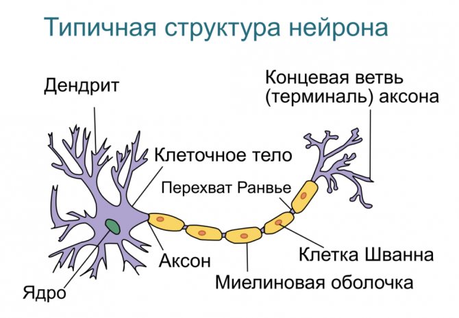
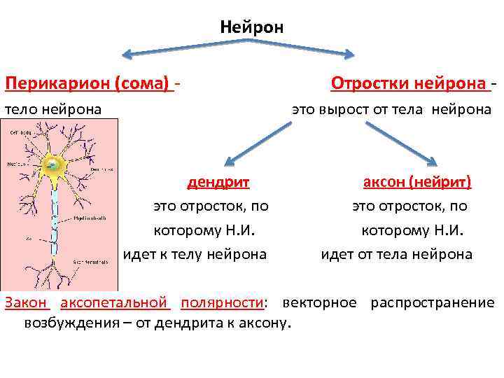
A dendrite is a short, dichotomously (bifurcating) branching process extending from the neuron body. According to the definition, the process forms synapses (electrical, chemical) with other elements of the nervous tissue, through which it receives signals from peripheral receptors and cells of the body in the histological structure of organs and systems. The word "dendrite" also has a different meaning if used not in the medical field, but in the field of materials science, in this case it refers to the crystalline structure of the material, regardless of its composition. The term is applicable to stones, metals, alloys, and artificial compounds that have a crystalline, tree-like structure.
What is a dendrite - functions and morphology
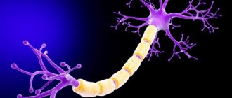
Dendrites are numerous thin tubular or round protrusions of the cell body (perikarya) of a nerve cell.
The term itself speaks of the extreme branching of these areas of neurons (from the Greek δένδρον (dendron) - tree). The surface structure of neurocytes can contain from zero to many dendrites. The axon is most often single. The surface of dendrites does not have a myelin sheath, unlike axon processes.
The cytoplasm contains the same cellular components as the nerve cell body itself:
- endoplasmic granular reticulum;
- accumulations of ribosomes - polysomes (protein-synthesizing organelles);
- mitochondria (energy “stations” of the cell, which, using glucose and oxygen, synthesize the necessary high-energy molecules);
- Golgi apparatus (responsible for the delivery of internal secretions to the outer layer of the cell);
- neurotubules (microtubules) and neurofilaments are the main components of the cytoplasm, thin supporting structures that ensure the preservation of a certain shape.
The structure of dendritic endings is directly related to their physiological functions - receiving information from axons, dendrites, and perikarya of neighboring nerve cells through numerous interneuron contacts based on selective sensitivity to certain signals.
Functions
The axon and dendrite, despite their different morphological structures, have similar functions - they serve as connecting elements, thanks to which interaction between all cells of the body is maintained, and all physiological processes are integrated. The main function of the dendrite is to perceive signals from other nerve cells and receptors of internal organs. Dendritic branches also perceive external stimuli.
As a result, synaptic connections of several types are formed - axonodendritic (dendrite-axon contact), dendro-dendritic (dendrite-dendrite contact), axospinous (axon-dendritic spine contact). The received impulses travel to the body of the neuron. The terminal segments of dendritic processes serve as a site of synaptic contact, from where inhibitory and excitatory stimuli arrive at the soma. Thanks to synaptic contacts, one neuron is connected to numerous (over 20 thousand) nerve cells.
Recent studies show that dendritic processes are capable of independently generating signals. Previously, it was believed that only the cell body generates impulses; the role of the processes is reduced to signal transmission. Scientists discovered that dendritic spines were much more active than somas when they studied the nature and strength of signals transmitted within nerve tissue. The signal passing along the trunk of the process may change.
Structure and types
The outer surface of the dendrites is covered with thin protrusions in the form of tiny spines measuring 2-3 microns. The number of such formations on the surface can vary from zero to tens of thousands. The shapes of the microspines themselves are varied, but the mushroom-shaped spine is considered the most common form.
The number of spines on the surface and their sizes can change quickly. The neuron’s response to signals from other cells depends on this.
The formation of protrusions-spines, their shape and development are influenced by internal and external circumstances: the age of the organism, the activity of synaptic connections, the information load of neural circuits, the organism’s lifestyle and much more.
The integrity and stability of the spine structure may be influenced by negative factors:
- pathophysiological factors (for example, neurodegenerative processes in nervous tissue mediated by severe heredity);
- toxicological agents (when using drugs, alcohol, poisons of various natures).
Under the influence of these negative factors, serious destructive transformations occur in the internal structure of microspines: destruction of the cisterns of the spine apparatus, accumulation of multivesicular bodies (proportional to the degree of destructive influences).
After a series of tests conducted with experimental mice, it was proven that not so much the dendrites themselves, but dendritic spines are the elementary units of memory storage and the formation of synaptic plasticity.
Branching
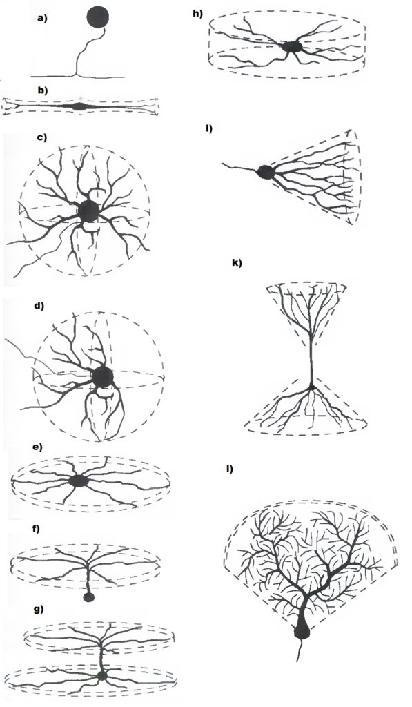
Dendritic structures are formed due to the tree-like branching of neuron processes. This process is called arborization. The number of branching points (or nodes) determines the degree of branching and complexity of the dendrite endings. Mitochondria are usually concentrated in the cytoplasm of branching nodes, since branching is an energy-consuming physiological process.
The structure of the dendritic tree determines the physical receptive area, that is, the number of input impulses that a neurocyte can receive and conduct in total.
One of the main purposes of dendrites is to increase the contact surface for synapses (increase the receptor field).
This allows the cell to receive and redirect more information that comes to the cell body of the neuron. The degree of branching determines how a neuron ultimately integrates electrical signals received from other cells: the larger and more complex the branching, the more closely the neurons fit together.
Due to the branched structure, the surface of the receptor membrane of the nerve cell increases 1000 times or more.
Diameter and length
Dendritic endings have different sizes, but are always characterized by a gradual decrease in the diameter of the preterminal branches. The length is usually from several microns to 1 mm. But, for example, some sensitive neurons of the spinal ganglia have very long dendrites - up to a meter or more.
General information
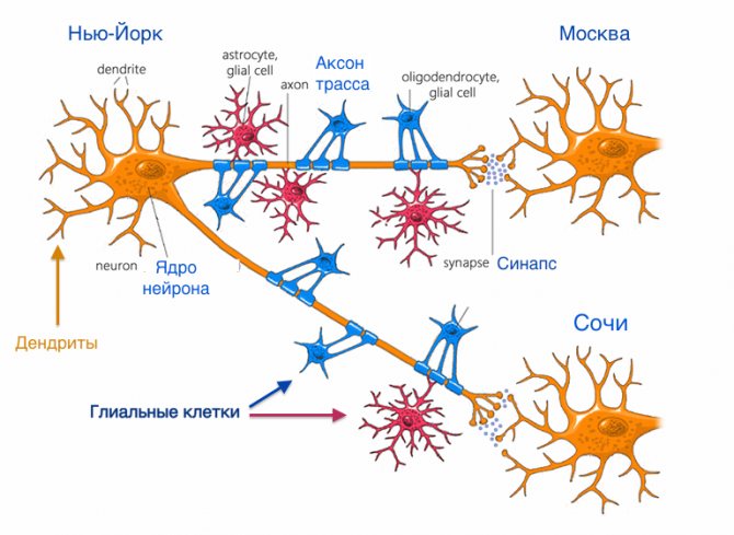
Neuron is the basic structural and functional unit of nervous tissue. A nerve cell is a formation with numerous processes measuring 4-130 microns. Several (less often one) dendrites and a single axon extend from a neuron. A dendrite in biology is a process that transmits excitation from peripheral receptors to the body of a neuron, which determines its leading role in the perception of external stimuli. Features of dendritic branches that are observed during microscopic studies:
- Microtubule system.
- The presence of spines extending from a common axis.
- Presence of branch nodes.
- The presence of endoplasmic reticulum (an intracellular organelle represented by a system of tubules, cavities, vesicles surrounded by a membrane).
The processes located next to the soma (body), thickened, form a large number of synaptic contacts. The membrane of the process, like the membrane of the neuron itself, consists of a large number of protein molecules that play the role of chemical receptors. Receptor formations are endowed with specific sensitivity to certain chemical compounds.
The designated chemical substances - neurotransmitters of inhibition and excitation, are actively involved in the process when impulses spread through the nervous tissue and arrive at the soma. The structure of the dendrite suggests the presence of spines that form synaptic contacts with terminals - the end sections of hundreds of thousands of nerve cells. A huge number of spines are located on the processes of nerve cells that form the cortex of the cerebral hemispheres.
The dendritic spine is formed from a body and a head. The size and shape of the structural components of the spine vary significantly. Thanks to the spines, the area of the postsynaptic membrane significantly increases. Spines in the dendritic structure are labile formations that, under the influence of external stimuli, change configuration, degenerate (destroy), and regenerate (appear again).
The number of synapses determines the quality of impulse transmission and the speed at which they propagate. Several dendritic branches form a single branch. The totality of all dendrites is a dendritic tree - a surface that perceives external stimuli. Research shows that dendritic trees make up 90% of the brain matter.
Conduction of nerve impulses
The receptor membrane of the surface of the dendrites (as well as the body of the nerve cell) is covered with numerous synaptic plaques that transmit excitation to the receptive area of the surface membrane of the neuron, where a bioelectric potential is generated.
Information encoded in the form of electrical impulses is transmitted to the electrically excitable conductive membrane of the axon. In this way, the body's neural networks are formed.
Role in neural processes
A person is born with a genetically determined number of dendritic processes on each neuron. The gradual increase and complexity of brain structures and the construction of the nervous system, which occur during postnatal development, is realized through branching and an increase in the mass of dendrites.
According to numerous studies, at the peak of development of the nervous system, dendrites occupy about 60-75% of the total mass of nerve cells.
According to fundamental theories describing the principles of operation of the nervous system, dendrites have always been considered the part of the neuron that receives an impulse and conducts it to the body of the nerve cell.
However, modern research by neuroscientists using the latest technologies such as microelectrodes has revealed greater electrical activity of dendrites compared to the cell body.
These studies confirmed the fact that dendritic endings are capable of generating electrical impulses themselves - local action potentials.
Structure and functions of dendrites
.
Functions of dendrites.
I would like to note that the main difficulties that a researcher faces when studying the function of dendrites is the lack of information about the properties of the dendrite membrane (as opposed to the membrane of the neuron body) due to the impossibility of introducing a microelectrode into the dendrite.
Assessing the overall geometry of dendrites, the distribution of synapses and the special structure of the cytoplasm at the sites of dendritic branching, we can talk about special neuron loci with their own function. The simplest thing that could be attributed to dendritic sites at branching sites is a trophic function.
From all of the above, it follows that the cytoplasm of dendrites contains many ultrastructural components capable of providing their important functions. There are certain loci in the dendrite where its work has its own characteristics.
The main purpose of the numerous dendritic branches of a nerve cell is to provide communication with other neurons. In the mammalian cerebral cortex, a large proportion of axodendritic connections occur in contacts with special specialized processes of dendrites - dendritic spines. Dendritic spines are phylogenetically the youngest formations in the nervous system. In ontogenesis, they mature much later than other nervous structures and represent the most plastic apparatus of the nerve cell.
As a rule, the dendritic spine has a characteristic shape in the mammalian cerebral cortex. (Fig. 2). A relatively narrow stalk extends from the main dendritic trunk, which ends in an extension - the head. Probably this form of dendritic appendage (presence of a head) is associated, on the one hand, with an increase in the area of synaptic contact with the axon terminal, and on the other hand, it serves to place specialized organelles inside the spine, in particular the spine apparatus, which is present only in the dendritic spines of the mammalian cerebral cortex. In this regard, it seems appropriate to draw an analogy with the shape of the synaptic axon terminal when
a thin preterminal fiber forms an extension. This extension (synaptic plaque) forms extensive contact with the innervated substrate and contains inside a large set of ultrastructural components (synaptic vesicles, mitochondria, neurofilaments, glycogen granules).
There is a hypothesis (which, in particular, is shared and developed by Nobel laureate F. Crick) that the geometry of the spines can change depending on the functional state of the brain. In this case, the narrow neck of the spine can expand, and the spine itself becomes flattened, as a result of which the efficiency of the axo-spine contact increases.
If the shape and size of dendritic spines in the cerebral cortex of mammals can vary somewhat, then the presence of a specific spine apparatus is most constant in them. It is a complex of interconnected tubules (cisterns), located, as a rule, in the head of the spine. This organelle is probably associated with very important functions inherent in the phylogenetically youngest brain formations, since the spinous apparatus is found mainly in the cerebral cortex, and only in higher animals.
Despite everything, the spine is a derivative of a dendrite, it lacks neurofilaments and dendritic tubules, its cytoplasm contains a coarsely or finely granulated matrix. Another characteristic feature of spines in the cerebral cortex is the obligatory presence of synaptic contacts with axon terminals. The cytoplasm of the spine has special components that distinguish it from dendritic shafts. One can note a peculiar triad in the cytoplasm of the spine: subsynaptic specialization of active zones - spiny apparatus - mitochondria. Given the variety of complex and important functions that mitochondria perform, one can also expect complex functional manifestations in “triads” during synaptic transmission. It can be said that the cytoplasm of the dendritic spine and the spine apparatus may be directly related to synaptic function.
Dendritic spines and dendritic tips are also very sensitive to extreme factors. With any type of poisoning (for example, alcoholic, hypoxic, heavy metals - lead, mercury, etc.), the number of identified spines on the dendrites of cerebral cortex cells changes. In all likelihood, the spines do not disappear, but their cytoplasmic components are disrupted, and they are less impregnated with heavy metal salts. Since spines are one of the structural components of interneuronal contacts, malfunctions in them lead to serious disorders of brain function.
In some cases, with short-term exposure to an extreme factor, a seemingly dorsal situation may occur, when the number of identified spines on the dendrites of brain cells does not decrease, but increases. Thus, this is observed during experimental cerebral ischemia in its initial period. In parallel with the increase in the number of identified spines, the functional state of the brain may improve. In this case, hypoxia is a factor that promotes increased metabolism in nervous tissue, better utilization of reserves not used in normal conditions, and rapid combustion of waste accumulated in the body. Ultrastructurally, this is manifested in a more intense development of the cytoplasm of the spines, proliferation and enlargement of the cisterns of the spine apparatus. Probably, this phenomenon of the positive effect of hypoxia is observed when a person, experiencing great physical exertion under hypoxic conditions, conquers mountain peaks. These difficulties are then compensated by more intense productive work of both the brain and other organs.
Formation of dendrites.
Dendrites and their interneuronal connections are formed during the ontogenetic development of the brain. Moreover, the dendrites, in particular the apical ones, in young individuals remain free for some time for the formation of new contacts. Areas of the dendrite located closer to the cell body are possibly associated with stronger and simpler - natural conditioned reflexes, and the ends are left for the formation of new connections and associations.
In adulthood, there are no longer areas on the dendrites that are free from interneuronal contacts, but with aging, it is the ends of the dendrites that first suffer, and in terms of their saturation with contacts
in old individuals they resemble the dendrites of childhood. This occurs both due to the fact that transport protein-synthesizing processes in the cell are weakened, and due to disturbances in the blood supply to the brain. Perhaps this is where the morphological basis lies for such a fact, widely known in neurology and in everyday life, when old people have difficulty mastering something new, often forget current events and remember the past very well. The same thing happens with poisoning.
As already noted, the increase and complexity of the dendritic tree in phylogenesis is necessary not only for the perception of a large number of incoming impulses, but also for preliminary processing.
The dendrites of neurons of the central nervous system have synaptic function along their entire length, and the terminal sections are in no way inferior in this to the middle ones. If we are talking about the distal (terminal) sections of the apical dendrites of the pyramidal neurons of the cerebral cortex, then their share in the implementation of interneuronal interactions is even more significant than the proximal ones. There, in addition to a larger number of terminal synaptic plaques on the trunk itself and the branches of the apical dendrite, contacts on dendritic spines are also added.
By studying this problem using electron microscopy, the researchers also became convinced that the terminal portions of dendrites are densely covered with synaptic plaques and, thus, are directly involved in interneuronal interactions. (Fig. 3) Electron microscopy also showed that dendrites can form contacts with each other. These contacts can be either parallel, to which most authors attribute electrotonic properties, or typical asymmetric synapses with clearly defined organelles that ensure chemical transmission. Such dendro-dendritic contacts are just beginning to attract the attention of researchers. So, the dendrite performs a synaptic function along its entire length. How is the surface of the dendrite adapted to provide contacts with axon endings?
The surface membrane of the dendrite is designed to maximize its use for interneuronal contacts. The dendrite is all pitted with depressions, folds, pockets, has various irregularities such as microprotrusions, spines, mushroom-shaped appendages, etc. All these reliefs of dendritic trunks correspond to the shape and size of incoming synaptic endings. Moreover, in different parts of the nervous system and in different animals, the relief of the dendritic surface has specific features. Of course, the most remarkable outgrowth of the dendritic membrane is the dendritic spine.
Dendrites are very sensitive to the action of various extreme factors. Disturbances in them lead to many diseases, such as mental disorders.
Conclusion.
If we take into account the ultrastructural features of the cytoplasm of the nodes of dendritic branching, dendritic spines, which I described above, as well as the evolutionary trends towards an increase and complexity of the dendritic tree, then it becomes clear what truly colossal and important work is performed by these nervous structures. often in some autonomy from the nucleus-containing region of the neuron. It is not always likely that the nerve cell body plays a decisive role in the processing of nerve impulses and the generation of axonal responses. Sensitive neurons of the intervertebral ganglia, where the impulse is generated in the receptors at the first node of Ranvier, the neuropil of the invertebrate ganglia, where the axons extend away from the cell bodies located on the periphery of the ganglion - all this indicates the possibility of processing and response generation of impulses in the extranuclear zone of the neuron.
Such areas of neurology as higher nervous activity, psychiatry, neuropharmacology, problems of memory and learning urgently require accurate information about the structure of interneuronal connections, in which dendrites play a primary role. The need for a multifaceted, targeted approach to the study of geometry, ultrastructure, cytochemistry and molecular
organization of dendrites and axodendritic synapses. Functions
dendrites are so complex and multifaceted that they still remain completely unclear.
BIBLIOGRAPHY:
- N.S. Kositsyn: Nerve cell - healthy and sick; publishing house "Knowledge"; 1987/89
- A.G. Khripkova: Developmental psychology; M; Education; 1987
- Edited by R. Soper: Biology; publishing house "Mir"; 1990
- Edited by A.V. Korobkov: Normal physiology; M; graduate School; 1980