Pituitary microadenoma is a benign neoplasm in the pituitary gland, the size of which does not exceed 10 mm.
Microadenoma can be hormonally active or hormonally inactive. It is formed from the cells of the anterior pituitary gland and is found, as a rule, in the area of the sella turcica. For a long time the disease is asymptomatic. As the symptoms of pituitary microadenoma develop, they manifest themselves as endocrinological disorders and signs of compression of the pituitary gland and adjacent brain structures.
Treatment methods for pituitary microadenoma are conservative and radiation therapy, surgery.
What is the pituitary gland?
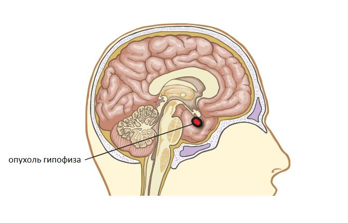
The pituitary gland is a gland located on the undersurface of the brain. It is located in the process of the base of the skull, which is called the “sella turcica”. The pituitary gland is divided into two lobes: posterior and anterior, each of which produces certain types of hormones.
The anterior lobe of the pituitary gland synthesizes the following hormones:
- thyroid-stimulating. Affects the functioning of the thyroid gland, reducing or, conversely, increasing the production of its hormones;
- adrenocorticotropic. Stimulates the activity of the adrenal cortex;
- gonadotropic hormones that affect the activity of the gonads;
- somatotropic. Regulates the growth of the body in childhood and adolescence, and is also “responsible” for the absorption of proteins, fats and carbohydrates at the cellular level;
- prolactin. Produced during lactation and pregnancy. It is believed that prolactin plays a huge role in the formation of attachment to offspring.
Pituitary adenoma in women and men develops only from the anterior part of the gland, which is called adenohypophysis.
The posterior lobe of the pituitary gland produces vasopressin and oxytocin. Vasopressin acts on the muscular layer of arteries and arterioles, thereby regulating blood pressure. In addition, the hormone has an antidiuretic function, that is, it reduces diuresis, increasing the absorption of water in the renal capillaries. Oxytocin takes part in the process of childbirth and lactation, affects sexual behavior and even plays a role in the formation of trusting relationships with others.
What is a microadenoma?
Adenoma is not one disease, but a whole group of pathologies that can have significant differences in clinical course and manifestations.
Microadenoma is an adenoma whose size does not exceed 1 centimeter. In most cases, adenoma is benign. The malignant variant is extremely rare, accompanied by a rapid increase in symptoms, as well as neurological disorders caused by the effect of the tumor on nearby brain structures.
Prevalence of adenoma
Adenoma is considered one of the most common brain tumors. It occurs in 2 people per 100 thousand population. However, we are talking only about tumors that were diagnosed. Doctors believe the disease is much more widespread. This is especially true for microadenoma, which in many cases occurs without any noticeable symptoms and is discovered by chance.
Diagnosis of microadenoma is also complicated by the fact that it can “masquerade” as diseases of many endocrine glands, for example, the adrenal glands or the thyroid gland.
The main symptoms of pituitary microadenoma in women
There are several types of adenoma, each of which manifests itself with a specific set of symptoms. However, there are general signs that suggest the presence of a tumor. These include:
- visual impairment. Most often, with pituitary adenoma, the lateral visual fields are lost. Of course, with microadenoma this phenomenon is observed quite rarely due to its small size. Visual impairment is associated with the fact that the pituitary gland is located near the optic chiasm. As the tumor grows, the nerves can be compressed, which disrupts the transmission of information from the analyzers to the cerebral cortex;
- headaches caused by the impact of the tumor on surrounding tissues and increased intracranial pressure;
- dizziness, increased fatigue.
Fortunately, adenomas in most cases are benign, so they do not cause depletion or intoxication.
Types of treatment for pituitary adenoma
Any brain tumor requires treatment, and the sooner it is started, the greater the patient’s chances of maintaining working capacity and completely getting rid of the consequences of the pathology. Conservative treatment of pituitary adenoma is often ineffective, since the tumor itself is usually hormonally active and has a negative impact on the entire human endocrine system. Currently, three main treatment methods are used:
- traditional surgery (or craniotomy), which may accidentally damage brain tissue;
- stereotactic radiosurgery/radiation therapy;
- endoscopic surgery to remove pituitary adenoma is the least traumatic method that is well tolerated by patients.
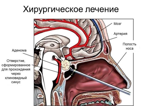
Since the pituitary gland is located in the so-called. sella turcica, it can be accessed through the sphenoid sinus. This is a transsphenoidal approach, which is implemented in three ways:
- Direct - through a puncture in the back of the nasal cavity.
- Nasal - through an opening in the nasal septum.
- Through an incision in the oral cavity (approximately under the upper lip).
To visualize a brain tumor, a binocular microscope is used in conjunction with a fiber optic endoscope. During the operation, the adenoma is separated from the tissues of the pituitary gland of the brain and scraped off. For large adenomas that extend beyond the boundaries of the sella turcica, transnasal treatment is carried out in two stages - initially the tumor is excised, and after it has descended into the sella during repeated intervention, it is completely removed. With all the advantages of this method of treatment, it should be noted that there is a drawback, which is the fairly high cost of transnasal surgery - for Moscow this amount is about 70 thousand rubles.
Endoscopic removal of pituitary adenoma
Radiosurgery is a highly effective method of treatment that does not harm healthy brain tissue, which means there are no serious consequences. The essence of the method comes down to irradiating the tumor with a directed beam of radiation - each time from a different angle. In this way, an almost complete absence of negative effects on the brain tissue surrounding the tumor is achieved. Depending on the indications, methods of influence such as cyber-knife, gamma knife, and Novalis can be used. The irradiation process is carried out under MRI/CT control.
There is no need to delay surgery for pituitary adenoma - it leads not only to severe hormonal imbalance, but also to complete blindness. The longer the period passes from the moment of tumor formation, the less chance there is of maintaining vision even after its successful removal.
Prolactinoma
Prolactinomas are diagnosed when the tumor interferes with the production of prolactin. In women, such tumors manifest themselves with the following symptoms:
- menstrual irregularities. The hormone prolactin in women regulates the menstrual cycle. If its production is disrupted, there may be delays in menstruation, amenorrhea, that is, a complete absence of menstruation, or, on the contrary, prolonged bleeding that lasts more than a week. If such symptoms appear, you should immediately consult a doctor: they may indicate not only the presence of an adenoma, but also a serious gynecological pathology;
- inability to get pregnant without gynecological pathology. It is this reason that often becomes the reason to consult a doctor;
- sudden discharge of colostrum or milk from the nipples in the absence of pregnancy;
- decreased libido or complete loss of sexual desire. The normative hormone prolactin is largely responsible for sexual behavior, in the absence of which libido practically disappears;
- body hair growth according to the male pattern (appearance of hair on the chest, face and abdomen).
Symptoms of the disease
Almost no patient encounters symptoms of pituitary microadenoma at the very beginning of its development, so this pathology is most often discovered completely by accident. The patient can consult a neurologist with headaches for reasons that are not related to the microadenoma, and have a computed tomography scan, where the doctor will detect the tumor.
With pituitary microadenoma, the symptoms will depend only on how hormonally active the tumor is. Since the neoplasm does not lead to compression of surrounding tissues, the patient does not have headaches or disturbances in visual activity. In addition to the fact that most adenomas of this type are not hormonally active, they often do not lead to the production of secretions. Therefore, symptoms of microadenoma are absent or appear to a minor extent.

Most often, microadenomas that are marked by hormonal activity are prolactinomas. In the body, prolactin promotes lactation, suppresses ovulation and weight gain. With prolactinoma, the symptoms in women are infertility and menstrual irregularities. If the microadenoma is highly active, then a liquid similar to colostrum will be released from the mammary glands. In men, such an adenoma can cause active breast growth, nipple discharge, and problems with potency. With a tumor, weight gain occurs, up to obesity.
If the formation of a tumor is provoked by the hormone somatropin, which is responsible for body growth, such a microadenoma is called somatropinoma. In children, adenomas of this type most often lead to an accelerated increase in body length. In adult patients, this sign is absent due to closed bone growth zones.
Women and men may develop a pathology such as acromegaly. It makes the hands and feet longer, the facial features rougher, the brow ridges larger, the fingers thicker. With acromegaly, the timbre of the voice decreases, in addition, this pathology leads to secondary diabetes mellitus and arterial hypertension.
With corticotropinoma, a large amount of adrenocorticotropic hormone is produced, resulting in Itsenko-Cushing's disease. Adrenocorticotropic hormone promotes increased production of cortisol by the adrenal glands, which leads to a change in the patient’s appearance. Fat tissue is redistributed, and muscle tissue partially atrophies, so the limbs become thin. Fat begins to be deposited on the abdomen, leading to bright, thick stretch marks (striae). The face becomes rounder, the cheeks become constantly rosy.
There are practically no other symptoms of microadenoma in the pituitary gland. But since, along with hormonal imbalances, disturbances occur in almost the entire body, microadenoma often leads to severe fatigue of the patient even in the absence of physical activity. The entire body weakens, which should be a reason for immediate examination and initiation of therapy.
Somatotropinoma
Somatotropinomas, or adenomas, in which the synthesis of somatotropin is disrupted, manifest themselves in the same way in men and women: acromegaly develops. Acromegaly is a disease in which in adults bone and cartilage tissues begin to rapidly grow and thicken, which leads to noticeable changes in appearance.
Acromegaly manifests itself as follows:
- enlargement of feet and hands. Often the first sign of the disease that patients notice is the inability to wear shoes that previously fit;
- an increase in the spaces between the teeth associated with the growth of the upper and lower jaws;
- coarsening of facial features;
- pain in the joints. Pain syndrome in late stages of the disease can be so severe that the patient loses the ability to move and needs to constantly take analgesics;
- sudden weight gain;
- fatigue, lethargy and irritability;
- disappearance or decrease in libido;
- persistent arterial hypertension, which is difficult to control with drugs to lower blood pressure.
Causes of pathology
Many people experience microscopic adenoma in the pituitary gland, but researchers have not been able to establish the exact causes of microadenoma in the pituitary gland.
However, factors have been identified that can cause various disorders in the pituitary gland and the development of microadenoma:
- hereditary predisposition,
- disruption of the activity of the endocrine system glands,
- infectious or traumatic lesions of the central nervous system,
- frequent abortions,
- long-term use of hormonal contraceptives,
- increased load on the pituitary gland during pregnancy, childbirth and lactation,
- malfunction of the peripheral glands, which leads to stimulation of the pituitary gland.
Microadenoma of the pituitary gland of the brain is more common in women, but cases of hormonally active adenomas are also found in the male half of the population. Many people wonder whether microscopic pituitary adenoma is transmitted from one person to another. No, this pathology is a disease of one specific person and is not contagious, like other tumors.
Corticotropinoma
Corticotropinomas disrupt the production of adrenocorticotropic hormone. As a result, Itsenko-Cushing syndrome is formed, which has the following manifestations:
- persistent arterial hypertension;
- obesity. The bulk of fat in Itsenko-Cushing syndrome is deposited on the face (it becomes round, moon-shaped) and in the abdominal area. In this case, the limbs may remain thin, which is why the patient’s figure takes on the appearance characteristic of the syndrome;
- the appearance of numerous pustular rashes on the skin;
- in areas of increased fat deposition, in particular on the abdomen, stretch marks (striae) appear;
- decreased libido;
- the appearance of male pattern hair in a woman;
- increased bone fragility (osteoporosis). With severe osteoporosis, even a minor injury can lead to a fracture;
- decreased immunity. With Itsenko-Cushing syndrome, patients experience more frequent infectious diseases that take a protracted course.
Thyrotropinoma
Thyrotropinomas cause an increase in the blood level of thyroxine, a hormone produced by the thyroid gland. In this case, the woman develops the following symptoms:
- sweating: patients may sweat even with minor physical exertion;
- irritability and increased emotional lability;
- increased heart rate;
- a feeling of interruptions in the work of the heart;
- rapid loss of body weight in the absence of diets and increased exercise;
- exophthalmos, that is, bulging eyes. Eyes with thyrotoxicosis become shiny and appear larger; for this reason, patients in the initial stages of the disease often report that they feel more attractive.
Types and symptoms of pituitary microadenoma
Pituitary microadenomas can produce a certain type of hormones, or they can be hormonally inactive.
In turn, all hormonally active pituitary microadenomas, depending on the hormone they produce, are divided into the following types:
- Somatotropin-producing adenoma. This type of tumor produces somatotropin - growth hormone. Tumors that originate from cells that secrete this hormone lead to the development of gigantism in children (excessive increase in body length) and acromegaly in adults (increase in the size of the feet and hands, thickness of the fingers, coarsening of facial features, growth of the brow ridges, decreased timbre of the voice). Acromegaly can lead to the development of secondary diabetes mellitus, arterial hypertension, and an increased risk of cancer;
- Prolactin-secreting adenoma (prolactinoma). Synthesizes prolactin, which helps suppress ovulation, stimulate lactation and increase body weight. At the same time, in women, symptoms of pituitary microadenoma, which produces prolactin, can manifest as menstrual irregularities, infertility, decreased libido, and discharge from the mammary glands. If a man has prolactinoma, he may develop impotence;
- Adrenocorticotropin-producing adenoma. Produces adrenocorticotropic hormone, which stimulates the activity of the adrenal cortex. As a result, patients develop Itsenko-Cushing's disease, in which a person's appearance undergoes significant changes: due to the redistribution of adipose tissue and muscle atrophy, the limbs become thinner, and excess subcutaneous fat is localized mostly in the abdominal area, which leads to the appearance of anterior abdominal wall stretch marks more than 1 cm thick; the person’s face takes on a moon-shaped shape, and a blush is always noticeable on it. Changes in behavior and mental reactions may also occur;
- Thyrotropin-producing adenoma. Produces thyroid-stimulating hormone, which stimulates the activity of the thyroid gland. It is quite rare and leads to the development of thyrotoxicosis;
- Gonadotropin-producing adenoma. Synthesizes gonadotropic hormones that activate the sex glands. In women it causes uterine bleeding and menstrual irregularities, in men it causes impotence and gynecomastia.
Quite often, the symptoms of pituitary microadenoma do not manifest themselves in any way, and the detection of this type of tumor is, as a rule, accidental.
In some cases, with hormonally inactive microadenomas, the patient may experience headaches and ophthalmological symptoms (decreased vision, optic nerve atrophy).
X-ray symptoms of this disease are expressed in changes in the size and shape of the sella turcica, thinning (sometimes destruction) of bone structures.

For what reasons can a microadenoma appear?
Until now, scientists have not been able to find out for what reasons microadenoma develops. At the moment there are two main hypotheses. According to the first, pituitary tumors develop due to dysfunction of the hypothalamus: the gland that produces releasing factors (hormone-like substances that regulate the functioning of the pituitary gland). The second hypothesis explains the development of microadenoma by the patient’s genetic characteristics and hereditary predisposition to the formation of tumors of the nervous tissue. However, no convincing evidence was found for any of these hypotheses.
It is believed that risk factors for the development of microadenoma are:
- previous inflammatory diseases of the central nervous system, for example, meningitis or encephalitis;
- suffered traumatic brain injuries;
- constant use of hormonal contraceptives;
- intrauterine hypoxia. During hypoxia, brain cells are the first to suffer, so people who survive this condition are more likely to develop various tumors of the nervous system, including microadenoma;
- increased emotional stress and frequent stress.
It is important to remember that the listed factors do not cause microadenoma, but can only provoke its development.
Causes of pituitary microadenoma
In the vast majority of cases, it is not possible to establish the cause of pituitary microadenoma.
The following factors can be the causes of pituitary microadenoma:
- Female;
- Hereditary predisposition;
- Functional overload of the pituitary gland in the form of pregnancy, abortion, childbirth, breastfeeding, use of oral contraceptives;
- Infectious processes in the central nervous system;
- traumatic brain injuries;
- Pathologies of pregnancy and childbirth.
Diagnosis of pituitary microadenoma
The most reliable methods for diagnosing pituitary microadenoma are radiological techniques.
Microadenoma can be determined using X-ray only when it reaches a size exceeding the size of the sella turcica. In this case, it will be noticeable in the picture that the sella turcica has become thinner.
A more reliable method is MRI - magnetic resonance therapy. The following changes will indicate the presence of a tumor to the doctor:
- swelling in the area being examined;
- displacement of brain structures adjacent to the pituitary gland and deformation of its ventricles;
- changes in the structure of the sella turcica.
The most informative method for identifying microadenoma is PET (positron emission tomography). Thanks to PET, it is possible not only to determine the presence of a tumor, but also to determine the degree of its hormonal activity. Unfortunately, PET is a rather expensive method. In addition, it is not available in all cities.
Computed tomography can also help in making the correct diagnosis, but CT has its own specifics: this method is better suited for visualizing dense structures, such as bones. Using CT, you can determine the deformation of the sella turcica, thanks to which the doctor may suspect the presence of a microadenoma. Computed tomography has another drawback. This method involves radiation exposure, so it cannot be used if the patient is pregnant.
Additional research methods
Additional research methods may be prescribed:
- blood test for hormones. If the tumor has affected the pituitary gland, the treatment of which will be described below will affect hormonal levels. Naturally, the choice of a specific analysis will depend on the clinical picture of the disease. For greater accuracy, hormone tests are performed two or three times;
- Ultrasound of the thyroid gland. The cause of hyperthyroidism can be not only an adenoma, but also neoplasms of the thyroid gland. Therefore, this analysis is important for making a differential diagnosis;
- Ultrasound of the pelvic organs. The doctor must make sure that the patient does not have ovarian neoplasms, such as cysts or malignant tumors, which can give a clinical picture similar to prolactinomas.
Pituitary microadenoma
Pituitary microadenomas represent a minority of all pituitary tumors but may pose some imaging challenges due to their size and associated clinical manifestations. By definition, a microadenoma is a tumor of the pituitary gland measuring less than 10 mm.
Clinical picture
Pituitary microadenoma is limited to the sella turcica, so symptoms associated with mass effect are not observed. This is the most common finding in cases of hormonal imbalances (excessive production of one or more hormones). More rarely, microadenomas are an accidental discovery, since they can only be detected with a targeted examination of the pituitary gland.
Diagnostics
Plain radiography and CT. Before the advent of MRI, the pituitary gland was visualized using a lateral radiograph of the skull (remodeling of the walls of the sella turcica was assessed) and CT. Despite the fact that on CT in 80-90% of cases it is possible to visualize microadenomas measuring 5-10 mm, smaller tumors are more difficult to detect
MRI
MRI is the main method for imaging pituitary microadenomas and requires special sequences (small slice thickness, small field of view, dynamic contrast). MRI with CT has a sensitivity of up to 90%. Post-contrast images and especially images after dynamic coronary angiography are an integral part of the MR examination protocol for the pituitary gland and significantly increase diagnostic accuracy. However, sometimes morphological changes can be visible in non-contrast images. These include changes in the gland from the side of the adenoma, thinning and remodeling of the lower wall of the sella turcica, as well as deviation of the pituitary infundibulum from the adenoma.
- on T1 are usually isointense to the pituitary gland;
- T1-KU:
- on dynamic sequences, a rounded area of reduced enhancement compared to the rest of the gland;
- On delayed images, the pattern ranges from hypoenhancement (the most common pattern) to isointensity compared with the rest of the gland to hyperintensity.
- T2 – the picture is different, but in general – slight hyperintensity.
It is very important to remember the following fact when performing an MR examination of the pituitary gland: small incidentalomas of the pituitary gland are quite common, and in approximately 2-30% of cases, small asymptomatic microadenomas are detected at autopsy.
Catheterization of the inferior petrosal sinus
Catheterization of the inferior petrosal sinus is performed in two cases when a microadenoma is suspected with a normal MRI picture:
- Confirm the presence of a microadenoma not located in the pituitary gland, especially in the case of Cushing's disease, when there are many sources of ACTH outside the pituitary gland (for example, some tumor diseases of the lung);
- Tumor lateralization – to determine surgical tactics.
Therapy methods
There are many ways to treat microadenoma. Usually the doctor selects several methods, based on the characteristics of the tumor, the degree of its hormonal activity, the patient’s health status and other factors.
The following approaches to adenoma treatment have been developed:
- observation. If the tumor is small, does not grow in size, and does not affect the patient's hormonal levels, observation may be the optimal strategy. The patient should regularly undergo an MRI or X-ray of the skull, as well as donate blood for hormones. If during follow-up there is no negative dynamics, other treatment methods can be abandoned;
- drug treatment. Taking special medications is recommended if the patient is diagnosed with somatotropinoma or prolactinoma. The doctor selects drugs that block the production of hormones, as a result of which the patient’s condition returns to normal. Additionally, symptomatic treatment is prescribed, for example, immunomodulators and calcium supplements for Itsenko-Cushing syndrome, painkillers to relieve pain in acromegaly, etc.;
- radiosurgery. This method is relatively new. Thanks to radiosurgery, it is possible to destroy a tumor, while avoiding both extensive surgical intervention and destruction of tissues adjacent to the tumor, which is extremely important when performing brain surgery. Removal of microadenoma is carried out through targeted exposure to radiation;
- surgical intervention. This method is the most traumatic. It is used only if other methods are ineffective or are not suitable for a particular patient. The pituitary gland is accessed in one of two ways: through the nasal passages and by opening the skull. For microadenoma, the first option is safer, and therefore the most preferable. Surgical treatment is often used if a microadenoma is detected in a pregnant woman for whom radiosurgery and drug treatment are contraindicated.
Endoscopic removal of pituitary adenoma (video)
A 45-year-old woman contacted us. She complained of an increase in the size of her hands and feet, changes in facial features, an increase in the size of her tongue, irregular periods, etc. Until that day, she had been ill for 15 years, and during all this time, a diagnosis had not been established for her at her place of residence. During the consultation, our neurosurgeons suspected hormonal problems in the patient, after which they performed an MRI study and immediately confirmed the diagnosis of pituitary macroadenoma.
The patient underwent endoscopic surgery to remove a pituitary macroadenoma. After the operation, on the third day, the patient herself, subjectively, noted the normalization of facial features and a decrease in the tongue. By the time of discharge, the hormonal levels had completely returned to normal.
Pituitary adenoma is a group of tumors that are located in the area of the so-called “sella turcica”. They differ in size and degree of hormonal activity. Most often they manifest themselves either as visual disturbances (the patient's visual fields are lost) or changes in hormonal status (enlarged limbs, uncontrolled growth, disruption of the female menstrual cycle, the phenomenon of diabetes insipidus, etc.).
According to the most modern methods, the removal of most pituitary adenomas and other tumors located in the area of the sella turcica is performed using the transnasal transsphenoidal method, i.e. through the natural nasal passage. These operations are performed using an endoscopic stand in a minimally invasive manner. Previously, the removal of such tumors was carried out using a major traumatic operation accompanied by craniotomy.
The essence of the operation is as follows. Through the right nasal passage the base of the anterior cranial fossa is accessed. To do this, a small hole is made to allow access to the bottom of the sella turcica. Then, using endoscopes with different viewing angles (0, 30 and 45 degrees), the adenoma is removed. After which the hole is closed using special synthetic materials: a small fragment of the bone is placed and sealed with special glue. According to current modern methods, no tampons are placed in the nose so that the patient can breathe freely.
Activation of the patient is recommended on the first day after surgery. In some cases, there is a need for subsequent correction of hormonal levels. Those. After tumor removal, hormonal therapy is selected to eliminate the clinical manifestations that the patient had. In the normal course of the disease, discharge from the hospital is possible on the fourth day after surgery.
Complications
The most common complications of pituitary adenoma include the following:
- cystic degeneration. The development of this complication is indicated by symptoms such as severe headaches, progressive visual impairment, frequent increases in blood pressure in the absence of diseases of the cardiovascular system. Occasionally, patients experience mental changes, for example, increased irritability, causeless decrease in mood, etc.;
- hemorrhage in the adenoma tissue. Prolactinomas are especially often complicated by hemorrhage. Hemorrhage is a rather dangerous, but usually not fatal complication. Its development is indicated by symptoms such as a sharp headache, sudden blurred vision, as well as pituitary insufficiency. Sometimes there are cases where, after hemorrhage into the adenoma, spontaneous self-healing occurs.
It is important to remember that if the patient has a diagnosed microadenoma, if the above symptoms appear, you should immediately contact a medical facility.
Causes of microadenoma
With regard to tumor diseases, the reasons for the formation of pituitary microadenoma have not been fully identified. There are supposed factors that cause cell division:
- heredity;
- failure of the hypothalamus to regulate the functionality of the pituitary gland;
- damage to the central nervous system as a result of infections, injuries;
- being female and the associated consequences - pregnancy and childbirth, abortion, hormonal pills;
- failure of the functions of the peripheral glands, which stimulates the pituitary gland, as a result, its cells grow, forming microadenomas in the future.
The tumor is classified according to its structure into cystic and homogeneous. The first is considered to be a consequence of hemorrhages in the tumor tissue, which does not affect the prognosis.
Are there ways to prevent microadenoma?
It is believed that the prevention of pituitary microadenoma, like other tumors of the central nervous system, is primarily the protection of the fetus during pregnancy from adverse environmental influences. It is important for the expectant mother to regularly attend antenatal clinics, not take medications without a doctor’s recommendation, and try to lead a healthy lifestyle even at the stage of pregnancy planning.
Due to the fact that the connection between microadenoma and infectious diseases is considered proven, it is important to treat infections in a timely manner. If the doctor has prescribed a course of antibiotics, you should never stop taking them ahead of schedule: because of this, a focus of infection can form in the body, which not only negatively affects the immune system, but can also cause the development of adenoma. To prevent infections during epidemics, it is recommended to take multivitamin complexes and immunomodulators.
It is equally important to try to avoid emotional overload. The pituitary gland is sensitive to stress, so constant stress can provoke hormonal imbalance and, as a result, the development of microadenoma.
Women should not uncontrollably take hormonal contraceptives. Most modern drugs to prevent unwanted pregnancy have a direct effect on the hypothalamus, and through it, on pituitary activity. Therefore, you should choose contraception under the guidance of a doctor. The same applies to other hormonal drugs. Their uncontrolled use is unacceptable: the endocrine system reacts very sensitively to any outside interference.
Another measure to prevent pituitary adenoma is to avoid abortion. It is important to protect yourself if a woman does not want to have children. Abortion is a brutal intervention that inevitably affects hormonal levels.
A reliable measure for preventing adenoma is regular medical examination. By monitoring your health, tumor development can be noticed at an early stage.
Currently, diagnosing microadenoma is not difficult despite the small size of the tumor. It is important to remember that the earlier the disease is detected, the easier it is to treat. Unfortunately, some types of adenoma can lead to irreversible health changes, for example, deformation of the skull bones due to acromegaly. Only paying attention to your health and following all doctor’s recommendations will help you avoid the negative consequences of microadenoma.
Post published: 03/23/2019
0