Cochlear neuritis (sensorineural hearing loss, auditory neuritis) is a lesion of the auditory nerve, which results in hearing loss and constant subjective noise in the affected ear (or both ears). According to statistics, 6-8% of the population suffer from hearing impairment to one degree or another, and in most cases it is cochlear neuritis that leads to disruption of the auditory analyzer. The disease is polyetiological, that is, it has several possible causes. Let's talk about the causes and symptoms of cochlear neuritis.
Causes of pathology
The vertebral artery of the cervical spine is surrounded by the spinal column and muscle tissue. Any changes in them will affect the condition of the artery.
All causes of the development of pathology can be divided into three groups:
- Congenital abnormalities in the structure: bends along the course, tortuosity.
- Some diseases leading to a decrease in lumen: thrombosis, atherosclerosis, arteritis.
- Pressure on the artery from the outside:
- osteochondrosis;
- injuries;
- rachiocampsis;
- tumors;
- muscle spasm.
Most often, compression of the cervical vertebral artery develops against the background of several provoking factors.
In addition to the listed reasons, predisposing factors for the development of the disease can be noted. These include:
- Chronic diseases of the ENT organs.
- Serious infections.
- Traumatic brain injuries.
- Features of the constitution.
- Receipt of additional impulses from pathological foci in the upper part of the body: from the heart, gall bladder, lungs, skin.
- Developmental anomalies.
- Poor circulation in the body due to heart failure, surges in blood pressure.
- Blood clotting disorder.
- Diseases of the bronchopulmonary system.
- Smoking.
- Anemia.
Long-term static-dynamic loads, jerky head movements, stress and negative emotions negatively affect the artery.
Benefits of cochlear implantation in Belgium
- The ability to use binaural cochlear implantation to maximize hearing and speech outcomes in both children and adults.
- Hybrid implantation for patients with residual hearing using short implants that contact the basal or high-frequency part of the cochlea, leaving the low-frequency (apex) part intact.
- Using the latest models of external units from the range of companies Cochlear Corporation, MED-EL Corporation and Advanced Bionics Corporation. The patient receives the most advanced design with minimal weight, the most powerful processor and the largest battery that currently exists.
- The doctor has no commercial interest in promoting an implant of a certain brand or model to a patient.
Symptoms
The disease can manifest itself with various signs depending on the stage of development, and there are two of them:
- Functional (dystonic).
- Organic (ischemic).
Symptoms of the functional stage
Symptoms of vertebral artery syndrome at this stage include the following:
- Headache.
- Disorders of the autonomic nervous system.
- Deterioration of vision.
- Dizziness.
- Hearing loss.
- Upon examination, an ophthalmologist detects decreased vascular tone in the fundus.
If the causes are not eliminated, the disease progresses and the next stage begins.
Organic
Persistent foci of ischemia appear in the brain. The pathology is manifested by the following symptoms:
- Everything is swimming before my eyes.
- Nausea.
- Speech problems.
These manifestations are aggravated by sudden turns of the head. If you lie down during such an attack, the symptoms practically disappear.
Need rehabilitation!
The patient can hear the first sounds after activation of the speech processor, that is, 20–30 days after surgery. At this time, a long and difficult period begins for the patient, as you need to be patient and constantly follow the recommendations of specialists - a teacher of the deaf, an audiologist, a speech pathologist.
The recovery period is especially difficult for parents of children who have had surgery. Typically, mom and dad want to get results as quickly as possible, and the first fruits of cochlear implantation usually appear 6–9 months after surgery. All this time you need to work, study and train. Sometimes this is difficult to explain to parents, but without cooperation with doctors and constant visits to the center where cochlear implantation was performed, there will be no result.
If your ear goes deaf, is it deaf? How to avoid hearing problems Read more
Clinical forms of the syndrome
Pathology may not occur in the same way, so several clinical varieties are distinguished.
Barre-Lieu syndrome
It manifests itself against the background of compression of the nerve endings around the artery. Main manifestations:
- Throbbing headache.
- Noise in ears.
- Nausea.
- Floaters before the eyes.
In the absence of effective treatment, sleep is disturbed, irritability appears, and memory deteriorates. In severe cases, there may be fainting.
Autonomic change syndrome
With the development of this syndrome, the following are observed:
- Either fever or chills.
- Increased sweating.
- Cold extremities.
- Heart pain.
- Migraine.
Transient ischemic attacks
Patients report the following symptoms:
- Sensitivity disappears.
- Vision problems.
- Speech is impaired.
- Movement disorders are observed.
- Unsteady gait.
- Dizziness.
- Difficulty swallowing.
Unterharnsteidt syndrome
This syndrome often develops against the background of osteochondrosis, as a provocateur of spinal artery syndrome. It appears like this:
- Headache.
- Visual and hearing impairment.
- Dysarthria.
In severe cases, loss of consciousness may occur.
Drop attack episodes
This syndrome is manifested by a sudden fall of a person. The head falls back, movements are difficult. Consciousness is not impaired during such attacks; after a few minutes everything returns to normal.
Basilar migraine
An attack with this syndrome begins with visual impairment, and then additional symptoms appear:
- Unsteady gait.
- Noisy in the ears.
- Speech is impaired.
- The headache is accompanied by dizziness and vomiting.
Often the attack ends with loss of consciousness.
Vestibulocochlear syndrome
The manifestations are influenced by the position of the human body. The patient complains:
- For hearing problems.
- Swaying sensation.
- Unsteady gait.
Ophthalmic syndrome
Its manifestations begin with the appearance of disturbances in the organ of vision:
- Burning and pain.
- Feeling of sand in the eyes.
- Increased tear production.
- Reduced image clarity.
- Fields of vision may be lost.
The syndrome is especially acute during visual stress.
A little more apparatus
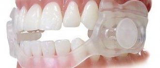
The inner part of the cochlear implant system is located in the inner ear, so it cannot be removed. But the speech processor is removed, but it is on it that the microphones are located, and as soon as a person removes the processor, he stops hearing. Therefore, patients, especially children, after they get used to the speech processor, wear it constantly.
The speech processor is larger than conventional hearing aids. This is due to large batteries, which must power not only the processor itself, but also the electrode located in the inner ear. However, despite this, wearing a cochlear implant does not cause problems in either adult patients or children.
Diagnostics
It is important to make an accurate diagnosis, then the effectiveness of therapy can be guaranteed. Diagnostics is carried out in two stages.
On the first:
- The doctor listens to the patient's complaints and clarifies the symptoms.
- Anamnesis is studied to identify diseases that can lead to vertebral artery syndrome.
- A survey on the presence of genetic abnormalities in the structure of blood vessels and the musculoskeletal system.
- Conversation about lifestyle, professional activities.
At the second stage of diagnosis, various methods are used:
- MRI. The resulting image reveals the cause of the circulatory disorder.
- CT scan. Allows you to study the pathological area in detail.
- X-ray of the cervical spine. The picture is taken in different projections.
- Doppler ultrasound. The vertebral artery is analyzed after the injection of a special substance to detect an area with problematic blood circulation.
After making an accurate diagnosis, the specialist prescribes therapy.
Contraindications
Absolute contraindications for cochlear implantation
- aplasia of the cochlear nerve (confirmed on tomography)
- congenital absence of the cochlea
Relative contraindications
- significant intracochlear ossification or fibrosis
- birth abnormalities of the inner ear
- active chronic otitis media
- inability to tolerate general anesthesia or psychiatric conditions that may prevent the use of a cochlear implant
- poor psychosocial environment
In addition, chronic infections of the middle ear and mastoid process or perforation of the eardrum may be contraindications.
Hematological, pulmonary or cardiac diseases may also be contraindications.
Treatment
Treatment of vertebral artery syndrome requires complex treatment. It is being carried out in several directions:
- Treatment of the cervical spine.
- Normalization of the artery lumen.
- Additional therapy.
Treatment can be carried out on an outpatient basis, but in the presence of acute cerebrovascular accidents, hospitalization is required. The treatment regimen is selected for each patient individually, taking into account concomitant pathologies and the severity of the syndrome.
Medication
Drug therapy involves prescribing the following groups of drugs:
- To relieve swelling of soft tissues, Troxevasin and Detralex are prescribed.
- Non-steroidal anti-inflammatory drugs: Meloxicam, Nimesulide.
- In order to normalize cerebral circulation, it is recommended to take Vinpocetine and Cinnarizine.
- To normalize metabolic processes in the brain and to prevent ischemic disorders, the following are prescribed: Mexidol, Actovegin, Mildronate.
If osteochondrosis is the trigger for the development of the syndrome, then the following drugs are prescribed:
- Chondroprotectors. These medications help restore damaged cartilage tissue.
- Medicines to reduce pain: Ibuprofen, Diclofenac.
Auxiliary treatment involves taking:
- Antispasmodics and muscle relaxants.
- Multivitamin complexes.
- Medicines to lower cholesterol levels.
- Sedative medications if the psycho-emotional state is disturbed.
- To relieve migraine attacks and headaches: “Antimigren”, “Sumatriptan”.
It is important to understand that the therapy will be long-term. Only a full course will significantly reduce the manifestations of the syndrome and improve the patient’s quality of life.
Neuroprotective therapy
This type of treatment is aimed at selecting drugs that will be able to prevent the development of ischemia at the cellular level.
In the medical practice of treating vertebral artery syndrome, doctors consider the use of nootropic drugs that will have an antioxidant effect as promising therapy.
Domestic scientists have developed such a drug, Phenotropil, and it is widely used in neurological practice. The medicine has the following effects:
- Increases concentration.
- Increases the resistance of brain tissue to oxygen deficiency and toxic effects.
- Has an anticonvulsant effect.
"Phenotropil" has a positive effect on all metabolic processes in the brain, which has a positive effect on treatment.
Exercise therapy
After the acute period has passed, you can begin to perform a special exercise complex, which is selected by the doctor. It includes the following exercises:
- The doctor puts his hand to the patient's forehead, and he presses on it.
- The same thing, but the hand is placed on the back of the head.
- Slowly turn your head to the sides.
- Nods.
- Side bends.
- Back pressure on the sides of the head.
At first, it is better to perform the complex under the supervision of a specialist, and then you can do it yourself at home.
Massage and manual therapy
After the attacks have stopped, the doctor recommends massage and a visit to a chiropractor.
The procedure will relieve muscle tension and reduce vascular compression. But only a specialist should perform massage. Inept execution is fraught with serious complications: compression of blood vessels or even a stroke.
Surgical methods
If drug therapy and physical therapy do not give the desired result and the risk of ischemic brain damage is high, then it is advisable to use surgical intervention. The following methods are used:
- Reconstruction of the vertebral artery. During surgery, the surgeon removes the wall from the affected area of the artery.
- Surgical decompression of the spinal artery.
- Removal of osteophytes.
- Expansion of a vessel with an inert gas.
- Replacement of a pathological area of a vessel with a prosthesis.
- Periarterial sympathectomy. The procedure involves making a parallel incision along the artery, exposing it and cutting the outer lining of the vessel. As a result, blood circulation improves.
Successful surgery provides a chance for a full recovery.
Treatment of dizziness in osteochondrosis of the cervical spine
Cervical osteochondrosis is a rather insidious disease. Many people of working age experience its symptoms, but not everyone knows what caused it. And if you can guess the origin of the pain, then few people associate the appearance of dizziness, nausea and depression with osteochondrosis. But in vain, because the source of such problems often lies in the spine.
Causes and mechanism of development
As is known, the development of osteochondrosis is mediated by dystrophic-degenerative processes in the intervertebral discs and surrounding tissues.
Under the influence of excessive load and metabolic disorders, the height of the cartilaginous plates decreases, their protrusion, the formation of bone growths (osteophytes) and pathological uncovertebral joints. It not only disrupts the biomechanics of the entire cervical spine, but also affects the nerves and vessels passing through here.
Dizziness with osteochondrosis is precisely caused by a lack of blood supply to the brain by the vertebral artery passing in the canal of the transverse processes. In turn, this becomes the result:
- Direct compression by pathological formations.
- A spasm that occurs when one’s own nerve plexus is irritated.
- Concomitant formation of atherosclerotic plaques in elderly people.
Hypoxia of various parts of the central nervous system causes functional disorders in internal organs. This is why chondrosis has so many faces and is often hidden behind the masks of other diseases. Based on this, differential diagnosis should become an important component of the clinical examination.
If you feel dizzy or worried about depression, then there is no need to panic right away - such phenomena are quite typical for cervical osteochondrosis. But only a doctor can find out whether this is really so.
Treatment
Treatment of dizziness with cervical osteochondrosis consists of eliminating the cause-and-effect relationship between structural disorders in the spine and cerebrovascular manifestations.
This helps to get rid of many problems that are directly related to degenerative processes in the axial skeleton.
The conservative treatment program is formed by the doctor and may include the following methods:
- Drug therapy.
- Physiotherapy.
- Therapeutic gymnastics exercises.
- Massage and manual therapy.
- Folk remedies.
First of all, it is necessary to treat osteochondrosis, while simultaneously eliminating its neurological and vascular complications.
Drug therapy
For chondrosis, drugs with multidirectional effects are used. Many patients are interested in what medications can relieve neck pain and dizziness. But these are not the only indications for the use of medications. The following drugs are commonly used:
- Non-steroidal anti-inflammatory drugs (Xefocam, Airtal).
- Muscle relaxants (Mydocalm, Tolperil).
- Vitamins (Vitaxon, Milgamma, nicotinic acid).
- Vascular (Trental, Actovegin, Cavinton).
- Neuroprotectors (Instenon, Cerebrolysin).
- Sedatives (Glitsed, Persen).
- Histamine-like (Vestibo).
It is recommended to use injectable forms first and then switch to tablets. But under no circumstances should you take medications on your own.
Correctly selected treatment can quickly relieve any signs of osteochondrosis, including dizziness and depression.
Physiotherapy
Along with the use of drugs, physiotherapy helps treat dizziness with cervical osteochondrosis. They improve blood flow in blood vessels, eliminate muscle spasms and reduce pain. The following procedures are useful for the spine:
- Electrophoresis.
- Laser treatment.
- Magnetotherapy.
- Sinusoidal currents.
- Paraffin and mud therapy.
- Reflexology.
- Hydrotherapy.
This is a general list of techniques, and the physiotherapist selects those that are suitable for a particular patient.
Physiotherapy
With cervical osteochondrosis, it is impossible to do without physical therapy exercises. By contracting, muscles promote normal blood flow; during movements, their fitness increases, ligaments are strengthened, which helps stabilize the spine. Isometric gymnastics has a good effect:
- Having fixed your head, try to bend in different directions and turn.
- Pull your shoulders back, bringing your shoulder blades closer and feeling the tension in your back muscles.
- Sitting on a chair, grab the seat with your hands, trying to lift yourself.
In this case, static muscle tension should alternate with their complete relaxation. If you are worried about dizziness, then first use a set of exercises performed while lying down. And after the vestibular disorders subside, they move on to sitting and standing exercises.
Therapeutic gymnastics exercises are performed under the supervision of an instructor or physical therapy doctor and at a slow pace, avoiding sudden turns and head rotations.
Massage and manual therapy
Treatment of dizziness with cervical osteochondrosis includes methods of manual manipulation of the spine. During a massage of the collar area, muscles relax and blood flow improves. This helps reduce cerebral disorders. And compression of neurovascular structures can be reduced using manual therapy methods: traction, mobilization, torsion.
Folk remedies
When dizzy due to cervical chondrosis, some people prefer alternative treatments. Basically, they are aimed at the same mechanisms as classical methods: eliminate inflammation, muscle spasm, improve blood circulation. The following folk recipes are used:
- Compresses made from horseradish root or leaves, mustard, honey and mumiyo.
- Rubbing from a decoction of mint leaves, plantain, birch buds, chamomile flowers, burdock root and dandelion; tinctures of calendula flowers.
- Applications from bread cakes, blue clay.
- Balm made from camphor, juniper and fir oils.
You can use such products only after consulting a doctor, remembering that it is impossible to replace traditional tablets or injections with them.
To cure dizziness due to osteochondrosis, you need to consult a doctor in a timely manner. The sooner therapy is carried out, the higher the likelihood of complete restoration of motor function and elimination of cerebrovascular disorders.
Source: //MoySkelet.ru/spina/shejnyj-otdel/lechenie-golovokruzheniya-pri-osteoxondroze-shejnogo-otdela-pozvonochnika.html
Prevention
Pathology can be prevented; for this it is important to follow the following recommendations:
- Perform exercises to strengthen your neck muscles.
- Wear the Shants collar for several hours a day.
- For osteochondrosis, the mattress and pillow should be orthopedic.
It is much easier to prevent any pathology than to engage in long-term therapy later. Vertebral artery syndrome requires a serious approach to therapy to avoid complications, including disability. An effectively selected course of treatment will allow you to return to a normal lifestyle.
Source: columna-vertebralis.ru
Cost of implantation
For an operation such as cochlear implantation, the price is determined by the model of the external unit plus the cost of the surgeon.
At the same time, the real cost of the operation can be determined only after examination by the surgeon, as well as all the parameters of the upcoming operation.
But in Belgium, the cost of medical services themselves—doctor’s work and hospital stay—is 40-60% lower than in neighboring EU countries (France, Germany) and Israel.
Therefore, the final price for the patient will be approximately 30% lower in this country.
Causes
A large number of factors can provoke the appearance of cochlear neuritis. call the following diseases the most common
- Age. The older a person gets, the more diseases begin to bother him. Therefore, it is not surprising that at this age many people develop pathology of the auditory nerve. Most often it occurs against the background of other diseases - cerebrovascular accident, stroke or arterial hypertension.
- Harmful production. People who have to work in enterprises with high levels of noise and vibration have a high risk of developing cochlear neuritis. More than others, machine operators, miners, miners, builders and workers in other professions expose themselves to this danger.
- Injury. Pathology can occur as a result of a traumatic brain injury, which causes serious changes - cerebrovascular accidents, edema, local hemorrhages.
- Long-term use of medications. Cochlear neuritis is most often found in patients who have been taking antibiotics for a long time.
- Toxic substances. Problems with the auditory nerve can occur as a result of toxic chemicals entering the body, such as mercury, arsenic and others. Alcoholic drinks, if consumed in large quantities, as well as smoking, also cause great harm to the tissues of the auditory nerve.
- Infections. Quite often, the diagnosis of cochlear neuritis is made to people who have previously been treated for diseases such as influenza, meningitis, brucellosis, ARVI, scarlet fever and others.
Contact with any of these factors can cause serious harm to the tissues of the auditory nerve , and this can create favorable conditions for the development of the inflammatory process, which will subsequently lead to the death of some cells.
At the same time, a patient with a similar diagnosis may experience disturbances in the functioning of the auditory nerve, which ultimately ends in hearing impairment.
Sensorineural hearing loss
The auditory analyzer is a system consisting of 3 sections: the outer, middle and inner ear. Each department has a number of constituent elements. The outer ear is the pinna and the external auditory canal.
The middle ear is a cave with a mastoid process, a tympanic cavity containing the auditory ossicles (incus, malleus, stirrup), auditory (Eustachian) tube and tympanic membrane.
The inner ear consists of the cochlea (the sound-receiving part) and the semicircular canals (the peripheral part of the vestibular analyzer). This complex structure is responsible for a wide range of ear diseases.
Sensorineural hearing loss (sensorineural hearing loss, cochlear neuritis, auditory neuritis) is a non-infectious ear disease that affects the auditory nerve and, accordingly, sound perception.
The main symptom is hearing loss, accompanied by subjective tinnitus. Most often they talk about acute and subacute sensorineural hearing loss .
This disorder affects people of all age groups, but with age, hearing decreases to one degree or another in all people. This condition is called presbycusis (age-related decrease in hearing acuity).
The main cause of these conditions is atrophy of the sound-perceiving nerve endings of the organ of Corti. chronic sensorineural hearing loss is diagnosed , hearing, unfortunately, cannot be restored.
CLINICAL FORMS (the criterion is the duration of the disease):
- up to 4 weeks – acute sensorineural hearing loss – treatment effectiveness is 70-90%,
- from 1 month to 3 months – subacute sensorineural hearing loss – treatment effectiveness – 30-70%,
- more than 3 months – chronic sensorineural hearing loss – the effectiveness of treatment is questionable.
SYMPTOMS
The main symptom of sensorineural hearing loss is hearing loss. Often occurs after an acute respiratory viral infection, psycho-emotional stress, or intoxication. It can affect either one ear or both at the same time.
A very common symptom of this disease is noise in the ear: it can be either high-frequency (ringing, squeaking, “buzzer,” “hissing”), which is more common, or low-frequency (hum).
Such phenomena require immediate medical attention.
CAUSES
- infectious viral diseases: influenza and ARVI, infectious parotitis;
- trophic disorders of nervous tissue: vascular disorders, insufficient blood supply (vascular atherosclerosis, hypertension, vegetative-vascular dystonia);
- emotional stress;
- traumatic brain injuries;
- acoustic injuries: short-term but excessively strong sound - a gunshot, a scream, a beep;
- exposure to a number of chemicals that have ototoxic effects (industrial and household substances);
- long-term use of ototoxic drugs - these are antibiotics (primarily aminoglycosides), antimalarials, salicylates.
There is such a form as idiopathic sensorineural hearing loss , when the cause of the development of damage to the hearing organ cannot be identified.
PREVENTION
The rules of prevention are simple - avoid risk factors. It is necessary to promptly treat upper respiratory tract infections and take all medications only as prescribed by a doctor. A special risk group consists of people working in toxic, noisy industries; Vibration is also harmful.
In such cases, it is necessary to observe all safety measures and working conditions (work with headphones when there is noise, take more frequent short breaks from work). These people need to change jobs when the first symptoms of sensorineural hearing loss appear.
If this is not possible, it is necessary to be observed by an otolaryngologist every 3 months and take courses of vascular medications (Tanacan, Trental, etc.).
COMPLICATIONS
Without treatment and prevention, the disease progresses and leads to complete deafness.
DIAGNOSTICS
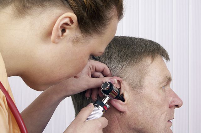
A doctor who suspects sensorineural hearing loss needs to perform tuning fork tests . To verify the diagnosis and accurately determine the degree of hearing loss, a hardware test is performed - pure tone threshold audiometry .
TREATMENT
On the first day of the appearance of noise in the ear and hearing loss, you must immediately consult a doctor for a comprehensive course of therapy.
Source: //www.pirogov-center.ru/specialist/diseases/detail.php?ID=540
Symptoms and signs
To diagnose cochlear neuritis , it is enough to know how exactly this disease manifests itself:
- if the neuritis was caused by an infection, the patient will have a fever, weakness and malaise, as well as a cough and runny nose.
- Improper functioning of the cerebral circulation can cause floaters, increased blood pressure, and loss of coordination.
- If a patient complains of acute pain in the ears during loud noise, then most likely this is a consequence of a previous noise injury.
- Signs of intoxication. In cases where the disease has developed under the influence of toxic substances, the patient will complain of headaches, attacks of nausea, dizziness, weakness and malaise.
- Continuous noise in the ears. This symptom may manifest itself differently in each patient and have different intensity and frequency of occurrence. In particularly severe cases, a person’s condition may deteriorate to such an extent that he is no longer able to perceive any noise.
- Hearing impairment. At first, when the inflammatory process begins to develop, mild hearing loss develops. If treatment is not started in time, then over time a person may completely lose his hearing.
Stages of the disease
The form and stage of the disease can affect the clinical picture of collinear neuritis. Experts distinguish three main stages:
Spicy. The disease is highly pronounced. The patient complains of partial hearing loss. There are no other symptoms, which is why this problem in most cases is explained by the presence of wax in the ears. The duration of this stage is approximately 1 month.
Subacute. The disease begins to manifest itself approximately a month after the onset of inflammation. The stage of subacute neuritis lasts approximately 3 months, and at this time the patient experiences a partial impairment of sound perception, and a buzzing occurs in the ears. It is the hum that prevents the patient from correctly recognizing sounds that are too loud and too quiet. A similar problem at the subacute stage often forces the patient to ask the interlocutor again the words he spoke.
Chronic . At this stage the disease manifests itself most clearly. The person completely loses hearing and is no longer able to recognize noise. At the same time, he may experience other symptoms - deterioration in intellectual abilities, problems with coordination, and a depressed emotional state. If the disease was diagnosed at this stage, then even doctors will not be able to help the patient return hearing sensitivity to its previous level.
Keeping in mind that cochlear neuritis negatively affects not only the hearing organ, but also individual brain centers, this can cause certain changes in human behavior.
He can no longer express his thoughts clearly, as before, he begins to avoid the company of other people, his circle of friends is reduced to a minimum.
Treatment of cochlear syndrome in osteochondrosis
What are osteochondrosis syndromes? Most people are familiar with a discomforting sensation in any part of the body, when the pain covers the neck, back or arms. At the same time, it does not allow you to function normally and unsettles you.
The process of pain syndrome can be caused by various diseases and practically has many varieties, which are distinguished by location.
The appearance of pain tells a person that the condition of the body needs to be checked and, if necessary, treatment.
Then, from a variety of syndromes, an accurate diagnosis can be made, which will have a specific method for eliminating pain.
Establishing diagnosis
To make an accurate diagnosis of a person who comes in with suspected cochlear neuritis of the auditory nerve, the otolaryngologist prescribes the following tests:
- electrocochleography. During diagnosis, cochlear neuritis is very easy to confuse with Meniere's disease, since these diseases have similar symptoms. This procedure allows us to differentiate these diseases.
- Microscopy. This examination is prescribed in order to differentiate between defects of the eardrum and disease of the external ear.
- Acoustic impedance measurement. Using this examination, abnormal hearing impairments can be differentiated.
- Threshold pure-tone audiometry. A procedure that can be used to determine the degree of hearing loss.
- Audiometry. The main procedure that is carried out when making a diagnosis allows you to determine hearing indicators.
But even the studies described above do not always help make an accurate diagnosis. In this case, the doctor may decide to conduct additional examinations:
- study of thyroid hormone levels;
- radiography of the cervical spine and skull;
- urine and blood analysis.
Hearing impairment
The world around us is full of beautiful sounds: the singing of birds, the roar of the surf, the murmur of a stream, the first sounds and words spoken by your children.
We hear these sounds. We rejoice in them. It is important for every person to have good hearing - one of the main sources of information about the world around us. Weakening, and even more so loss of hearing, is a serious illness that affects a person’s performance and emotional state. There are examples in the literature that indicate the painful condition of people suffering from hearing loss and tinnitus.
Thus, Ludwig van Beethoven suffered from hearing impairment from a young age. This made him an irritable and withdrawn person. The great composer had amazing willpower, struggled with his illness and created, while deaf, the ninth symphony, the leitmotif of which is the theme of “joy.”
The most dangerous are weakening and loss of hearing at an early age, since later the disorder and complete loss of speech develops - the child grows up deaf and mute. There are many known diseases, the development of which leads to weakening and loss of hearing.
One such disease is cochlear neuritis (neuritis of the auditory nerve or VIII cranial nerve), characterized by decreased hearing (hearing loss).
The reasons for this are various. More often, neuritis develops as a result of influenza, epidemic cerebrospinal meningitis, mumps, and syphilis. Hearing loss also occurs under the influence of ototoxic drugs. Such drugs include: antibiotics (gentamicin, kanamycin, neomycin, streptomycin), diuretics (arbitrary ethacrynic acid), antitumor drugs, salicylates, quinine. As a result of exposure of the body to salts of heavy metals (mercury, lead), phosphorus, arsenic, petroleum products, alcohol, and tobacco, hearing loss develops. Vascular disorders caused by hypertension, circulatory disorders in the vertebrobasilar region, and sclerosis of the cerebral vessels lead to the occurrence of auditory neuritis. Hearing loss can also be of a traumatic nature: traumatic brain injury, acoustic impact (shot, explosion). There is damage to the auditory nerve after 60-70 years, presbycusis, as a result of the aging process of nervous tissue. The harmful effects of industrial noise and vibration have been known for more than 400 years. The German physician Paracelsus described deafness among miners in 1567 in his book “On Consumption and Other Diseases of Miners.” The Italian professor Romazzini wrote in 1700 about a disease of the auditory nerve among workers involved in ore processing.
Patients complain of hearing loss and tinnitus. When examining the ear, the otolaryngologist finds no changes. “The doctor sees nothing, and the patient hears nothing.” - they joke about acoustic neuritis.
Tinnitus occurs both with ear diseases and damage to other organs and systems of the body. According to the literature, 32% of the US adult population complains of tinnitus, more often in women aged 40-50 years.
This symptom is most pronounced in diseases of the ear - otosclerosis, Meniere's disease, chronic purulent otitis, sensorineural hearing loss, tumor of the VIII cranial nerve.
Tinnitus accompanies diseases of older people: cerebral atherosclerosis, hypertension, osteochondrosis of the cervical spine. The cause of this symptom can also be arthrosis of the temporomandibular joint, poor circulation in the great vessels of the neck and skull. Ear noise can be one-sided or two-sided, constant or periodic, pulsating, and varying in pitch of sound color. Otosclerosis occupies a special place among ear diseases, the main manifestations of which are decreased hearing and tinnitus. The disease is based on a focal lesion of the capsule of the labyrinth, a part of the inner ear located in the temporal bone. The essence of the disease is as follows: healthy hard bone is replaced by porous, spongy, enriched with blood vessels. The disease is hereditary and occurs mainly in women. The first manifestations are often observed during puberty, pregnancy and childbirth. In patients, along with weakened hearing and painful tinnitus, there is an improvement in hearing in noisy environments (the phenomenon of paracusis Willisii), a deterioration in speech intelligibility when swallowing and chewing (the phenomenon of deprecusis Scheer). Otosclerosis is bilateral.
This category of patients must undergo a hearing test using equipment - audiometry .
Treatment of patients suffering from hearing loss and tinnitus should be comprehensive and carried out by an otorhinolaryngologist with the participation of a neurologist or general practitioner. For some forms of chronic hearing loss, it is possible to select a hearing aid.
– cochlear implantation – restore hearing in deaf children and adults In this case, an implant is implanted into the inner ear, and then postoperative auditory-speech rehabilitation is carried out. Kohler implantation is performed for people with bilateral deafness or degree 4 hearing loss.
Authors:
Sedykh M.I. otorhinolaryngologist of the highest qualification category, candidate of medical sciences, Zavalko T.A. head Department of Otorhinolaryngology, otorhinolaryngologist of the highest qualification category, Ph.D.
Contact phone:
ATTENTION! IMPORTANT! The information is provided for informational purposes only and should not be used as a guide to self-medication. Self-medication can be dangerous to your health!
16.07.2019
Source: //gkb-8.ru/news/art578.html
Treatment methods
Currently, the level of medicine has reached such a level that the diagnostic methods used by specialists make it possible to identify cochlear neuritis at the initial stage of development.
Therefore, treatment of the syndrome carried out at this stage provides a 50% guarantee that the person’s hearing will return. But the longer a person delays contacting a specialist, the less likely his chances of recovery become.
The measures that are included in the complex therapy of cochlear nerve syndrome are designed primarily to restore auditory sensitivity. But before the doctor begins to choose the most appropriate treatment method, he needs to determine the cause that caused the neuritis and eliminate it.
Considering that in each person the syndrome can be caused by various factors, the choice of treatment method is carried out individually, taking into account all the features.
Drugs for the treatment of cochleoneuritis
If a patient seeks medical help with acute neuritis, he is prescribed treatment in a hospital , where to restore hearing he is prescribed diuretics in combination with drugs that have a stimulating effect on metabolism and improve blood circulation in the brain.
As part of the treatment of auditory neuritis, which has an infectious nature, patients are most often prescribed:
- from the group of antibacterial drugs Amoxicillin;
- Ingavirin as an antiviral agent;
- to relieve inflammation Ibuprofen or Ortofen;
- as maintenance therapy, vitamin complexes containing vitamin C and antioxidants.
In addition to the measures described above, cleansing the body of accumulated toxins is prescribed. The patient should increase the amount of fluid he drinks and also undergo a course of treatment with diuretics .
To reduce the negative impact of pathogenic microorganisms on the hearing organ, the patient is recommended to undergo strict bed rest and a special diet.
In cases where the cause of the syndrome is exposure to toxic substances, the patient is prescribed antidotes - barbiturates, activated carbon or methenamine. These medications act on toxic substances, accelerating their removal from the body.
After completing a course of detoxification, the patient is prescribed physical therapy . It involves procedures such as mud therapy, mineral baths, balneotherapy, as well as treatment in sanatoriums.
To avoid the development of complications after neuritis, the patient is prescribed drugs that have a stimulating effect on cerebral circulation, supplemented with drugs that eliminate edema and painkillers.
After this stage of treatment, the patient is prescribed vitamin complexes and medications that will help restore proper blood circulation in the vessels of the brain - Nicergoline or Xanthinol nicotinate.
Patients whose syndrome arose due to the specifics of their professional activity are strongly recommended to change their field of work.
If the cause of the syndrome is an age factor , then it is best to prescribe the patient medications to normalize blood pressure and cholesterol levels, and if there is a tendency to form blood clots, medications that reduce blood clotting.
If a hereditary factor contributed to the development of the syndrome, then the use of medications alone may not be enough for recovery. With their help, you can only stop further progression of the disease.
Such patients can only hope for hearing aids. Other indications for surgery include debilitating ringing in the ears and a chronic form of the disease.
Patients in whom the syndrome has developed to a similar stage may be prescribed surgery to remove the neuroma or cochlear or stem implantation .
Classification of cochlear neuritis
Conventionally, a sudden form of cochlear neuritis is distinguished, acute and chronic. Sudden cochlear neuritis develops over several hours and is usually detected in the morning, after a night's sleep.
Acute cochlear neuritis takes several days to form. Without treatment, it becomes chronic with a stable or progressive course.
A stable course of neuritis is considered favorable; noise in the ear and hearing loss do not cause significant discomfort, disturbances in night sleep, and do not affect the socialization of the patient.
Progressive hearing loss leads to isolation of the patient, depression, and is often accompanied by disorders of the vestibular apparatus. Dizziness, unsteady gait, and imbalance are noted.
The most favorable form of cochlear neuritis is considered to be a reversible form of the disease, in which the lesions are temporary. This type of course of cochlear neuritis occurs after hearing impairment due to an infectious disease.
Acquired unilateral damage to the auditory nerve is more common. The congenital form of cochlear neuritis is less common and is caused by trauma during childbirth or genetic disorders.
Possible complications
Patients who seek medical help at the initial stage of development of the syndrome most often manage to restore hearing sensitivity. Although, to assess the likelihood of such an outcome, it is necessary to take into account the cause of the neuritis.
If the cause of the development of the syndrome was trauma, an infectious disease or toxic poisoning, then such patients have a very high chance of regaining their hearing.
If the neuritis managed to move to the third stage or the patient turned to the doctor for help too late, when a lot of time had passed since the first symptoms appeared, then he will have to come to terms with complete hearing loss.
The only solution for such patients may be surgery to install implants. For this reason, you need to consult a doctor as soon as the first symptoms of the disease are detected.
Source: lor.guru
This article describes the clinical picture of the disease, which is caused by impaired blood supply to parts of the brain due to changes in the structure and functioning of the vertebral artery, as well as the symptoms of this syndrome and methods for its treatment.
Vertebral artery syndrome is a disorder of the brain due to insufficient blood supply due to malfunction of the vertebral artery. Doctors-scientists Lieu and Barre were the first to describe this syndrome in 1925. The basis for the development of vertebral artery syndrome can be diseases associated with vascular dysfunction. In practice, there are vessels with an abnormally changed shape and structure; they may be too convoluted or bent. It is also not uncommon for blood vessels to be blocked from the inside and compressed from the outside by various pathologies of bone and muscle tissue. All this leads to the occurrence of vertebral artery syndrome.
Unfortunately, this disease is difficult to diagnose due to the many factors that cause this disease. First of all, with this course of the disease, attention is paid to changes in blood flow in the system, because this can become the basis for the appearance of acute pain. But the main reason for determining vertebral artery syndrome is disturbances in the activity of the cervical spine, specifically the unstable functioning of the atlas vertebra, which entails disruptions in blood saturation of the brain and its components.
The vertebral artery consists of two constituent elements, extracranial and intracranial. Disturbances in the blood supply to parts of the brain are primarily caused by the fact that at the level of C1-C2 of the spinal column, the vertebral arteries are protected only by soft tissue, which is not enough to completely isolate the arteries from the action of the adjacent elements of various systems. What could happen in this case? Since the extracranial section of the vertebral artery has made its way through the cervical spine, which is mobile relative to the spinal column, it may be subject to the development of reflex vascular spasm due to compression of surrounding tissues. Consequently, the vessels are compressed, and thereby the flow of blood to the parts of the brain is hampered.
Very often, the cervical vertebrae are exposed to diseases that precede the development of vertebral artery syndrome: reflex-muscular syndromes, herniated vertebral discs, spondyloarthrosis, metabolic disorders in intervertebral cartilage, facet syndrome, uncoarthrosis, loss of the VDS, osteophytes (bone growths). Also, a serious reason for the development of vertebral artery syndrome can be the presence of vessels near the uncovertebral joints of the spine, in other words, this is called uncovertebral syndrome. With this syndrome, mechanical constriction of the vessels occurs, in which the lumen of the arteries becomes narrower, which impedes blood circulation and reduces blood flow to the brain. Uncovertebral syndrome is most often observed at the level of the fifth and sixth vertebrae, and at the level of the sixth and seventh, fourth and fifth it occurs quite less frequently. Also, factors causing vertebral artery syndrome are anomalies associated with the cervical vertebrae, for example, Kimmerle’s anomaly - an additional bony arch of the first cervical vertebra, can compress the arteries of the spine and thereby reduce the lumen of blood vessels.
Symptoms
Vertebral artery syndrome is determined by two stages of the clinical development of the disease: functional and organic.
The first stage is characterized by the appearance of systematic headaches, accompanied by malfunctions in the visual and cochleovestibular apparatus. Pain in this case appears when turning the head and spreads from the back of the head to the frontal part, has a pulsating nature of pain or aching, especially during long hard work. Disturbances in the functioning of the visual organs are accompanied by darkening in the eyes, the appearance of sparks and an unpleasant sensation, as if there is sand in the eyes. Disturbances in the activity of the cochleovestibular apparatus are accompanied in some cases by decreased hearing and systematic dizziness, characterized by an unstable position in space and swaying when walking.
The second stage, organic, appears with prolonged disruption of the vertebral artery, with its systematic compression, which leads to more significant consequences in terms of pain. In addition to dizziness and pain in the head, nausea followed by vomiting, dysarthria, ischemic attacks when turning the head, drop attacks are attacks of falling, while the person is conscious, syncope episodes, characterized by a fall in an unconscious state, which can last up to ten minutes . The general condition of a person is characterized by general fatigue, malaise, and may also cause tinnitus. All these symptoms are inherent in transient brain disorders.
Also in the second stage, differences in blood circulation are distinguished through the vessels of the brain stem, which in turn are divided into several types of vertebral artery syndrome:
- The irritative form occurs when there is severe irritation or excitation of sympathetic nerve endings or fibers, leading to a reflex spasm of the artery;
- The compression form is characterized by mechanical action (compression) of the blood artery;
- The angiospastic form is similar to the irritative one, since it is reflexive in nature, differing only in that excitation occurs during the movement of the cervical vertebrae;
- The combined form is caused by a combination of symptoms of compression and irritation forms.
Clinical types of the syndrome
Basilar migraine. It is characterized by acute headache in the occipital part of the head, accompanied by nausea and vomiting, dysarthria and ataxia, in some cases there is loss of consciousness and disturbances in the functioning of the visual organs. Migraines are caused by tightness (stenosis) of the vertebral artery and are difficult to diagnose.
Posterior cervical sympathetic syndrome (Barré-Lieu) is accompanied by headaches in the cervical-occipital region and radiating to the frontal, parietal and temporal parts of the head. The nature of the pain is mainly pulsating, piercing and intensifies with head movements, and may be accompanied by disturbances in the visual system and vestibular apparatus. Headache occurs as a result of long walking, horseback riding and driving a car, and pain can also appear in the morning with an uncomfortable body position during sleep or an uncomfortable bed and bedding.
Syndrome of autonomic changes. This syndrome mainly manifests itself in conjunction with some other syndrome, and diagnosing it separately from the other is very difficult, since it does not manifest itself in any way. A person may feel a general malaise of the body, accompanied by a local change in skin color, restless sleep, increased sweating, and a feeling of hot or cold hands and feet.
Vestibulocochlear syndrome occurs with dizziness and is accompanied by a prolonged buzzing in the ears and head, which makes it difficult to perceive quiet speech. When the position of the head changes, the nature of the noise also changes.
Ophthalmic syndrome is associated with disruption of the visual apparatus. Diseases such as conjunctivitis, photopsia (false vision of foreign objects), and ocular migraine appear. In some cases, partial loss of the field of vision is possible, which is associated with a change in the body in space.
Drop attack episodes are the fall of a person. Mainly caused by a dysfunction of the blood supply to the brain, and in particular its components (brain stem and cerebellum), which are responsible for coordination and body position in space. This syndrome is diagnosed as tetraplegia when the patient throws his head back. In this case, a person’s motor ability is restored quickly.
Transient ischemic attacks, or also called transient, are characterized by such symptoms as disturbances in sleep, vision, speech, nausea and vomiting, dizziness, problems when swallowing food or water, and double vision. Most often, these signs appear at the ischemic stage of vertebral artery syndrome.
Syncopal vertebral syndrome (Unterharnscheit Syndrome) occurs when there is a pathological disruption of the blood supply to the reticular formation of the brain, and is dangerous when turning the head sharply, since at this moment a person may fall unconscious.
Diagnostics
To determine vertebral artery syndrome, it is necessary to put in a lot of effort, knowledge and use the necessary equipment. Most often, patients with this syndrome complain of acute headache and blurred vision, which does not give a complete picture of the disease and causes difficulties for the doctor in making a diagnosis.
In order to correctly determine vertebral artery syndrome, the doctor must identify one of the nine clinical variants of the syndrome or possibly a combination of them in the patient’s presentation. It is also necessary to assign the patient one of the examinations, either an MRI or a multislice computed tomography, thanks to this examination it is possible to determine external changes in the cervical vertebrae leading to the development of this syndrome. Also, the main criteria for determining vertebral artery syndrome are the results of ultrasound of blood flow after dynamic work of the cervical vertebrae and changes in body position in space.
Treatment
After establishing the motive for the occurrence of vertebral artery syndrome, it is necessary to prescribe treatment, which has two ways to eliminate the syndrome. Firstly, it is necessary to establish the functioning of the bloodstream in the blood supply to the organs, and secondly, it is necessary to eliminate the sources that resulted in compression of the vessels.
When the vertebral vessels are compressed, swelling occurs, which must be eliminated with medications that regulate venous outflow, these are troxerutin, ginkgo biloba, diosmin and non-steroidal anti-inflammatory drugs, such as Celebrex, lornoxicam, celecoxib. This therapy is aimed primarily at eliminating the primary signs of tissue inflammation and swelling, both local and extremities.
Further therapy aimed at maintaining blood vessels in tone and improving their function is represented by the following list of medications: trental, vincamine, vinpocetine, caviton forte, calcium antagonists (nimodipine), alpha-blockers (nicergoline), instenon, sermion. These drugs help improve blood flow through the vessels leading to the brain. Thanks to ultrasound examination, it is possible to accurately determine the hemodynamics of blood vessels, which makes it possible to prescribe effective drugs. Patients with vertebral artery syndrome always have disturbances in the blood supply to parts of the brain, and this should be the first thing to pay attention to.
Neuroprotective therapy
Nowadays in medicine it is important to treat with medications to increase the level of energy metabolic processes in parts of the brain, which helps to quickly restore damaged nerve cells and endings when they are insufficiently supplied with oxygen and other necessary substances. Such medications are called neuroprotectors, here are some of them:
- Nootropics - improve mental activity (piracetam, mexidol);
- Cholinergic drugs that increase the reflex activity of cells are citicoline and gliatilin;
- Actovegin, Cerebrolysin - medications that accelerate the process of tissue and cell regeneration;
- Metabolic therapy drugs that affect metabolism in the body as a whole - mildronate, thiotriazoline, trimetazidine.
Therapeutic physical education, massage, acupuncture, manual therapy are used to eliminate symptoms of degenerative diseases. When eliminating the symptoms of vertebral artery syndrome, the following drugs are used to reduce skeletal muscle tone, suppress migraines and block free histamine in the body.
Most often it is necessary to use mixed treatment for the patient to achieve the desired result. The use of drugs with physiotherapy or other non-drug means can shorten the course of the disease and improve the outcome when the disease ends.
Sometimes surgical intervention is necessary, for example, if vertebral artery syndrome is caused by mechanical compression of the vessels from a herniated disc or osteophyte. As a result, after removing the cause surgically and restoring it with medications, vertebral artery syndrome recedes, and the brain receives full oxygen and necessary nutrients.
Source: www.spina.ru
What is cochlear implantation?
Cochlear implants are small implantable devices that restore hearing to those who do not use conventional hearing aids.
A cochlear implant is not a hearing aid. It is a neural prosthesis that helps restore hearing to people with moderate to profound hearing loss by transforming acoustic waves into electrical signals. While conventional hearing aids simply amplify sounds, a cochlear implant bypasses damaged structures in the inner ear and stimulates the auditory nerve.
As of December 2012, approximately 324,000 patients worldwide have received cochlear implants. The use of cochlear implants to treat profound hearing loss has been a revolutionary treatment and is recognized as one of the most effective medical interventions of our time.
For whom is surgery recommended?
Surgery is not indicated in all cases. Implantation is recommended for patients when the hair elements of the internal hearing are severely damaged or absent. This condition can be congenital or acquired as a result of an injury, complicated infectious or other disease.
Implantation is indicated for patients in the following cases:
- short-term hearing loss – up to 70 dB;
- sensorineural hearing loss;
- developed speech skills;
- when hearing aids are ineffective;
- with a functioning auditory nerve;
- no absolute contraindications.
Elderly patients are not recommended to undergo such an operation, since the unusual load on the brain can disorient the user. Rehabilitation will take too long and may not be effective.
Implant installation
The procedure includes several stages:
- Markings are applied to the area behind the ear and the location of the implant is determined.
- A small incision is made to allow access to the mastoid process and middle ear.
- A small depression is created in the bone tissue, the implant is introduced and secured.
- A small hole is made in the cochlea to connect the electrodes.
- An electrode is placed near the layer of neurons.
- The device is checked and postoperative sutures are applied.
The operation is performed under general anesthesia and lasts about 6 hours. In the postoperative period, the patient needs care, which is supervised by doctors.
Recovery period
The sound processor is connected only after the implant is completely implanted. This usually happens a month and a half after the operation. It is very important to correctly configure it for each specific user so that he can comfortably hear and perceive audio information without experiencing difficulty or discomfort.
An audiologist and a teacher of the deaf are involved in the development of an individual rehabilitation program. They explain the principles of operation of the implant, show independent exercises and help them perform them, and conduct initial classes. For two years after implantation, patients undergo regular rehabilitation consultations. In the future, their number decreases, but the quality of the device must be checked at least once a year.
Postoperative complications
When cochlear implantation is performed in a specialized clinic by qualified surgeons, the operation will be absolutely safe. But there is a risk of complications. In some cases it is possible:
- headaches and dizziness;
- noise and ringing in the ears;
- facial nerve damage;
- loss of coordination;
- pain and discomfort in the installation area.