Neurolysis
- an operation during which the nerve is freed from the tissues compressing it. These can be adhesions, scars, bone calluses. Neurolysis surgery helps restore nerve conduction that has been disrupted for one reason or another. The main reason for the formation of obstructive tissues is injury and inflammation. In this case, the process affects the surrounding elements, damaging nerves, blood vessels, and soft tissues. The result is the formation of hard fibrous scars that compress the nerves and prevent impulses from passing normally through the tissues.
In what cases is it performed?
Neurolysis of the nerve is aimed at releasing it and restoring the impaired function of the limb. There is external and internal neurolysis:
- External is used when the scar has not penetrated deep into the nerve trunk.
- Internal is necessary when scar tissue grows between the fibers of the nerve that needs to be released.
There are many options depending on which nerve needs to be worked. Neurolysis of peripheral nerves is required after injuries, household or industrial injuries:
- Ulnar Nerve Area – Problems in this area are common and cause serious problems for the patient.
- The area of the median nerve under the knee greatly affects sensitivity.
- Radial nerve area - eliminating the problem of a hanging hand.
- Peroneal nerve area - to eliminate paralysis that prevents extension of the foot.
- Operations are also performed in the areas of the facial nerve, recurrent laryngeal nerve, etc.
How does ulnar nerve neuropathy manifest itself or when the elbow gets an electric shock?
Among lesions of individual nerves, ulnar nerve neuropathy is the second most common and this is primarily due to its location.
Paresis, loss of sensitivity, pain - this is not a complete list of symptoms characteristic of this disease.
In this article we will look at what neuropathy (neuritis) of the ulnar nerve is, treatment and symptoms of this disease.
Anatomy of the ulnar nerve
The anatomy of the ulnar nerve (topography of the ulnar nerve) is not the most complex of all the nerve processes in the human body, but it also has its own characteristics.
The ulnar nerve itself performs a conductive function, like other types of nerves, but its anatomical structure is special.
So, the ulnar nerve takes its origin from the spinal nerve, and to be precise, it comes out of the medial bundle of the brachial plexus, then passes along the shoulder girdle to the human shoulder.
The structure of nerves It is worth noting that during the passage from the spinal cord to the shoulder, not a single branch departs from the nerve.
Further, from the shoulder it runs along the back side to the elbow, where it runs along the back of the arm. On the back surface of the elbow, the nerve is located as close to the skin as possible.
After the elbow bend, the nerve enters the cubital canal and it is the entry point that is one of the dangerous ones, from the point of view of pinching or compression (in the cubital groove, the nerve is located in the joint as close as possible to the ulna bones and can be compressed by muscles or the ulna bone, which will cause pain and accompanying symptoms.)
Further, after passing through the cubital canal, the nerve passes along the forearm and divides into two branches: the dorsal and palmar branches.
The innervation of the skin and muscles of the wrist lies on the dorsal branch. The palmar branch runs along the back of the hand and innervates the skin of the palm and the sensitivity of the little finger, ring finger and partly the middle finger.
In addition, it is divided into deep and superficial branches. The superficial one is responsible for the innervation of the skin, and the deep one is responsible for muscle sensitivity.
The deep branch enters Guyon's groove (Guyon's canal) where it can also be compressed or squeezed due to the close proximity of the small carpal bones.
The topographical feature of the nerve structure is important in determining the cause of the disease.
Classification
Neuropathy (neuropathy) of the ulnar nerve (also known as corporal tunnel syndrome of the ulnar nerve) has its own classification, which is not as extensive as other ailments. So, depending on the cause of the occurrence, inflammation of the ulnar nerve occurs:
Primary is associated with the presence of the disease, regardless of the factors that can provoke pinching or neuropathy (prolonged leaning on the elbow, etc.). Secondary, in turn, depend on the underlying disease or external factor (trauma, infectious disease, etc.).
What happens inside when pinched
Depending on the location, the disease may also vary:
- Cubital lesion.
- Pinching in the Guyon canal area.
More than 70% of all cases of pinched nerve occur either in the cubital region or in Guyon's fissure, due to the peculiarities of the nerve's passage.
Carrying out the operation
The operation is carried out in several stages:
- Introduction of anesthesia.
- Opening the connective tissue with a scalpel along the entire length of the nerve.
- Detection of fiber bundles, excision of the scar between them.
- Access to the nerve is open, it is isolated from the surrounding tissues.
- The scarring area is determined by palpation. The scar is found and removed.
- Excision can be carried out only over the nerve, bypassing the sides, to avoid damage to blood vessels and other structures.
- Application of miniature sutures.
- Placement of the nerve into the resulting muscle space.
- After these manipulations, the tightness disappears, and previously lost sensitivity appears.
Rehabilitation after neurolysis
Recovery after the intervention begins with the restoration of sensitivity and the cessation of painful sensations. This happens by relieving pressure on the nerve. All other functions are restored somewhat more slowly. The sooner the operation is performed, the easier the rehabilitation will be. After the operation, gradually:
- The functionality of the nerve improves or is completely restored.
- The pain disappears.
- The ability to feel is restored.
- Sweating returns to normal.
Neurolysis of the ulnar nerve, what it is, the course of the operation, rehabilitation are described on the website
Nerve neurolysis
Neurolysis is a surgical operation during which excision of growths consisting of connective tissue is performed. They can compress the nerve and cause disruption of the innervation of the limbs.
Most often, the need for neurolysis arises as a result of traumatic injuries, stretching of the nerve, or its compression by scar tissue.
Neurolysis can be external (the pathological process affects the nerve trunk) and internal (the connective tissue has managed to grow inside the nerve).
Nerve neurolysis: what is it?
This operation is performed for injuries, sprains or compression (compression) of nerve nodes. Neurolysis is performed under local anesthesia or pain relief.
During the operation, the necessary conductivity of the damaged nerves is restored. To do this, interfering factors are eliminated: scars, adhesions, tissue structures that could cause compression of the nerve.
The progress of the procedure is controlled through the use of a microscope and other microsurgical instruments. After completing the manipulations, mandatory testing of the nerve for electrical excitability is carried out to check whether it will respond and work normally. If functionality is restored, the wound is sutured.
Then, in the postoperative period, rehabilitation is required, which includes physiotherapeutic techniques and physical therapy.
Indications
This surgical intervention is prescribed for any damage to the peripheral nerves to preserve their anatomical integrity. Indications for such surgical intervention:
- traumatic stretching of nerve trunks;
- deep location of the branches of the ulnar nerve;
- carpal syndrome;
- combined bone and nerve injuries;
- traumatic aneurysms where nerves are involved.
Today, this operation is used for injuries of the brachial plexus, pseudoneuromas, partial nerve lesions, after unsuccessful nerve suturing, and for various injuries to the nerves of the digital branches.
Neurolysis is used to restore the functions of peripheral nerves in cases of:
- lesions of the median popliteal nerve, which occur with pronounced disturbances of sensory ability;
- damage to the radial nerve, which causes drop hand syndrome;
- injuries to the ulnar nerve that lead to disruptions in motor functionality;
- damage to the peroneal nerve, which is characterized by paralysis of the extensors of the foot, muscles and fingers of the lower extremities, ensuring its rotation outward;
- pinching of the sciatic nerve, which causes pain and inability to fully move.
Neurolysis of the ulnar nerve
Damage to the ulnar nerve most often manifests itself as impaired motor activity of the hand. Bending, bringing together, spreading the little finger, ring finger and half of the middle finger is difficult or impossible. Removing scar tissue allows nerve conduction to be restored. The use of microsurgical techniques helps to avoid damage to nerve bundles.
Neurolysis: progress of the operation
The neurolysis operation includes several stages:
- anesthesia of the nerve below and above the club-shaped thickening;
- opening of the epineurium along the pathological process;
- isolating individual bundles from scars;
- scar excision;
- the integrity of the epineurium is restored;
- a preserved muscle bed is created;
- nerve laying;
- after such manipulations, the constriction disappears and previously lost sensitivity appears.
Rehabilitation after surgery
Recovery from neurolysis begins with sensitivity and pain relief. This occurs by relieving pressure on the nerve. The restoration of other functions is somewhat slower. Rehabilitation will be easier if the surgery is performed sooner. After surgery, gradually:
- The functionality of the nerve improves or is completely restored.
- The pain disappears.
- The ability to feel returns.
Neurolysis of the median nerve
The median nerve innervates the thumb, index, middle and half of the ring finger. Damage to the median nerve causes impairment of their sensitivity and motor activity. This allows us to achieve excellent results without the formation of significant cosmetic defects (scars and scars).
If you contact a specialist in a timely manner, neurolysis allows you to restore the conductive ability of the nerve in most cases.
11-05-2018
Source: //medicina-top.ru/nevroliz-nerva/
Where to do it?
Neurolysis requires extreme care, deep knowledge, high qualifications and experience. Neurolysis can be done at the clinic of the Central Clinical Hospital of the Russian Academy of Sciences in Moscow. Experienced specialists perform operations with a quality guarantee. To get a preliminary consultation and learn more about prices and terms of service, call the phone numbers listed on the website.
The disease neuropathy of the median nerve is often encountered in the practice of a neurologist. Proper movement of the arms and hands depends on the health of the radial, median, and ulnar nerves. The slightest damage to them leads to problems and discomfort. Disturbance in the functioning of nerves is accompanied by a disease called in neurology neuropathy of the upper extremities.
According to human anatomy, the median nerve (from the Latin nervus medianus) is the largest in the brachial plexus. It innervates almost the entire upper limb.
The median nerve responds:
- for flexing the muscles of the forearm;
- for motor activity of the thumb, middle and index finger;
- wrist sensitivity;
- abduction and adduction of the left and right hand.
Causes of defeat
Neuropathy of the median nerve is considered to be damage to a section of the median nerve. The cause of the disease is often swelling of the soft tissues due to any mechanical damage or disease.
Damage to the median nerve is caused by the following factors:
- Injuries. Sprains, dislocations, fractures, bruises provoke dilation of blood vessels, fluid accumulates in the soft tissues. The nerve is compressed. The situation can be aggravated by bone damage and improper fusion.
- Arthritis. With this disease, the soft tissues of the body swell and pressure occurs on the nerve. Chronic disease often leads to disastrous results, hand deformation. This occurs due to the fact that the tissues begin to wear away, and the surfaces of the joints undergo fusion, exposing the bone.
- Fluid in soft tissues also accumulates due to other diseases, such as: nephrosclerosis, problems with the kidneys, problems with thyroid hormones, pregnancy, menopause, ischemia, as well as some other pathologies.
- Genetic predisposition. If parents or grandparents suffered from joint problems, then sometimes this is inherited.
- The risk group includes people suffering from diabetes. Due to impaired glucose metabolism and oxygen starvation of cells, nerve fibers are destroyed.
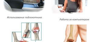
- . This disease belongs to diseases of the peripheral nervous system. Blood circulation is disrupted when the hands do not change their position while in a static state. This causes compression of the nerve. The syndrome often develops during prolonged use of the mouse and keyboard.
- Due to certain activities, compression-ischemic neuropathy of the median nerve occurs. It is associated with long-term macrotrauma to the nerve. This is facilitated, for example, by heavy physical labor with overload of the forearm and hand.
External causes of neuropathy of the median nerve of the arm also include:
- intoxication of the body;
- alcohol abuse;
- past infections (for example, HIV, diphtheria, herpes).
Classification
Neuropathy (neuropathy) is a disease characterized by damage to nerve fibers. When only one nerve becomes inflamed due to an illness, this is called mononeuropathy; two or more are called polyneuropathy.
Neuropathy is divided into 3 forms:
- (when nerve fibers and blood vessels are affected due to high blood sugar);
- toxic (infectious diseases, chemicals - all this affects the condition of nerve fibers);
- post-traumatic (this type of illness develops after damage to the myelin sheath of the nerve. Most often the sciatic, ulnar and radial nerves are injured);
Neuritis develops under similar conditions as median nerve neuropathy, but this disease is characterized by inflammation.
Based on the type and location of the pathology development zone, neuropathy has the following classification:
- damage to the lower extremities;
- sciatic nerve neuropathy;
- median nerve;
- peroneal nerve;
- facial nerve;
- tunnel neuropathy;
- sensorimotor neuropathy.
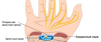
N medianus approaches the hand through the carpal tunnel. Here it innervates the muscles responsible for the opposition and abduction of the thumb, the lumbrical muscles, and the muscles that flex the finger. Its branches also supply nerve fibers to the wrist joint.
Median nerve neuropathy is associated with carpal tunnel syndrome, as the disease develops from constant compression in the wrist area.
From a surgical point of view, lesions of the median nerve are divided into open and closed. Open ones, in addition to the nerve, affect the tendons, blood vessels and muscles of the patient. Closed injuries include bruising, squeezing or spraining. Damage to the median nerve can develop along with plexopathy—damage to the cervical or brachial nerve plexuses.
Complex lesions (eg, trauma) often extend to the ulnar nerve. Cubital syndrome occurs (when the nerve of the cubital canal is compressed).
Median nerve neuropathy
Median nerve neuropathy
- defeat n. medianus on any part of it, leading to pain and swelling of the hand, sensitivity disorder of the palmar surface and the first 3.5 fingers, impaired flexion of these fingers and opposition of the thumb.
Diagnosis is carried out by a neurologist based on the results of a neurological examination and electroneuromyography; Additionally, musculoskeletal structures are examined using radiography, ultrasound and tomography. Treatment includes painkillers, anti-inflammatory, neurometabolic, vascular pharmaceuticals, exercise therapy, physiotherapy, and massage.
Surgical interventions are performed according to indications.
Median nerve neuropathy is quite common. The main contingent of patients are young and middle-aged people.
The most common places of damage to the median nerve correspond to the areas of its greatest vulnerability - anatomical tunnels, in which compression (compression) of the nerve trunk is possible with the development of the so-called. tunnel syndrome.
The most common tunnel syndrome is n. medianus is carpal tunnel syndrome - compression of the nerve as it passes to the hand. The average incidence in the population is 2-3%.
The second most common site of damage to the median nerve is its section in the upper part of the forearm, running between the muscle bundles of the pronator teres. This neuropathy is called “pronator teres syndrome.” In the lower third of the shoulder n.
medianus may be compressed by an abnormal process of the humerus or Struther's ligament. Its lesion in this place is called Struzer's band syndrome, or supracondylar process syndrome of the shoulder.
In the literature you can also find a synonymous name - Coulomb-Lord-Bedosier syndrome, which includes the names of the co-authors who first described this syndrome in 1963.
Symptoms of the disease
Neuropathy of the median nerve of the hand (or neuritis) refers to diseases of the nervous system. When the disease begins to develop, the patient experiences difficulty clenching the first, second and third fingers of the hand into a fist. It is also difficult for him to simply move the second and third fingers. Other symptoms:
- Inability to oppose the thumb to the rest.
- Poor sensitivity in the palm and fingers.
- The appearance of the "monkey's paw". This is due to the fact that atrophy of the hand muscles occurs. As a result of this, the first finger of the hand is installed with the second in the same plane.
- The main symptom is acute pain, manifested in the area from the forearm to the fingers of the affected hand.
- Numbness of the hand, muscle weakness, tingling in the forearm.
Massage for radial neuritis
Massage of the radial nerve develops after injury or cooling in hockey players. The motor fibers of the radial nerve innervate the extensors of the forearm, hand and fingers, the supinator muscle of the forearm, and the abductor pollicis muscle. At the same time, the back of the hand is somewhat swollen, the stretched tendons are inflamed, it is impossible to straighten the hand at the wrist joint and the fingers at the metacarpophalangeal joints, as well as to abduct the thumb.
The main goals of massage are:
acceleration of restoration of nerve conduction and sensitivity;
providing an analgesic effect.
Diagnostics
To diagnose median nerve neuralgia, the doctor performs a series of procedures. As the disease develops, the patient cannot perform certain actions. For example, an attempt to scratch the surface of a table with the index finger (with the palm pressed against the table) fails. The patient is unable to clench his hand into a fist, or to place his thumb against the rest.
Another diagnostic method is to ask the patient to show the “mill”. To do this, with your arms crossed, you need to rotate the sore finger of your healthy hand around the thumb of the injured one. If the nerve is affected, the person will not be able to do this.
With median nerve neuropathy, the patient’s thumb cannot be moved to the side enough to form a right angle with the index finger. Also, the index finger of one hand cannot scratch the healthy hand if you put 2 palms together.
The doctor also diagnoses in the following ways:
- computed tomography of the hand;
- electroneuromyography;
- X-ray of the hand.
The examination will show which treatment is best. Diagnostic data will give the doctor the opportunity to study information about damage to the joint and bone canals of the nerve. The doctor will evaluate the reflexes, the condition of the muscles, and answer the question whether the disease is caused by the narrowness of the canal or the patient’s lifestyle. The doctor will determine whether it is possible to prescribe neurolysis to treat the disease - a surgical intervention during which the sensitivity of the nerves is restored.
Treatment
People with median nerve neuropathy rarely see a doctor at the first stage of the disease. Referral occurs when more alarming symptoms of neurological problems appear:
- spasms, convulsions;
- crawling sensation;
- problems with coordination;
- lack of sensitivity to temperatures.
For treatment of the median nerve of the arm to be successful, it is important to find the exact location of the lesion. It is equally important to establish the cause, which is done at the diagnostic stage.
For effective therapy, the doctor also needs:
- determine the degree of nerve damage;
- identify factors leading to this symptom;
- find a specific point of defeat.
Treatment happens:
- operative (using surgery);
- conservative (medicines). Often doctors turn to etiotropic therapy. This is treatment with antibiotics, antiviral agents, and vascular drugs.
The degree of damage is determined using a special examination - needle myography. If the nerve is compressed, treatment may include the following steps:
- Absorption therapy has a good effect in relieving nerve compression. It involves taking various medications and enzymes, agents that absorb and soften scar tissue. If the compression is not severe, manual therapy and special massage are often sufficient.
- Nerve restoration. Special medications prescribed by a doctor help “revitalize” the nerve.
- Muscle rehabilitation. The goal of therapy is to restore their muscle volume. Treatment procedures are prescribed by a rehabilitation doctor.
- Conservative treatment of the radial and ulnar nerves may include wearing special splints.
What other means are used?
- Demixidol in the carpal tunnel area.
- Acupuncture.
- Interstitial electrical stimulation.
- Therapeutic blockades in the carpal tunnel (diprospan plus lidocaine), intramuscular injections (movalis plus novocaine)
- Non-steroidal anti-inflammatory drugs, in addition to blockades (artrosilene).
During the diagnosis, a disease may also be identified - plexitis of the median nerve. It is caused by injury or infection.
Initially, medicinal, conservative methods of therapy are always used. If physiotherapeutic treatment is ineffective, surgery is performed in the clinic. The decision in favor of surgical intervention is made when the integrity of the nerve trunk is damaged and there is severe weakness in the fingers.
It is not recommended to treat the disease with folk remedies. During therapy, the patient should not overwork or subject himself to heavy physical activity. During the acute period of the disease, you need to lie down and rest more.
Exercise therapy and special exercises are usually prescribed in the postoperative period. Physiotherapy is carried out during conservative treatment or also after surgery.
For patients with the disease, sanatorium-resort treatment may be indicated. A contraindication to it is the acute period of the disease.
Methods and techniques of massage for overstrain of the neuromuscular system
In athletes, overstrain of the neuromuscular system manifests itself in the form of muscle spasms of the lower extremities, most often the calf muscles, muscle spasms, trembling, and twitching.
In these cases, massage is used for the following purposes:
have an analgesic, antispastic and trophic effect;
improve blood circulation and redox processes;
have a tonic effect on the body;
promote a speedy recovery of performance.
First, in a lying position, a preliminary massage is performed, using stroking, rubbing and kneading, above the sore spot. This massage relieves muscle tension and thereby reduces soreness, further making it possible to massage the painful area. Then, continuous and intermittent stroking, spiral and circular rubbing are used on the sore spot. Kneading can be applied in different directions. Chopping, tapping, etc. techniques are not used. A good effect is achieved by performing a massage in a warm bath at a water temperature of 36–38 degrees. The duration of the massage is from 5 to 10 minutes.
Prognosis and prevention
If there is no threat to health in the form of infections or injuries, sufficient attention should be paid to the prevention of neuropathy of the upper extremities, namely:
- Physical exercises for the arms. They include a simple warm-up for the hands.
- It is important to take breaks when working at a computer. When working with a computer mouse, you need to hold it in different hands alternately.
- Taking the vitamin is beneficial, as well as strengthening the overall health of a person. This reduces the risk of diseases of the neurology of the extremities.
It should be remembered that timely treatment begins to guarantee a good prognosis for future hand function. Restoring motor activity should begin as early as possible. Ignoring therapy or improper self-medication often causes disastrous consequences.
- | Email |
- | Seal
Rehabilitation of patients with nerve damage includes preoperative preparation, surgery (neurography, autoplasty, neurolysis) and postoperative restorative treatment, as well as conservative treatment for incomplete conduction impairment. Before nerve surgery, in the presence of deforming scars of the skin and soft tissues, skin grafting is performed to obtain adequate passive movements in the corresponding joints to a fuller extent.
Sometimes patients with nerve damage are admitted with varying degrees of joint contracture due to previous long-term inactivity as a result of limb immobilization and pain. In such patients, before surgery, passive movements in the joints are developed to overcome poor mobility.
To accelerate regeneration processes, postoperative treatment for distal levels of damage is carried out for at least a year, and for damage to the lower primary trunk of the brachial plexus and sciatic nerve - for more than 3 years.
The impact of a low-energy laser on a nerve with incomplete conduction impairment during surgery after neurolysis and in the immediate postoperative period accelerates the restoration of conductivity. A thin-walled transparent tube is placed along the nerve and parallel to it, the free end of which is removed from the wound. A flexible conductor is inserted through this end for laser exposure and daily 10-15 minutes of irradiation of the nerve are performed until the sutures are removed, when the entire tube is simultaneously removed by the free end.
The indication for direct electrical nerve stimulation is the persistence of reduced conduction through the intrastem neuroma. After neurolysis, metal electrodes, for example nichrome, are placed above and below the nerve, connected through insulated conductors removed from the postoperative wound to an electrical stimulator. 1-5 second sendings of rectangular pulses with a duration of each 0.1-1 ms and a frequency of 10-77 Hz are used. Use constantly subthreshold current or daily threshold current for 10-15 minutes up to 3 times a day.
UHF, alternating magnetic field (for example, with the Polyus-1 device) are prescribed from 2 days. to reduce postoperative swelling. The UHF electric field, using the longitudinal and transverse method, is applied through a bandage to the postoperative wound without feeling heat, for less than 15 minutes daily, for a course of 7 sessions. The wound is irradiated with an alternating magnetic field through a bandage in a continuous mode with an intensity of 2 or 3 for 15-20 minutes daily, 15-20 sessions per course. An elevated position of the limb is also used to reduce swelling.
After neurolysis 3 weeks. after suture of the nerve, healing of the wound and removal of the immobilizing splint, the patient is recommended to take baths at a temperature of 35-45°C for 30-40 minutes for 2-3 weeks. In warm water after neurolysis, active and passive therapeutic exercises are performed in the corresponding joints. After suture of the nerve for 1-2 months. movements, often passive, are aimed at bringing the sutured ends of the nerve closer together, but not at stretching them. After 2 months the range of motion can be complete in any direction. The postoperative scar is lubricated with Vaseline between procedures to remove crusts and soften the scars within 3-7 days.
To accelerate nerve regeneration, paraffin or ozokerite applications are applied to the nerve suture area and distally along its projection for 45 minutes - 10-15 sessions.
It is necessary to avoid applying applications in the anesthesia zone to avoid burns. Thermal application courses are constantly repeated with breaks of 15-20 days. until full range of motion and sensation are restored. Typically, for distal nerve injuries it is 1-1.5 years, for proximal ones - up to 2-3 years.
After the operation, when the first signs of regeneration appear (muscle soreness upon palpation and/or barely noticeable painful contractions, the manifestation of gross pain sensitivity in the anesthesia zone), they begin to stimulate neuromuscular transmission by prescribing anticholinesterase drugs (proserin, galantamine, oxazil in usual dosages every day, sequentially until 10 injections of each drug).
After the operation, until active movements appear, passive gymnastics are performed not only in joints with varying degrees of stiffness, but also without contracture phenomena to prevent it in the future.
To prevent the vicious position of a limb with articular contracture, immobilizing splints (plaster, plastic) are periodically applied for a long time.
To improve muscle condition, superficial and deep kneading massage is performed in courses of 15 sessions with a break of 1 month. within 1-3 years. Muscle soreness is not a contraindication to massage, as it is one of the signs of muscle reinnervation.
Electrical muscle exercises begin after the appearance of a bend in the intensity-duration curve or emerging potentials during electromyography. The duration of the current pulse is determined based on the data of this curve and pain tolerance. Find the shortest duration of the current pulse using the intensity-duration curve, which the patient can tolerate without significant pain with a single stimulation. The frequency of stimulation should not be increased above 77 pulses/sec. For electrical stimulation of muscles, cutaneous pseudo-unipolar and bipolar electrodes are used. Electro-gymnastics of muscles is carried out 15-20 minutes after the administration of anticholinesterase drugs.
In patients with a complete reaction of muscle degeneration after nerve suture, electrical stimulation is not indicated, since it does not lead to faster regeneration.
Impairment of the integrity or conduction of peripheral nerves
occurs as a result of domestic or work injuries, wounds (stabbing, gunshot) or sports training (sprains, fractures, dislocations).
There are frequent cases of birth trauma in newborns, aggravated by damage to the nerves of the limbs. Also complicated by impaired nerve conduction can be tumor diseases, hematomas, aneurysms, calluses and scars after suppuration. The nerve can be completely dissected, torn, compressed, or thinned. In the absence of proper surgical treatment at the site of injury, nerve cells are replaced by connective tissue. These scars that form impede nerve conduction. The limb loses sensitivity, loses functionality, and this may be accompanied by chronic pain. Surgical treatment of “neurolysis”
is used precisely in such situations. It is aimed at releasing the nerve from compression and restoring its conductivity.
Massage technique
Neuritis of the axillary nerve occurs due to infection or compression of the nerve (using a crutch), as well as dislocation in the shoulder joint. The patient complains of pain in the shoulder joint, limitation of arm movement, especially when raising the arm to a horizontal level. Objectively, there is paresis and atrophy of the deltoid muscle, decreased sensitivity, especially along the outer surface of the upper third of the shoulder and above the shoulder joint. The deltoid muscle is painful on palpation. Massage is used in the neck, shoulder girdle, scapula and deltoid muscle area (see method for brachial plexitis). The duration of the massage is 15 minutes daily. Course - 10-15 procedures.
Neuritis of the musculocutaneous nerve of the shoulder. This disease occurs as a result of infection or injury, as a result of which the flexion of the forearm from a supinated position is limited. There is atrophy of the biceps brachii muscle. Reflexes from this muscle are reduced or absent, and sensitivity along the outer surface of the forearm is reduced. For neuritis of the musculocutaneous nerve of the shoulder, massage of the neck, shoulder girdle, arm on the affected side is necessary (see the method for brachial plexitis) and selective massage of the biceps muscle. Before massaging the biceps brachii muscle, a general preparatory massage of the entire arm is used: 1. General grasping continuous stroking from the wrist to the acromial clavicular joint 2. Alternating or light double ring rubbing. 3. General grasping continuous stroking. After this, they begin selective massage of the biceps brachii muscle: 1. Forceps-like stroking. 2. Spiral rubbing with four fingers. 3. Pincer-like stroking. 4. Spiral rubbing with one thumb. 5. Pincer-like stroking. 6. Tong-shaped kneading. 7. Pincer-like stroking. 8. Mechanical vibration with a hemispherical ebonite vibratode. 9. Pincer-like stroking. After this, you can do a general light rubbing of the entire upper limb. The procedure is completed with general, enveloping, continuous stroking. Massage is done daily. The procedure lasts 15 minutes. Course - 12-15 procedures. Courses must be repeated until the function of the upper limb is completely restored.
Neuritis of the median nerve of the hand. The cause of neuritis of the median nerve of the hand can be infection, trauma, or occupational hazards. In this case, it is difficult or impossible to oppose the thumb, bend the fingers into a fist, or abduct the thumb. There is atrophy of the muscles of the thumb, causing the hand to take on the appearance of a monkey's paw. There is pain in the fingers and along the inner surface of the forearm. Sensitivity is impaired in the area of the first, second, third and half of the fourth fingers on the palmar side and on the terminal phalanges of the first, second, third fingers on the dorsal surface. Autonomic disorders are expressed in thinning of the skin, brittle nails, hyperkeratosis, cyanosis, and hyperhidrosis. Patients with median nerve neuritis are prescribed massage of the forearm, hand and fingers. First, massage the forearm: 1. Enveloping pressure stroking the palmar surface of the forearm from the base of the fingers to the middle of the shoulder. 2. Alternate rubbing. 3. Stroking. 4. Spiral rubbing with four fingers. 5. Stroking. 6. Spiral rubbing with one thumb along the projection line of the median nerve. 7. Stroking. 8. Mechanical vibration with a spherical rubber vibratode along the median nerve. 9. Stroking. The same techniques are used on the back of the forearm, with the exception of mechanical vibration and rubbing with the thumb. On the palmar surface of the hand, the interosseous muscles and thenar muscles are massaged. On the interosseous muscles the following is used: 1. Smoothing with the pad of the thumb.2. Spiral rubbing with thumb.3. Smoothing with thumb.4. Thumb pressure. 5. Smoothing with your thumb. In the thenar area the following is used: 1. Forceps-like stroking. 2. Spiral rubbing with the thumb. 3. Pincer-like stroking. 4. Tong-shaped kneading. 5. Pincer-like stroking. 6. Mechanical vibration with a hemispherical ebonite vibratode. Then they begin to massage the fingers: 1. Longitudinal and transverse stroking. 2. Alternate rubbing. 3. Longitudinal and transverse stroking. 4. Spiral rubbing with the thumb. 5. Longitudinal and transverse stroking. The entire procedure is completed by stroking the hand and forearm. The massage procedure lasts up to twenty minutes daily. Course - 12-15 procedures.
Radial neuritis. The cause of radial nerve neuritis can be infection, intoxication, or trauma. The patient has weakness of the extensors of the forearm, hand and fingers, atrophy of the triceps brachii muscle and muscles that extend the hand and fingers. Hypoesthesia is noted on the outer surface of the forearm and the back of the hand, first, second and third fingers. The hand droops, swells, fingers are bent. There are no sharp pains or vegetative disorders. There is a loss of reflexes from the triceps brachii muscle. For radial nerve neuritis, it is advisable to prescribe massage of the collar zone and selective massage of the upper extremities. Massage of the collar zone can be done as with brachial plexitis, and on the upper limb the main attention is paid to massage of the triceps brachii muscle, extensors of the hand and fingers: 1. General grasping continuous stroking of the entire arm. 2. Double ring rubbing of the entire hand. 3. General grasping continuous stroking. After this, the following is applied to the triceps brachii muscle: 1. Grasping continuous stroking with one hand. 2. Spiral rubbing with four fingers. 3. Embracing continuous stroking with one hand. 4. Tong-shaped kneading. 5. Grasping continuous stroking with one hand. 6. Semicircular kneading. 7. Grasping continuous stroking with one hand. 8. Transverse kneading. 9. Embracing continuous stroking with one hand. 10. Mechanical vibration with a hemispherical ebonite vibratode. 11. Embracing continuous stroking with one hand. The massage can be completed with a general grasping continuous stroking from the wrist joint to the acromial clavicular joint. Then they move on to massage the extensors of the hand and fingers: 1. Enveloping continuous stroking. 2. Spiral rubbing with four fingers. 3. Embracing continuous stroking. 4. Spiral rubbing with the thumb. 5. Embracing continuous stroking. 6. Tong-shaped kneading. 7. Embracing continuous stroking. 8. Mechanical vibration with a hemispherical or ebonite vibratode. Vibration can also be produced with a spherical rubber vibratode along the radial nerve. 9. Embracing continuous stroking. Massage of the triceps muscle and extensors of the hand and fingers should be quite energetic and deep. It should be dominated by kneading and vibration techniques combined with deep stroking. The duration of the procedure is 15-20 minutes daily. The course consists of 15 procedures, which must be repeated regularly after three to four weeks until the function of the affected limb is fully restored.
Ulnar nerve neuritis occurs due to infections and injuries (fractures, bruises). In this case, the flexion of the main phalanges of all fingers and the terminal phalanges of the fourth and fifth fingers is impaired. Difficulty in adducting fingers. Atrophy of the interosseous muscles develops. The pain syndrome is sharply expressed. The pain is localized along the ulnar edge of the hand. In addition, there is hypo- and anesthesia of the skin of the forearm, the palmar surface of the hand, the fourth and fifth fingers and the back of the fifth finger. Autonomic disorders are manifested by cyanosis, hyperhidrosis, and thinning of the skin. Massage should begin immediately after acute symptoms subside. For ulnar nerve neuritis, massage of the forearm, hand and fingers is used. The massage should be quite energetic, deep in the area of the palmar surface of the forearm and interosseous muscles. But it is necessary to take into account the presence of pain. To eliminate pain from the very first procedures, it is necessary to use mechanical vibration using a spherical rubber vibratode in the area of the ulnar groove between the internal epicondyle of the humerus and the process of the ulna, as well as along the course of the ulnar nerve. A similar massage scheme is used for neuritis of the median nerve. The duration of the massage is 15-20 minutes daily. The course must be repeated regularly after three to four weeks until the function of the affected limb is fully restored.
Ulnar nerve damage
The ulnar nerve is massaged with the arm slightly bent at the elbow joint in the area between the internal epicondyle of the humerus and the olecranon process of the ulna with the patient sitting. They start from the proximal sections, then at the exit site of the nerve and along its course (shao-hai point). The procedure time is 15–20 minutes, the course is 12–15 procedures, daily.
This text is an introductory fragment.
Read the whole book
Diagnosis of peripheral nerve injuries
For serious extremity injuries, initial examination may reveal peripheral nerve damage. When providing first aid, you need to pay close attention not only to the muscles and bones, but also try to identify or eliminate damage to the integrity of the nerves. Peripheral nerve injury may be indicated by:
- movement disorders;
- loss of sensation;
- weakening or loss of motor reflexes.
A thorough examination, palpation, analysis of the severity and depth of damage is carried out. Nerve fiber breaks may be visualized as whitish particles among the damaged tissue.
Massage for neuromyositis
Neuromyositis is a joint disease of muscles and peripheral nerves and is characterized by a chronic course with periodic exacerbations. Dystrophic processes develop in muscle tissue. In athletes, neuromyositis occurs when prolonged physical overexertion is combined with cooling.
The goals of massage for neuromyositis are:
providing anti-inflammatory, trophic, analgesic effects that affect metabolism;
improving blood and lymph circulation in the damaged area;
stimulating regenerative processes in the affected nerve.
When performing a massage, the main attention is paid to the techniques of stroking, rubbing with fingertips, the base of the palm, the phalanges of bent fingers and light vibration. In addition, the corresponding segments of the spinal column are massaged. Chopping techniques are not used. A combination of thermal procedures and massage gives a good effect. The massage is performed for 10–15 minutes.
Neurolysis of peripheral nerves of the extremities
Surgical treatment of nerve damage can be carried out at different times after injury: primary, delayed and late operations.
Neurolysis
is an effective technique for restoring damaged nerves of the limbs, allowing the limbs to return functionality and sensitivity.
In a number of situations, during neurolysis, a nerve is transposed
, that is, it is displaced or a fragment is transferred from other parts of the body for implantation at the site of the rupture.
The essence of neurolysis
– in isolating nerve fiber and eliminating factors that disrupt its conductivity. This is, first of all, compression due to scars or calluses. This kind of disruption of nerve conduction simultaneously causes loss of a number of functions of the limb and increased irritation of the nerve, manifested by pain.
Surgical manipulations for neurolysis require a significant tissue incision so that the surgeon can isolate the nerve from the surrounding tissue. Projection, non-projection, roundabout and angular access on bends is possible. Particular attention is paid to the preservation of the connection between muscle tissue and nerve fiber.
During the operation, damaged sections of the nerve can be removed, after which the ends are sutured or the fragments are transposed. The surgical technique here has a number of features; special suture materials and micro-instruments are used.
Massage for acute muscle or neuromuscular spasms
Acute muscle or neuromuscular spasm occurs at the moment of rapid and sudden movement. A sudden convulsive contraction of individual muscle bundles is accompanied by acute local pain. The contraction gradually weakens, the acute pain turns into dull pain, but when you try to move, it worsens.
Complete relaxation of the contracted muscle is extremely difficult or impossible. If there are residual effects of spasm, training cannot be resumed. In the future, with rational training, it may not be repeated, and, conversely, with systematic overexertion, it can be repeated quite often.
In this case, the main goals of massage are:
providing analgesic and resolving effects;
strengthening of redox processes;
promoting the speedy recovery of the athlete’s performance.
When performing a massage, the muscles should be as relaxed as possible. First, a preliminary massage is performed, that is, the area that is located above the painful area is massaged. When performing a massage, the following techniques are used: stroking, rubbing, kneading and light vibration.
Massage should be performed two to three times a day without causing increased pain at the site of injury. The techniques of chopping, beating, and patting are not used. A good effect is achieved by performing a massage in a warm bath with a water temperature of 36–38 degrees, using stroking and kneading techniques. Massage time is 5 – 10 minutes.