Microangiopathy is a pathology characterized by damage to small blood vessels (primarily capillaries). Most often it is a symptom of other independent diseases. One of its varieties is considered to be pathologies of the vessels of the retina and capillaries in the kidneys. The disease develops in vascular systems and organs against the background of various types of infectious or oncological diseases, diabetes mellitus, liver disease, as well as hemolysis (a pathological condition accompanied by the destruction of red blood cells and the release of hemoglobin from them).

In most cases, microangiopathy is a consequence of:
- Necrosis and death of tissues and cells of the body (necrosis);
- Thrombosis is a process that is accompanied by the formation of blood clots inside blood vessels, disrupting normal blood flow;
- Hyalinosis (or hyaline dystrophy) is a condition that is one of the types of protein dystrophy and is characterized by the deposition of hyaline in tissues;
- Fibrinoid (or fibrinoid swelling) is an irreversible condition characterized by a sharp increase in vascular permeability and manifestations of deep disorganization of connective tissue, which is based on the destruction of its main structural substance and fibers.
The clinical picture of microangiopathy completely depends on the specifics of the affected tissue, on the anatomical and morphological characteristics of the affected organs, as well as on the impact of individual external factors on the body. Its main manifestations are: damage to the integrity of the walls of small blood vessels and dysfunction of the blood coagulation system (hemostasis).
The disease is often accompanied by renal failure, purpura (subcutaneous hemorrhages - a medical symptom that is a characteristic sign of pathology of any part of hemostasis) and damage to red blood cells.
Reasons for the development of microangiopathy
The development of microangiopathy is caused by:
- Pathologies caused by a hereditary genetic factor, which are accompanied by a violation of the tone of the walls of blood vessels;
- Certain diseases affecting blood and plasma;
- Intoxication of the body;
- Various types of physical injuries.
It is believed that microangiopathy can be provoked by excessive smoking and addiction to alcohol, as well as hypertension, general weakness of the body, which is caused, for example, by working in hazardous industries or certain age-related changes.
Cerebral angiopathy: main causes, symptoms and treatment features
- June 22, 2018
- Neurology
- Voloshchuk Natalya
The brain today continues to remain a big mystery for biology. Despite the successes and achievements in medicine, no one can clearly answer the question of how a person thinks.
The brain is the main organ that is responsible for all processes occurring in the body. Sometimes malfunctions occur in its operation, which are manifested by various symptoms and signs.
Today we will talk about what cerebral angiopathy of the brain is, how and why it occurs, and in what ways and methods it is treated.
Description
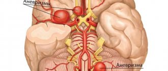
Angiopathy is a process in the capillary walls that leads to disruption of their patency, resulting in a deterioration or cessation of blood supply to brain cells. This results in healthy neurons being replaced by glial neurons, which proliferate to replace the missing cells. A scar appears at the site of damage to brain tissue.
The disease can affect small vessels, which increases the risk of complications and even death. Cerebral angiopathy refers to cerebrovascular diseases that lead to pathological changes in cerebral vessels with impaired blood circulation in the brain.
The disease often progresses in old age and is accompanied by diseases of the cardiovascular system.
Kinds
Angiopathy is caused by damage to small vessels; it comes in two types:
- The walls of the hyaline arteries thicken.
- There is a thickening of the capillary walls.
In both cases, the processes occur due to a lack of cerebral blood supply on an ongoing basis, which leads to damage to neurons.
Over time, this can provoke the appearance of lacunar infarction, non-inflammatory brain diseases that are caused by diffuse damage to the organ.
Thus, diffuse cerebral angiopathy occurs - damage to all brain vessels in alternating places of healthy and diseased tissue.
Etiology
This disease most often affects older people, as well as those who have diabetes, abnormal vascular structure, trauma, autoimmune diseases, body intoxication, poor diet and physical activity. Dysfunctions of nervous regulation also often lead to the disease. Sometimes cerebral angiopathy leads to stroke - the second disease after a heart attack, resulting in death.
Forms
Depending on the symptoms, the following forms of the disease are distinguished:
1. Cerebral amyloid angiopathy occurs most often in those who suffer from Alzheimer's disease. Amyloid (a substance made from proteins and saccharides) collects on the walls of the veins; this disease cannot be cured.
2. Lenticulostriate angiopathy occurs in childhood and does not require therapy.
3. Diabetic angiopathy - disruptions in blood circulation appear due to diabetes mellitus.
4. Hypertensive – disruptions in blood circulation appear due to hypertension.
Clinic
At the initial stage of development, the disease does not show any symptoms, since small areas with deformed vessels are fed with blood from neighboring vessels. When a large part of the bloodstream is already affected, neurons begin to die and the following symptoms occur:
– excruciating pain that occurs intensely and practically does not subside with the use of medications;
– disturbances in the functioning of the vestibular apparatus, which manifests itself in dizziness, impaired coordination and orientation in space;
– astheno-neurotic syndrome, accompanied by a decrease in the emotional background, irritability and nervousness, and the manifestation of depression;
– sleep disturbances, which are characterized by increased sleepiness during the day and insomnia at night.
Cerebral angiopathy can manifest different symptoms depending on the location. They are all nonspecific; the same signs can be observed with other brain ailments.
Further manifestation
If the disease continues to progress, this can lead to the following consequences:
– absent-mindedness and memory lapses;
– weakened vision and changes in pupil size;
– bleeding from the nose and inside the stomach;
– loss of coordination of movement.
All this is the result of impaired blood flow in the brain tissue.
Forecast
Vessels are means of transporting blood in the human body. The condition of all organs and systems depends on them. Any disruptions in the brain lead to dangerous pathologies that cannot be cured.
One of them is dyscyclic encephalopathy, which is a complex of changes in the blood vessels of the brain. It is important to take timely prevention from this disease. The disease cannot be completely cured.
But it is quite possible to significantly slow down the process of tissue scarring.
The diagnosis of “cerebral angiopathy” today often occurs during hospitalization of patients with cardiovascular diseases. It is necessary to be able to understand what this pathology is, what its symptoms are, diagnosis and treatment methods.
Then you can achieve good results in eliminating symptoms, since it is impossible to completely recover from the disease. You can also slow down the spread of pathological processes in the brain by adhering to the right diet and lifestyle.
Source: https://SamMedic.ru/330632a-tserebralnaya-angiopatiya-osnovnyie-prichinyi-simptomyi-i-osobennosti-lecheniya
Forms of microangiopathy
Modern medicine distinguishes two types of disease:
- Cerebral microangiopathy (or cerebral microangiopathy);
- Diabetic microangiopathy.
A characteristic symptom of cerebral microangiopathy is damage to microscopic blood vessels located in the brain. It develops most often against the background of disruption of normal blood circulation in the brain, atherosclerosis and persistently elevated blood pressure (hypertension).
Diabetic microangiopathy is one of the symptoms accompanying diabetes mellitus and is the cause of the development of tissue (or histotoxic) hypoxia, a condition resulting from impaired tissue use of oxygen supplied to them.
Symptoms of cerebral microangiopathy
The insidiousness of cerebral microangiopathy lies in the fact that in the initial stages of development it manifests itself and the person is not aware of the presence of a problem. Small isolated areas with damaged vessels are supplied with oxygen and nutrients from neighboring capillaries. Thanks to this, there are no symptoms of the pathology at first.
Over time, when a large volume of the microvasculature is affected and the blood supply from neighboring vessels cannot replace the damaged capillaries, many brain neurons die, and signs of microangiopathy appear.
The first symptom that all patients complain about is pain in the head, which cannot be eliminated with painkillers. Patients note the presence of a pronounced astheno-neurotic syndrome, which is expressed in the following:
- being in a bad mood;
- irritability and depression;
- fast fatiguability;
- sleep problems;
- feeling overwhelmed and empty;
- significant decrease in performance;
- fatigue.
Microangiopathy of the brain is manifested by symptoms:
- dizziness, confusion;
- violation of movement coordination;
- drowsiness during the day and insomnia at night;
- memory problems;
- absent-mindedness, forgetfulness;
- pupils of different diameters;
- disturbance of temperature and pain sensitivity in some parts of the body;
- bruises, subcutaneous bruising;
- blood from the nose;
- dry and flaky skin on the legs;
- visual impairment.
Microangiopathy of the brain
Cerebral microangiopathy, accompanied by damage to small blood vessels and their branches in the brain, is divided into two types, one of which is characterized by thickening of the walls of arteries of a hyaline nature, and the other by thickening of the walls of capillaries. In both the first and second cases, cerebral microangiopathy is considered as a result of chronic insufficiency of blood supply to the brain and leads to serious damage to its white matter - an accumulation of nerve fibers extending from neurons, forming wire pathways. In turn, dysfunction of the brain provokes the development of:
- Lacunar infarction (or cerebral stroke) is a condition that occurs against the background of occlusion of one of the penetrating branches of the brain vessels;
- Non-inflammatory diseases of the brain (encephalopathies), characterized by diffuse brain damage.
Treatment
Conservative treatment is used to improve the rheological properties of blood. It is aimed at improving microcirculation in the brain. If you have diabetes, insulin therapy is adjusted to normalize glucose levels. Patients without critical stenoses go to the hospital 1-2 times a year for observation and “dropping”. Doses of medications are selected taking into account the degree of ischemia. It is better to use folk remedies in the intervals between hospitalizations, and in no case replace medications with them.
Surgery:
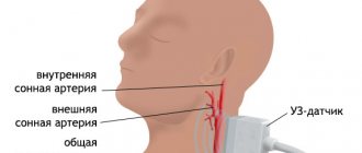
When the vessels of the neck are affected, when there is macroangiopathy, traditional open surgery, such as carotid endarterectomy, is used. After diagnosis and establishment of the exact location of the narrowing site, access to the carotid artery is performed. The plaque is removed through a longitudinal incision and a patch is sewn into the vessel wall to prevent future stenosis. It is also possible to completely transect the vessel, with the installation of a prosthesis sewn either into the intact common carotid artery or into the aorta.
A more modern method is X-ray endovascular techniques - balloon angioplasty and artery stenting. Under X-ray control, a thin catheter is passed through the puncture into the artery. The tip of the catheter is equipped with a special balloon, which is inflated at the site of narrowing. Often, in addition to expansion, a thin spring-stent is installed inside the wall. It is completely intact (not rejected by the body), and will prevent further development of the disease. This operation is easier to tolerate by the patient and is gradually gaining more and more popularity.
Diabetic microangiopathy
This form of microangiopathy develops due to an increase in blood glucose levels above normal levels. Against this background, patients experience damage to the walls of small-caliber vessels (venules, capillaries and arterioles) located in close proximity to the body tissues. In this case, the clinical picture completely depends on the location of the lesion.
Microangiopathy of the diabetic type is one of the most dangerous manifestations of diabetes mellitus. This is due to the fact that the pathology provokes a disruption in the processes of tissue nutrition and the removal of various metabolites from them. As the disease progresses, vasoconstriction occurs, signs of oxygen starvation of tissues become more pronounced, and the course of the disease worsens significantly.
Irreversible narrowing of the walls of blood vessels is a consequence of:
- Damage to the membranes of small arteries;
- Endothelial proliferation;
- Thickening of basement membranes;
- Accumulations of mucopolysaccharides in the walls of arteries.
At the same time, as a result of an increase in osmotic pressure caused by an increase in the level of fructose in the blood and excessive water intake, patients experience an increase in edema in the cells, and due to impaired metabolism of proteins and fats, the condition of the blood vessels suffers.
The most characteristic manifestations of this type of microangiopathy are:
- Diabetic nephropathy (occurs in about a third of patients and is characterized by impaired renal function, swelling of the kidneys, and the presence of large amounts of protein in the urine);
- Diabetic angioretinopathy (damage to the vessels of the retina of the eyeball);
- Microangiopathy of vessels located in the lower extremities.
Clinical manifestation
In the early stages, the disease has no specific symptoms and therefore remains undetected. The patient lives his normal life, occasionally experiencing mild headaches. If left untreated, irreversible pathological changes occur in the vessels, which leads to the appearance of severe symptoms. The nature of the signs of cerebral angiopathy depends on the location of the affected area. The most commonly observed symptoms are:
- short-term attacks of headache (over time, the discomfort becomes permanent);
- decreased concentration of attention associated with oxygen starvation of brain tissue;
- loss of sense of time and space (attacks have a short duration);
- memory deterioration (a person begins to forget events that happened before the onset of the disease);
- changes in behavior and personality structure;
- increased nervous excitability, aggression, irritability;
- uncontrollable outbursts of anger;
- intolerance to loud sounds and bright light;
- frequent mood changes;
- decreased muscle tone of the limbs up to paralysis;
- decreased sensitivity of the skin;
- visual impairment caused by damage to the vascular walls of the retina;
- problems with speech and coordination of movement.
In severe cases of cerebral vascular angiopathy, realistic visual and auditory hallucinations, respiratory arrest, and problems with swallowing occur. With such a development of the clinical picture, even complex therapy does not completely eliminate neurological disorders.
Important information: How to treat multiple myeloma blood disease and what are the symptoms of Rustitsky Kaler
Diagnosis and treatment of microangiopathy
To diagnose microangiopathy, it is necessary to:
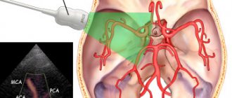
- Ultrasound examination of blood vessels;
- Ultrasound of the fundus;
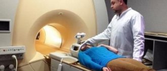
- Magnetic resonance imaging;
- X-ray examination;
- Computed tomography.
As for the treatment of microangiopathy, it depends on the localization of the pathological process and its etiology. In most cases, patients are prescribed drug therapy with the goal of improving the efficiency of blood microcirculation in tissues. In addition to this, physical therapy may also be prescribed.
Surgical intervention is required only when it is necessary to restore vascular patency. It is performed using cryosurgery or laser coagulation methods.
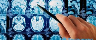
Diagnostics
Diagnosis begins with examination and questioning of the patient. The neurologist makes the patient perform simple actions, identifying dysfunction of the limbs, organs of vision and hearing. Taking an anamnesis helps to identify possible causes of cerebral vascular damage. After making a preliminary diagnosis, additional diagnostic procedures are prescribed:
- General blood and urine tests. Used to detect sources of inflammation and assess the condition of the body.
- MRI. The procedure helps to examine in detail the blood vessels and tissues of the brain. Using this method, signs of ischemia, hemorrhagic complications, vasoconstriction, benign and malignant neoplasms are detected.
- CT. Using computed tomography, the location and extent of the affected areas are determined.
- Ultrasound examination of the cervical and head areas. Prescribed for suspected narrowing or blockage of the arteries supplying the brain.
- X-ray examination. Refers to auxiliary methods aimed at identifying provoking factors (tumors, traumatic brain injuries).
- Angiography. Vascular examination with the introduction of a contrast agent is aimed at determining the patency of large vessels. The speed of blood flow is assessed by the nature of the distribution of the substance.
- Ophthalmoscopy. The doctor examines the fundus of the eye, identifying signs of damage to the veins and arteries of the retina that accompany cerebral angiopathy.