Agenesis (underdevelopment) of the corpus callosum (CAC) is one of the forms of cerebral dysgenesis (developmental disorder) caused by impaired fetal formation during the prenatal period. There are total and partial forms. In the first case, there are no commissural (nerve bundles connecting the hemispheres) fibers. In the second case, the caudal (posterior) and rostral (anterior) segments of the corpus callosum are underdeveloped. AMT often (about 70% of cases) occurs in combination with other developmental defects (usually pathologies of the cardiovascular, genitourinary and musculoskeletal systems) or in isolation.
Characteristic
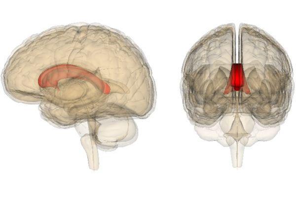
Agenesis is a pathology that is characterized by the loss of an organ’s ability to develop, which leads to a decrease in size and dysfunction. The corpus callosum is a part of the brain that connects the hemispheres; its agenesis is accompanied by a violation of the higher functions of the psyche. The corpus callosum is a plexus of nerve tissue fibers. The number of fibers exceeds 250-300 million.
Normally, the brain section is presented in the form of a strip about 8 cm wide, consisting of axonal processes, located under the cortex. Its fibers run transversely, connecting symmetrically located sections of the hemispheres. Some fibers connect areas located in different zones of different hemispheres, for example, the frontal zones of the right hemisphere with the parietal or occipital zones of the left.
The diagnosis of agenesis of the callosum means that connecting nerve fibers are partially or completely absent between the hemispheres. The prevalence of the disease is about 0.7% in the general population, about 3% among patients with diagnosed mental retardation. The presence of interhemispheric connections ensures the interaction of the brain structures of both hemispheres.
With segmental or complete absence of the corpus callosum in the newborn, interaction is disrupted. As scientific experiments show, sensory data partially processed in the cortical layer is transmitted through the commissures of the corpus callosum. The corpus callosum is less involved in the transfer of commands that control motor activity, and more involved in the interhemispheric transmission of verbal information and visuospatial data.
With increasing complexity of the intellectual tasks performed, the activity of the structures of the corpus callosum increases. An important function of the body is the inhibition of brain processes to better differentiate the work of the departments of different hemispheres, which ensures more efficient information processing. Research shows that the quality of interaction between the structures of different hemispheres affects the level of intelligence.
Aplasia (absence of part or all of an organ), thinning or thickening of a section of the corpus callosum are variants of abnormal formation. If a part of the brain is absent or the process of its normal development is disrupted, this can cause the occurrence of pathologies such as schizophrenia, autism, dyslexia (impaired ability to acquire reading and writing skills while maintaining the ability to learn), a syndrome manifested by lack of attention and hyperactivity.
For example, in early onset forms of schizophrenia, instrumental examination reveals thinning of body structures. In late-onset forms of schizophrenia with a relatively favorable prognosis, thickening of its structures is detected.
A direct connection between abnormalities in the formation of this part of the brain and the development of psychoses has not been established. However, it is believed that partial agenesis (underdevelopment) of the corpus callosum may play a role as a trigger in the development of psychotic disorders. Among the combined malformations of the formation of central nervous system structures, cystic formations (arachnoid, interhemispheric cysts), Arnold-Chiari and Dandy-Walker syndromes, and abnormal development of convolutions are often detected. In 80% of cases, AMT is combined with hydrocephalus.
Anatomy and topography of the corpus callosum.
The corpus callosum is a plate consisting of transverse fibers. The free upper surface of the corpus callosum has a gray covering, indusium griseum. On a sagittal section of the brain, one can distinguish the bends and parts of the corpus callosum: the knee, genu, continuing down into the beak, rostrum, and then into the terminal plate, lamina termindlis.
The middle part is called the trunk, truncus, of the corpus callosum.
Posteriorly, the trunk continues into a thickened part - the splenium. Transversely running fibers of the corpus callosum in each hemisphere of the cerebrum form the radiation of the corpus callosum, radiatio corporis callosi. The fibers of the anterior part of the corpus callosum - the knee - bend around the anterior part of the longitudinal fissure of the brain and connect the cortex of the frontal lobes of the right and left hemispheres. The fibers of the central part of the corpus callosum - the trunk - connect the gray matter of the parietal and temporal lobes. The roller contains fibers connecting the cortex of the occipital lobes.
Under the corpus callosum is the fornix, fornix. The arch consists of two strands connected in its middle part with the help of transversely running fibers - commissures, comissura. The middle part of the arch is called the body, corpus; anteriorly and downwards it continues into a rounded paired cord - a pillar, joint, arch. The column of the fornix ends in the right and left mastoid bodies. Posteriorly, the body of the arch continues into a paired flat cord - the pedicle of the arch, crus fornicis. The paired leg of the fornix on one side fuses with the hippocampus, forming the hippocampal fimbria, fimbria hippocampi. The fimbria of the hippocampus ends in the uncus, thus connecting the temporal lobe of the telencephalon with the diencephalon.
In front of the fornix in the sagittal plane there is a transparent septum, septum pellucidum, which consists of two plates lying parallel to each other. Between the plates of the transparent septum there is a slit-like cavity of the transparent septum, cavum septi pellucidi, containing a clear liquid. The lamina pellucida serves as the medial wall of the anterior horn of the lateral ventricle. In front of the columns of the arch is the anterior commissure, comissura rostrdlis.
Causes
Agenesis (impaired formation or absence) of the corpus callosum of the brain in a child is a congenital anomaly, which occurs with a frequency of about 30% among patients with genetically determined, hereditary syndromes. Among the causes of pathology, it is worth noting mitochondrial diseases, gene mutations, and cytogenetic syndromes.
AMT is found in the clinical picture of Huntington's disease (a chronic neurodegenerative disease characterized by the progressive death of nerve cells) and hereditary metabolic disorders. In most cases, hereditary syndromes involving AMT in the pathogenesis are multisystem (many organs and systems are affected). Provoking factors:
- Diseases suffered by the mother during gestation (sexually transmitted infections, respiratory viral diseases).
- Intoxication (chronic, acute).
- Unfavorable course of pregnancy (toxicosis in the first and second trimester of gestation, threat of miscarriage).
In the structure of the causes of congenital malformations of the callosum and other organs, random damaging factors are distinguished, which account for 60% of cases of developmental anomalies. In 10% of cases, the cause of anomalies is chromosomal diseases, in 20% of cases - teratogenic (impairing embryonic morphogenesis) effects of pharmaceuticals. Intrauterine infections (mostly viral) lead to birth defects in 10% of cases.
structure
The corpus callosum has a large number of structures. However, from an anatomical point of view, it consists of three main parts: the body or torso, the runner and the knee.
Each of these parts belongs to a separate area of the corpus callosum and has certain characteristics.
The body or trunk of the corpus callosum makes up the upper part of the structure. It has a convex shape at the back and is flat or slightly concave in the transverse region.
A longitudinal groove is observed in the body, which leads to the survival of the infundibulum of the corpus callosum. On each side of this groove there are two small cords known as longitudinal grooves.
The longitudinal grooves are connected to the middle tract by a thin curtain of gray matter called the industrial griseum. This gray veil is an extension of the cerebral cortex of the convoluted corpus callosum.
The lower side of the body is convex in the transverse direction and has a concave shape in the anteroposterior direction. In the midline it has a septum lucidum, and behind it it is in contact with the transverse fibers of the trine.
The impeller is the posterior end of the corpus callosum. It is a rounded area that is formed by folding the corpus callosum onto itself.
Between the impeller and the trigone there is a cleft connecting the hemispheres with the lateral ventricles.
Finally, the knee is the name given to the anterior end of the corpus callosum. This is the thinnest area and presents a downward and backward curve.
The knee is formed by reflected fibers that continue downwards with the sharp part of the beak. On the underside there are two whitish tracts called peduncles of the corpus callosum.
Clinical manifestations
An isolated form of agenesis (underdevelopment) of the corpus callosum is a pathology that can be asymptomatic, which makes early diagnosis difficult. Neurological deficits are often caused by concomitant pathologies. In the presence of combined pathologies, movement disorders are detected in 40% of patients.
30% of sick children experience delays in mental, speech and physical development. Mental retardation is diagnosed in 70% of patients with impaired formation of the callosum. The leading symptom in infants of the 1st year of life is convulsive syndrome. Other symptoms of agenesis (underdevelopment) of the corpus callosum, characteristic of newborns under 1 year of age:
- Impaired perception and sensory reactions.
- Deterioration in communication skills, lack of psychological connection with the mother.
- Insufficient modulation (change in strength, tone and other characteristics) of the cry.
In older children, signs are observed: a disorder of thermoregulation (hypothermia - a decrease in body temperature below 35°C, hyperthermia - a steady increase in body temperature), impaired motor coordination and spatial orientation, memory impairment (visual, auditory). For example, in the course of research in children 4-8 years old, a violation of tactile perception is discovered - the child finds it difficult to name an object that he feels with his subdominant (non-dominant) hand.
Patients experience difficulty in tactile (by touch) passage of the labyrinth, which is associated with a violation of the transfer of spatial information of an intermanual (between limbs) nature. Children with diagnosed pathology of the corpus callosum experience a deterioration in social adaptation and planning skills.
At the same time, symptoms are observed: poverty of emotions, emotional immaturity (inability to adequately respond to everyday problems and life situations, inability to act taking into account expediency). As adults, such people often suffer from loneliness.
Localization
Spatially, this part of the brain is located under the hemispheres along the midline. From anterior to posterior, the corpus callosum can be divided into several different zones: genu, midsection, body, posterior end, and splenium. The knee, curving downwards, forms the beak, as well as the rostral plate. On top, the corpus callosum is covered with a thin layer of gray matter.
Another structure of this part of the brain is radiance. Strands of fan-shaped neurons extend to the frontal, parietal, temporal and occipital lobes of the cerebral hemispheres.
Diagnostics
Underdevelopment of the corpus callosum is detected during an instrumental examination of the brain, which includes methods such as MRI, CT, and neurosonography. Diagnosis in MRI and CT format often shows the presence of a cystic formation in the interhemispheric zone, an increase in the diameter of the third ventricle, and a pathological change in the geometric shape of the lateral ventricles.
Agenesis (underdevelopment or absence) of the fetal corpus callosum is detected during prenatal ultrasound examination. In the presence of combined congenital malformations and chromosomal defects, the pathology can become a reason for termination of pregnancy. The period of prenatal screening is the II, III trimester of gestation.
Anatomy of the corpus callosum
The corpus callosum is a sheet of white matter that forms a quadrangular area and extends transversely from one hemisphere to the other.
This is a system of associations that unites the two halves of the brain through the connection of asymmetrical points in the cortex.
The width of the corpus callosum on the upper part of the face is about two centimeters, and on the lower part - up to 3-4 centimeters. The runner of the corpus callosum is 15 millimeters long.
The corpus callosum consists of approximately 200 million axons, which mainly come from the pyramidal cells of layers II and III of the cerebral cortex.
Sexual dimorphism
A number of Russian and foreign scientists believe that the difference in thinking and behavioral reactions between men and women is associated with the different structure and size of the corpus callosum. Thus, Newsweek published an article explaining the nature of women's intuition: women have a slightly wider corpus callosum than men.
After some time, a group of French scientists reported that, as a percentage of brain size, men have a larger corpus callosum than women, but scientists did not draw any clear conclusions. Be that as it may, all scientists only agree that the corpus callosum is one of the most important structural components that performs a number of vital functions.
Treatment
Modern medicine does not have effective methods for correcting the abnormalities that arise in a child when diagnosed with agenesis of the corpus callosum. Drug therapy for this condition is selected for the baby individually, taking into account the degree of agenesis, as well as the presence of concomitant developmental anomalies. To correct functional disorders, the following groups of medications can be used:
- Hormonal anti-inflammatory drugs, for example Prednisolone and Dexamethasone;
- Phenobarbital and benzodiazepines;
- Neuropeptide drugs, for example, Cerebrolysin and nootropics, for example, Piracetam, Semax;
- Diazepam drugs and antipsychotics.
In addition, children with a similar developmental anomaly are prescribed Mexidol, Diacarb and Asparkam.
In addition to the listed conservative treatment methods, some children are prescribed surgery to stimulate the fibers of the vagus nerve. This procedure is recommended for those children who have persistent functional disorders in the functioning of internal organs.
Taking into account the fact that agenesis of the corpus callosum is a potential cause of structural and functional abnormalities in the musculoskeletal system, such children very often develop S-shaped scoliosis.
That is why medical experts recommend that children regularly perform physical therapy and hardware physiotherapy sessions. Share: