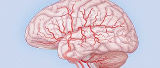
Dementia is pathological changes in mental functions resulting from some kind of brain damage. This is primarily expressed in the loss of cognitive abilities. Patients find it difficult to learn new knowledge and use old skills. The most common type of dementia is senile dementia, which begins at the age of 60. But a similar change in a mature or young person is also possible, which is most often associated with some kind of deviation, for example, taking alcohol or drugs. However, it is impossible to answer the question of what cerebral dementia is. This is a definition of the general condition of a person. The question of what dementia is is also inappropriate. This is not a disease, but dementia, which is a consequence of some kind of illness.
A fairly large number of reasons can lead to dementia. They themselves are divided according to the types of syndromes, the characteristic features of the disorders, and the areas of the brain that are affected by them. And all this division is associated with the age of the patients.
Dementia of the brain is a decrease in a person’s cognitive activity, loss of skills and knowledge previously acquired by a person
Causes and classification of vascular dementia
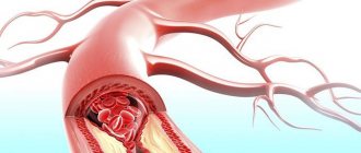
The incidence of vascular dementia is 612 cases per 1000 population over 70 years of age per year. According to statistics, vascular dementia (VC) accounts for 10-20% of all types of dementia. Various risk factors are involved in the development of vascular dementia, among which the most important are: arterial hypertension, pathology of intracranial vessels, hyperlipidemia, smoking, taking oral contraceptives, heredity, and age. Diabetes can be caused by lacunar infarctions, cortical and subcortical infarctions, diffuse lesions of the white matter of the cerebral hemispheres; brain, cardiogenic embolism, hemodynamic (hypotensive) infarction, parenchymal hemorrhage due to arterial hypertension, lobar hemorrhage, subarachnoid hemorrhage. Because different types of vascular dementia share the same risk factors, they often develop in combination. It is these types of combined options that are most often encountered in practice. At the same time, the severity of clinical disorders is determined by the increased mutual influence of various pathogenetic factors, and not simply by their summation.
Thus, DM in pathogenesis is classified as a mixed cortical-subcortical type. There are 8 subtypes of diabetes: Biswanger disease, lacunar status, genetically determined forms (CADASIL, familial amyloid angiopathy and coagulopathy). Despite the difference in pathological forms of the disease in the pathophysiological sense, there are certain conditions for the development of dementia in a patient with vascular pathology:
- loss of critical mass of brain matter with altered metabolism (50-100 ml);
- localization in strategically important areas (hippocampus, thalamus, deep temporal regions, corpus callosum);
- phenomenon of cortical-subcortical disconnection.
According to DSM-III-R criteria, there are 5 diagnostic signs of dementia:
- impairment of short-term and long-term memory;
- decrease in one of the cognitive functions;
- Quite noticeable impairment of daily activities;
- absence of confusion;
- absence of endogenous depression (pseudo-dementia).
In accordance with ICD-10, to make a diagnosis of diabetes, a defect in memory and cognitive functions must be observed for at least the last 6 months. A number of psychometric scales and tests are used to assess the state of cognitive functions and the severity of the patient's dementia. The MMSE brief mental status scale allows you to assess the state of the subject’s cognitive functions (short-term and working memory, ability to concentrate, understanding of spoken speech, listening and writing speech perception, praxis) and the level of their impairment. The criterion for establishing dementia is a score of 23 or lower on the MMSE scale. The Global Deterioration Scale (GDS) measures the severity of dementia. The criteria for the development of dementia are 4 points or higher on this scale. The Dementia Severity Rating (CDR) contains features that meet the criteria for dementia at stage 1 or more on the C'DR scale. The Frontal Dysfunction Battery is used to screen for dementia primarily affecting the frontal lobes or subcortical cerebral structures, where the sensitivity of the MMSE may be poor. The criterion for the presence of dementia is a score of 11 points or less. Clock drawing test: dementia corresponds to a quantitative score of 9 points or lower.
Symptoms of vascular dementia
Symptoms of vascular dementia are typical of emotional disorders in the form of decreased mood, depression or apathy, and loss of interest in the environment (cognitive impairment of the “frontal” type). Characterized by emotional lability (rapid mood swings, tearfulness or irritability), impaired memory and attention, slowness of thinking, difficulties in switching activities. In addition, patients with diabetes have a decreased ability to self-criticize and maintain a sense of distance, increased impulsiveness and distractibility, asociality, foolishness, and flat humor. Secondary neurodegenerative changes are manifested primarily by progressive impairment of memory (forgets recent events, but retains memories of distant events), spatial orientation and speech.
With vascular origin of dementia, epileptic seizures are observed more often (in 10-33% of cases) than with dementia of primary degenerative origin. Focal: 30% of patients have motor symptoms, 15% have dysarthria, 14% have sensory disorders, 10-21% have visual field impairment. At the same time, walking disorders are detected from 27 to 100% of cases of diabetes (in almost all cases of Binswanger's disease, CADASIL and other familial variants of vascular dementia). It is believed that walking disorders are an early and very specific clinical marker of dementia of vascular origin, as well as urination disorders of central origin, which are observed in almost 90% of patients.
In terms of its clinical manifestations, dementia of vascular origin with a predominance of frontal or frontal-subcortical disorders may resemble frontotemporal dementia or normal pressure hydrocephalus (Hakim-Adams triad). The cause of the development of a frontal defect in vascular dementia, in addition to pronounced changes in the white matter of the cerebral hemispheres, can be frontal infarctions, both cortical and subcortical, as well as bilateral infarcts in the region of the caudate nuclei and thalamus. in the latter case, patients may also exhibit choreoathetosis, which requires a differential diagnosis with Huntington's chorea. The occurrence of infarctions in the parietotemporal regions, especially the dominant hemisphere, can lead to a clinical picture reminiscent of Alzheimer's disease (Gustafson L., Passant U., 2004).
For diabetes, a fluctuating course (30% of patients), step-like progression and transient episodes of disorientation and confusion are considered very characteristic (Leys D., Englund E., Erkinjuntti T, 2002). Moreover, the severity of violations can vary quite significantly even within one day; It is also not uncommon that some patients may experience a short-term restoration of the cognitive defect to almost a normal level. All this indicates the complexity and variability of the state of cerebral hemodynamics, which determines clinical disorders in this category of patients. It is possible that the improvement is based on the processes of functional compensation due to the unaffected tissue surrounding the infarction zone. The cause of fluctuations in patients with vascular dementia, in addition to somatic disorders, may be psychological stress.
Stages of vascular dementia (dementia)
- the risk of developing vascular dementia – the presence of vascular risk factors;
- initial ischemic brain damage - vascular changes are detected during pathomorphological or neuroimaging studies, but there are no clinical symptoms (“silent” infarctions, leukoaraiosis);
- initial symptomatic stage - making a diagnosis of vascular dementia at this stage is difficult due to a less pronounced cognitive defect, revealed only by neuropsychological testing;
- clinical diagnosis of moderate cognitive impairment of vascular origin;
- clinical diagnosis of vascular dementia;
- severe vascular dementia;
- death.
The immediate cause of death is pneumonia, stroke (often repeated) or myocardial infarction.
In difficult cases, the Khachinsky ischemic scale is used to diagnose diabetes, which helps to differentiate multi-infarct dementia from Alzheimer's disease, but its value for diagnosing non-stroke forms of vascular dementia, as well as mixed dementia, remains low. Thus, using this scale it is possible to diagnose with relatively high accuracy only one of the variants of vascular dementia - multi-infarct dementia.
The Hachinski scale (Hachinski et al, 1975) includes the following criteria:
- sudden onset (2 points),
- step-like flow (1 point),
- presence of fluctuations (2 points),
- night confusion (1 point),
- relative safety of personality (1 point),
- depression (1 point),
- somatic complaints (1 point),
- emotional lability (1 point),
- Hypertension (in history or currently) (1 point),
- history of stroke (2 points),
- other (somatic) signs of atherosclerosis (1 point),
- subjective neurological symptoms (2 points),
- objective neurological symptoms (2 points).
A score of more than 7 points suggests a vascular cause of dementia; a score of 4 or less does not confirm the vascular etiology of the process. The most significant signs of the Khachinsky ischemic scale are: What distinguishes multi-infarct dementia from Alzheimer's disease is its acute onset, stepwise progression and fluctuating course of the disease, the presence of arterial hypertension, a history of stroke and focal neurological symptoms.
The most widely used criteria for vascular dementia are those proposed by the NINDS-AIREN working group of the National Institute of Neurological Disorders and Stroke - Association Internationale pour la Recherch et l'Enseignement en Neurosciences (Roman et al., 1993). These criteria, as well as the ICD-10 criteria, are based on the concept of infarction. To make a diagnosis of vascular dementia in accordance with the NINDS-AIREN criteria, three conditions must be met: the presence of dementia, manifestations of cerebrovascular disease (anamnestic, clinical, neuroimaging), and a cause-and-effect relationship between these two conditions. This requires the presence of one or more of the following signs: the development of dementia in the first 3 months after a confirmed stroke, sudden (acute) onset of cognitive impairment, fluctuating, step-like progression of a cognitive defect.
There are also “probable”, “possible” and “definite” vascular dementia. To diagnose “probable” vascular dementia, in addition to the above signs, it is necessary to have gait disturbances and pelvic disorders in the early stages of the disease, a history of staggering or frequent unprovoked falls, pseudobulbar palsy, and emotional and personal disorders. The criteria for the diagnosis of “possible” vascular dementia are focal neurological symptoms in the absence of objective evidence of vascular disease or a clear temporal relationship between dementia and stroke, subtle onset of the disease and a variable course (episodes of “plateau” or improvement) of cognitive defect in combination with manifestations of cerebrovascular disease. “Definite” vascular dementia is diagnosed when clinical criteria for probable vascular dementia and histopathological evidence of cerebrovascular disease are present at autopsy or biopsy.
In the ICD-10 criteria, diabetes is interpreted as “the result of cerebral infarctions due to vascular disease, including cerebrovascular disorders caused by arterial hypertension.” According to these criteria, the diagnosis of diabetes requires the presence of dementia, a disproportionate cognitive defect (impairments in some cognitive domains may be significant, while others may be spared), focal neurological symptoms (at least one of the following - central hemiparesis, unilateral increase in deep reflexes, pathological foot reflexes, pseudobulbar palsy) and signs of severe cerebrovascular disease, which is etiologically associated with dementia (history of stroke, signs of cerebral infarction). Depending on the disturbances in the patient’s daily activities, three degrees of severity of memory loss and cognitive impairment are distinguished – mild, moderate and severe.
ICD-10 identifies different types of vascular dementia: acute-onset dementia, multi-infarct dementia, subcortical dementia, mixed cortical and subcortical forms of vascular dementia.
Dementia with acute onset is characterized by the onset within the first month (but not more than 3 months) after a stroke, usually ischemic; in rare cases, the cause may be massive hemorrhage. Multi-infarct diabetes is predominantly cortical, the onset is gradual (over 3-6 months), develops after a series of minor ischemic episodes, between which there may be periods of clinical improvement. Cortical (usually multi-infarct) vascular dementia is characterized, in addition to a sudden onset, by the presence of asymmetric focal neurological symptoms (impaired visual fields, hemiparesis, asymmetry of reflexes). Impairments in the cognitive sphere in this type are quite diverse, which is due to the localization of the lesions. Patients with the subcortical type of vascular dementia, as a rule, have arterial hypertension and signs (clinical and instrumental) of vascular damage to the deep parts of the white matter of the cerebral hemispheres with preservation of the cortex. Clinically, the subcortical type is characterized by the presence of bilateral pyramidal symptoms, often in the legs; isolated hemiparesis, gait disturbances, urinary incontinence, dysarthria, positive axial reflexes, forced crying and laughter, parkinsonism and depression can be detected. Neuropsychological examination reveals slowing of mental processes, disturbances in executive functions, aspontaneity and apathy. The development of subcortical vascular dementia is associated with hemodynamic disturbances, damage to the vascular wall, and disruption of the blood-brain barrier. In older people, risk factors for this type of dementia, in addition to arterial hypertension, include metabolic disorders - obesity, diabetes mellitus and hypercholesterolemia, as well as smoking and a low level of education. It is possible that patients may simultaneously have clinical signs of both cortical and subcortical vascular dementia, which is confirmed by neuroimaging data.
Stages of development of dementia disorders
There are three clinical stages:
- Early: often goes unnoticed because it develops gradually and includes complaints of insufficient concentration, slowly increasing memory impairment, rapid fatigue, decreased range of interests and initiative, difficulties in performing complex everyday tasks (praxis) and orientation in space, loss of track of time , weakening of thought processes (abstraction, logic), low mood, general anxiety. Speech problems may develop - a person uses simplified phrases when speaking, has difficulty understanding complex expressive phrases (aphasia).
- Medium: the clinic becomes more obvious with strong limitations to the individual’s capabilities. This stage includes: patients forget recent events and the names of relatives and friends, the names of household items, it is impossible for them to work and the ability to navigate in a familiar environment (they may get lost within the apartment, cannot independently get to a nearby store), difficulty in communicating with others, the need for help to care for themselves increases (the ability to use household appliances, perform hygiene procedures, and dress is lost), behavioral disorders increase - they mindlessly walk around the room and ask the same type of questions.
- Late: with almost complete dependence on society and passivity. Memory disorders become significant, even to the point of agnosia - the inability to recognize information received from the outside, and physical signs and symptoms become more obvious. These include complete disorientation of a person in space and time, decreased motivation to perform everyday activities, neglect of personal hygiene, violation of social behavior, decreased criticism of one’s condition, difficulties in movement arise, behavioral personality traits inherent in him before the disease become sharpened to the point of aggressiveness or depression, for example, a previously cheerful, full of energy person may begin to fuss, expressing his anxiety, and in the past, a neat and thrifty person acquires traits of selfishness, greed, and becomes a slob. Shows some suspicion when communicating with people around him, often gets into quarrels with them, and gets offended over little things. Patients may leave home, begin to wander, and show interest in collecting unnecessary things. Men are most often subject to delusional jealous ideas and the occurrence of visual and auditory hallucinations.
The severity of cognitive functions, depending on disturbances in the patient’s daily activity and age-related changes in the brain, is distributed according to degrees of severity:
- Light - changes are almost invisible to the environment, but they are felt by the person himself. They are nonspecific and are associated with age. The memory of immediate events, surnames and names of previously familiar people is worse, but at the same time professional knowledge remains intact, there are no restrictions on daily activity. Assessment is based on frequent distractibility and slower performance on neuropsychological tasks;
- Moderate - have different genesis of their origin and are not limited by aging of the brain due to age; Changes in intellectual functions are determined on the basis of personal complaints and complaints of his relatives. They are compared with the previous ones, there is a clear deterioration, which is also confirmed by psychological tests. A person's daily activities do not change.
- Severe – stage of dementia signs.
Cognitive deficit at the stage of dementia is almost irreversible and indicates organic (structural) changes in the nervous system and correlates with the volume of damage to brain tissue; it can be used to judge the severity of the patient’s condition and further prognosis.
Neuroimaging of vascular dementia
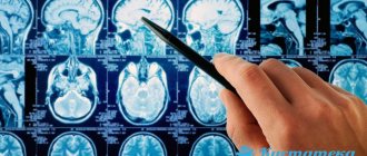
MRI indicators of vascular lesions of the brain are significant only in conjunction with clinical data. The absence of vascular lesions in the brain according to MRI data argues against a vascular etiology of dementia.
Neuroimaging MRI criteria for vascular brain damage are:
- the presence of single or multiple large or medium-sized infarcts in the cerebral cortex or subcortical region, or;
- the presence of multiple lacunar infarcts in the subcortical region or in the white matter or;
- quite common (at least 1/4 of the area) decrease in the density of subcortical white matter (leukoaraiosis).
Vascular dementia (dementia) is heterogeneous in its morphological substrate. The NINDS-AIREN criteria distinguishes several subtypes of diabetes.
- Multi-infarct dementia.
- Dementia due to infarcts in “strategic” areas.
- Dementia due to small vessel disease – subcortical (including Biswanger disease) and cortical.
- Hypoperfusion dementia.
- A combination of the above and other factors that have not yet been well studied.
In multi-infarct dementia, MRI reveals multiple large infarcts in the cortex and subcortical substance of the brain, caused by thrombosis of large cerebral arteries.
Dementia with infarctions in areas of the brain strategically significant for cognitive functions develops with damage to areas such as the frontal lobes, parieto-temporo-occipital regions of the brain, medio-basal parts of the temporal lobes, anterior and middle parts of the thalamus, globus pallidus, and dentate nucleus of the cerebellar hemispheres. Dementia in these cases is caused by disturbances in the integrative activity of the brain due to interruption of associative connections between different parts of the cortex, a decrease in the activating influence of the reticular formation and the development of symptoms of a focal disorder of higher mental functions.
According to the NINDS-AIREN criteria, the following are strategically important for cognitive functions:
- bilateral extensive strokes in the ACA basins;
- left-sided extensive stroke in the PCA basin, but only in the presence of a paramedian thalamic infarction, affecting at least 1/2 of the thalamus or a lesion of the medial temporal lobe, covering at least 1/2 of the temporal lobe;
- left-sided extensive stroke affecting one of the associative areas: parietotemporal or temporo-occipital, including the posterior region above the hippocampus, angular gyrus;
- left-sided extensive stroke in one of the watershed zones: superior frontal or parietal lobes;
- at least 2 lesions in two subcortical ganglia and at least 2 lacunae in the white matter of the frontal lobes measuring at least 2 mm;
- bilateral thalamic lesions measuring at least 1 cm or 2 or more lesions in the thalamus.
The subcortical variant of vascular dementia may resemble normal pressure hydrocephalus in some of its clinical and neuroimaging manifestations. MRI in both cases reveals a pronounced expansion of the ventricular system of the brain. However, diabetes is characterized by the presence of infarctions and severe subcortical leukoaraiosis; For normal pressure hydrocephalus, infarctions are not typical, and leukoaraiosis is much less common and is localized periventricularly. In diabetes, the expansion of the lateral ventricles corresponds to the severity of periventricular leukoaraiosis.
Pathological changes in the brain in Biswanger disease include extensive diffuse or patchy demyelination of the centrum semiovale, excluding U-fibers, astrocytic gliosis, microcysts (lacunar infarcts) in the subcortical white and gray matter. Single, cortical infarctions are possible. Characterized by pronounced thinning, sclerosis and hyalinosis of small cerebral vessels feeding the subcortical region, expansion of the perivascular spaces - “cribrosus status”. MRI images show changes in the white subcortical matter characteristic of encephalopathy; are found in the form of bilateral symmetrical zones of diffuse or patchy decrease in its density or increase in signal on T2-VI (so-called leukoaraiosis), not affecting the subcortical arcuate fibers and located periventricularly and/or in the semioval center, often in combination with lacunar infarctions. The expansion of the ventricles, primarily the lateral ones, is also revealed, and to a lesser extent - the expansion of the subarachnoid space of the cerebral hemispheres. In most patients, diffuse changes are combined with small ones; foci in the deep parts of the cerebral hemispheres (post-infarction and post-hemorrhagic cysts). Over time, leukoaraiosis increases, and in parallel with this, cognitive impairment increases.
Dementia caused by cerebral hypoperfusion occurs when the heart stops and blood pressure drops. It is based on global or limited cerebral ischemia with damage to areas of adjacent blood supply.
When describing the clinical picture of chronic forms of cerebral circulatory disorders, both with NPICM and DE, it is necessary to take into account the role of the vascular system, in which hemodynamic disorders predominate. Localization of vascular changes can be observed both separately in the carotid or vertebral-basilar basins, and simultaneously in all the great vessels of the brain.
Methods for diagnosing pathology
Timely diagnosis of vascular dementia in the initial stages gives a chance for recovery; in more complex cases, a correct diagnosis and selected treatment will help stop the development of the disease. For this purpose, modern neurologists use the following research:
- studying the history of life and illness;
- conducting psychological tests to identify cognitive impairment;
- blood pressure control;
- clinical blood test;
- determination of blood sugar;
- determination of lipid content in the blood and the level of cholesterol concentration in it.
Modern instrumental diagnostic methods that determine the degree of damage to the blood vessels of the brain and its tissues:
- radioisotope research of the brain;
- computer and magnetic resonance imaging;
- electroencephalograms;
- ultrasound examination of the brain;
- Dopplerography of cerebral vessels (determination of blood flow);
- angiography (x-ray examination of blood vessels);
- echocardiography.
Article on the topic: Severe cough with sputum without fever - causes and methods of treatment
Studying the research results, their analysis and comparison allows us to establish an accurate diagnosis.


