Spinal cord ependymoma is one of the types of tumors of the central nervous system. It is formed as a result of a mutation in the ependymal cells that line the spinal canal and ventricles of the brain. The tumor may differ in size and degree of malignancy - the prognosis for ependymoma depends on these factors, as well as the age of the patient and the presence of other chronic diseases. Treatment is selected individually and may include surgery and related techniques.
What is ependymoma
Ependyma is a thin membrane that lines the inner surface of the spinal canal and the ventricles of the brain. It is formed by nerve cells (ependymocytes), which belong to neuroglia. It is from them that ependymomas are formed. These tumors can be benign or malignant and differ in growth rate and other characteristics. Some of them grow through the nerve plexuses, while others entwine them. Ependymoma is a primary tumor and cannot be a metastasis - a secondary neoplasm in the presence of a main focus in other organs.
Ependymoma rarely forms in the spinal canal. It is most often localized in the ventricles of the brain and is diagnosed at the age of 45 years.
Causes of brain ependymoma
What serves as a prerequisite for the development of a tumor is unknown, but still there are several factors that can influence the development of pathology:
- heredity;
- long-term use of potent drugs;
- ionizing radiation;
- trauma to the skull or central nervous system;
- oncogenic viral diseases (human papillomavirus, herpes);
- poor environmental conditions;
- smoking and alcohol abuse.
The cancer trigger was initially thought to be the SV40 virus, which was discovered in ependymoma cells. However, subsequent studies did not confirm this theory, so the etymology of the disease is still unknown. As practice shows, cerebral ependymoma most often occurs in children and adolescents, less often in adults.
Possible reasons for development
It has not been possible to determine the exact reason why ependymomas form. However, doctors have several theories about the development of tumors, each of which has confirmation. The first of them explains their appearance by cellular mutations during embryonic development, namely due to the closure of the neural tube in the initial stages of embryogenesis.
There are also several main theories that can explain the mutation of ependymocytes and tumor formation.
- The role of exogenous factors - substances that enter the body from the outside, causing pathological growth and uncontrolled cell division. These include radioactive radiation, various food additives and dyes, heavy metals, and chemical compounds.
- Endogenous - all possible pathologies in the body that can lead to disruption of cell division processes. The role of the immune system in protecting against tumors has been proven, and when it decreases, the risk of tumors, including ependymomas, increases.
- Genetic predisposition is one of the possible causes of tumor growth. There are familial forms of cancer that are diagnosed simultaneously in several generations.
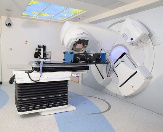
To diagnose epindema, modern equipment is required, which makes it possible to obtain the most accurate picture of the disease
The exact causes of ependymoma have no diagnostic value. For its treatment, it is important to determine the shape and size of the tumor, its exact location, tendency to rapid growth and degree of malignancy. Treatment is prescribed without taking into account the causes of the tumor and is aimed at its complete removal and preventing relapse.
Gamma Knife or Cyber Knife?
The CyberKnife has a special “tracking” system that prevents the flow of ionizing radiation from moving away from the tumor when the patient’s head or body moves. Therefore, the patient is not fixed inside the stereotactic frame, which was mandatory during radiosurgical treatment using the Gamma Knife method. Now the Gamma Knife is called the technology of the last century. In addition, anesthesia was required during Gamma Knife treatment.
Possibility of treatment on an outpatient basis
, hospitalization in a hospital is not necessary. Psychologically comfortable conditions are of great importance for the patient, because staying in a hospital is very stressful for any person.
(495) 740-58-05 — information on the Cyber Knife system
Development mechanism
The material for tumor formation is epencymocytes of the spinal canal. As a result of the processes of their uncontrolled division, neoplasms arise. Most ependymomas are benign, so with timely diagnosis, their complete removal and restoration without recurrence are possible. The disease develops in several stages:
- formation of ependymoma from cells of the inner layer of the spinal canal;
- compression (squeezing) of the tissues of the spinal cord, resulting in a disruption in the transmission of nerve impulses through the neurons of the affected area;
- compression of the pathways conducting nerve impulses is accompanied by paresis, paralysis of tissues that are located below the affected area;
- In the sacrum area, extradural growth of ependymoma is possible, as a result of which it grows into the subcutaneous tissue.
Malignant ependymomas also occur, but less frequently. As such tumors grow, they destroy spinal cord tissue and pose a danger to the patient’s life. Their growth rate is different - the prognosis for both benign and malignant neoplasms will depend on it.
Symptoms of subependymoma
The clinical picture of subependymoma is practically not much different from other glial brain tumors. Characteristic symptoms are severe and intense headaches, nausea and vomiting as a result of increased intracranial pressure, impaired coordination of movements and balance, paralysis and paresis, convulsions, behavioral changes, mental disorders, as well as visual impairment and disorders of consciousness of varying severity. Sometimes subependymoma may not have any clinical manifestations.
Classification
There are several classifications that are used to determine the type of ependymoma. The first of them takes into account the location of the tumor and distinguishes cervical, thoracic, lumbar types, as well as neoplasms in the cauda equina area. It is also customary to distinguish 4 types of ependyma depending on their main characteristics.
We advise you to read: Spinal cord contusion
- Subependymoma is a benign neoplasm that belongs to neoplasia of the 1st degree of malignancy (the lowest). It consists of ependymocytes and glial cells, grows slowly and does not spread to surrounding tissues. This type of tumor often forms in the brain, rarely in the spinal canal.
- Myxocapillary is another benign ependymoma that often forms in the cauda equina region and is characterized by extramedullary growth. It also belongs to neoplasms of the 1st degree of malignancy and is characterized by slow growth. It leads to damage to the inner lining of the spinal canal and the formation of cysts.
- True ependymomas may differ in histological characteristics, therefore their varieties are distinguished: cellular, papillary, mixed and others. They belong to the 2nd degree of malignancy - they grow slowly, but contain mutated atypical cells. After their removal, relapses are possible.
- Anaplastic ependymomas are the most aggressive type, which belongs to the 3rd degree of malignancy. Such tumors consist of immature, poorly differentiated cells and are prone to rapid growth and metastasis. Neoplasms can metastasize to other organs and tissues by transporting particles with the cerebrospinal fluid.
As a result of the initial examination, even after histological (cellular) analysis, it is not always possible to determine the degree of malignancy of the tumor. All features are studied in dynamics, observing the rate and nature of growth of the tumor.
Treatment of brain ependymoma
Modern oncology and neurosurgery has the latest equipment for the diagnosis and treatment of brain tumors. Research Institute of Neurosurgery named after. Burdenko is one of the few where complex operations to remove ependymoma are performed. The clinic employs a team of high-level doctors who, over several years, have performed many complex operations to remove tumors from brain structures. The oncology center is equipped with modern equipment for diagnosis and treatment. An integrated approach and an affordable price allow us to consider the Burdenko Clinic one of the best in Moscow.
To get a full consultation and detailed information about treatment at the Burdenko Clinic, you can go to the website of the neurological center, where all the necessary information for patients is provided. The cost of treatment at the oncology center is calculated individually for each patient. If a person is unable to pay for treatment, the clinic has a quota for free treatment.
Considering that brain ependymoma has a high degree of malignancy, therapy is most often complex, consisting of surgery and radiation therapy. Chemotherapy treatment is carried out only if surgery is not possible. This treatment of ependymoma does not give a good prognosis; it can only prolong the patient’s life by several months.
I consider surgery to be the optimal method for treating such a tumor. It is performed using microscopic technology, which allows intervention into brain structures with minimal risk to health and the highest accuracy. During the operation, there is often a need for craniotomy. This allows access to the tumor and its complete or partial excision. Removing brain ependymoma does not provide a 100% guarantee for the complete destruction of cancer cells, so surgery is often combined with radiation therapy. The procedure has a detrimental effect on cancer cells that could not be eliminated by surgery.
Stereotactic radiosurgery, which involves exposing tumor tissue to radioactive rays, is often used to destroy tumor cells. Thanks to this technique, it is possible to stop tumor progression and destroy cancerous tissue, without affecting the healthy membranes of the brain.
Treatment of ependymoma in the clinic takes a long time, since the patient, even after the operation, must be under medical supervision. After therapy, the patient requires rehabilitation, which includes diet therapy, consultation with a speech therapist, psychologist, and therapeutic exercises. The recovery period may take several months.
Considering the complexity of the operation and intervention in the structures of the brain and central nervous system, there are always risks of postoperative complications. They can manifest themselves in the form of neurological disorders - seizures, loss of memory, vision and hearing. Sometimes patients learn to speak, read and write again. They require constant care and examinations. Medical supervision after surgery allows you to monitor the dynamics of the disease, the appearance or absence of relapses. In case of complications, the patient is prescribed special restorative therapy to eliminate symptoms and improve the patient’s quality of life.
Any tumor in the brain, regardless of whether it is malignant or benign, requires immediate treatment, since even a small tumor can have a negative impact on the functioning of the body and a person’s well-being.
Tumor symptoms
All symptoms of spinal cord ependymomas are associated with compression of nerve tissue and disruption of their conductivity. Most neoplasms are benign and are not prone to rapid growth, so they can develop asymptomatically for a long time. The first signs may appear 1–1.5 years after the appearance of the tumor, but they become more pronounced only after 4–5 years.
The main one is pain in the back, which often gets worse when lying down. In addition, there are several symptom complexes that can indirectly signal the appearance of ependymoma:
- violation of the superficial sensitivity of the limbs, the appearance of sensations of heat or cold;
- large tumors cause unilateral or bilateral paresis of the limbs;
- disruption of the pelvic organs;
- deterioration of muscle function and decreased tone;
- decreased manifestation of tendon reflexes;
- when ependymoma is localized in the cervical region - headaches and dizziness, cough, shortness of breath, disturbances in chewing and swallowing.
Signs of spinal cord ependymoma gradually progress. It is important to start treatment in the first stages, since nerve conduction may not be restored even after the tumor is removed. Necrosis (death) of nerve cells, which develops with prolonged compression, can lead to irreversible changes in the functioning of internal organs, muscles and skin of the extremities.
Diagnostic methods
The initial examination is carried out by a neurologist. It is important to take into account the nature and intensity of pain, the location of the main focus and the time of its appearance. With ependymoma, the pain is constant, and neurological disorders appear symmetrically on the left and right sides. To clarify the type and size of the tumor, as well as to select the optimal treatment regimen, additional examination methods are prescribed.
Neurological examination - allows you to obtain basic data on the level of damage to the spinal cord by a neoplasm. At this stage, the neurologist analyzes various disorders, including central and peripheral, sensory and motor. However, the diagnosis needs to be confirmed with more accurate methods.
X-ray is prescribed to exclude diseases of the spinal column, injuries and pathologies of its structure. For the study of tumors of the spinal column, the method is not very informative.
Treatment of ependymoma involves its radical surgical removal.
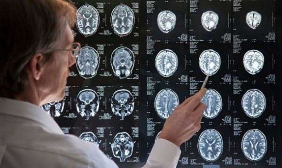
MRI of the spinal cord with the addition of a contrast agent is the main method for diagnosing tumors. As a result of the analysis, it is possible to obtain the most informative image, as well as evaluate the processes of nerve conduction. The images show even the smallest tumors and hematomas. MRI also makes it possible to differentiate tumors from edema and other pathologies that manifest similar symptoms.
Histological examination - analysis of the cellular structure of the tumor under a microscope. The technique is possible only after removal of the tumor. It is necessary to accurately determine the type of ependymoma, predict the further development of the disease and prescribe postoperative treatment.
Most patients are referred for diagnosis in the later stages, when the neoplasms have already become large in size and interfere with the normal innervation of organs and tissues. However, it is the early detection of ependymomas that is necessary for their safest removal, rapid recovery and the absence of dangerous consequences.
Prognosis after surgery
Life after treatment for ependymoma largely depends on the stage of the disease. If the disease is diagnosed in time and treated appropriately, the survival rate for more than 5 years is 70%. A less favorable prognosis is present with an anaplastic tumor, which can grow rapidly and metastasize. In this case, the chances of life are not great, and the treatment provided will prolong life by 1 - 2 years. If surgery is not possible, supportive care may increase life expectancy by just a few months.
If the operation fails to completely remove the tumor, there is always a risk of its recurrence. In such cases, the prognosis worsens, and the risk of its metastases to other organs and systems increases significantly. Unfortunately, there are no preventive measures that can protect against brain cancer. However, if you follow some rules, you can reduce tumor formation:
- proper and healthy nutrition;
- rejection of bad habits;
- eliminate the effects of radiation on the body;
- undergo regular medical examinations;
- monitor the state of the immune system.
By following basic rules, you can significantly reduce the risk of any cancer. Ependymoma is a type of brain cancer that requires timely diagnosis and quality treatment. By treating this pathology in the early stages of the disease, the chance of a positive prognosis is quite good, and the patient himself has the opportunity to return to a full life.
Treatment and prognosis
The only treatment for ependymoma is surgical removal. Benign neoplasms, which do not form for long and do not have time to reach large sizes, are easily removed without damaging surrounding tissues. For malignant neoplasms, the extended resection technique is used. It is aimed at preventing injury to the tumor and the entry of its fragments into the blood, lymph or cerebrospinal fluid. The operation is performed using microsurgical methods, with control of the conductivity of nerve impulses. After some time, a control MRI is performed.
Postoperative treatment depends on the type of tumor and its grade of malignancy. For grades 2 and 3, a course of radiation therapy is prescribed. It is aimed at preventing further relapse. In addition, some patients may require chemotherapy as a final treatment or in preparation for the next surgical step.
The prognosis for ependymoma depends on the degree of damage to the spinal cord, the size of the tumor and its tendency to malignant growth. Stage 1 ependymomas do not pose a danger to the patient if you seek treatment at the first symptoms. Malignant tumors can metastasize and recur. To treat them, courses of radiation therapy are carried out, but large tumors can compress the tissues of the spinal cord and disrupt the functioning of internal organs, skin and muscles in a certain area. Therapy is most effective in the first time after detection of a tumor - the prognosis in such cases is favorable.
Cost of spinal cord surgery
A neurosurgical procedure on the main organ of the central nervous system, located in the spinal canal, is a high-tech type of therapy of increased complexity. Prices for this category of medical care are quite high. The most accurate amount will be given to you during a consultation and examination of your specific diagnosis at your chosen medical institution. For your first acquaintance with approximate Russian prices for common medical services, you can use the data from our table.
| Operation | Cost, rub. |
| Elimination of SM fixation | 100-150 thousand |
| Ependymoma removal | 120-300 thousand |
| Excision of astrocytoma | 94-200 thousand |
| Glioblastoma removal | 150-340 thousand |
| Resection of an hourglass tumor | 250-500 thousand |
| Radiation therapy using the Cyber Knife installation (1 session) | from 150 thousand and more |