What happens if a person's brain is removed?
Until now, the human brain remains a mystery to science and medicine.
Of course, brain surgery is performed in different clinics, but no one can predict the outcome, which cannot be said for other parts of the body. Its importance is also proven by the fact that Mother Nature hid it in a reliable bone box - the skull, which protects it almost perfectly.
Of course, nature did not take care that people could reach a speed of 180 km/h and crash into a pole, but our brain is properly protected from most injuries.
Let's look at the paradoxical events with the brains of some people who subsequently, it would seem, should have died, but remained to live their previous lives.
1879: A woman's head was pierced by a bolt.
The woman worked at the mill. A large bolt hit the mill mechanism and flew out like a bullet, hitting the unfortunate woman in the forehead just above her right eye. The bolt settled at a depth of almost twelve centimeters.
Part of the brain was lost during the accident and during surgery to remove the bolt. No one expected a favorable outcome, and yet the victim not only did not lose consciousness at the time of the incident, but also did not feel any pain.
Two years later, only a small scar on his forehead reminded him of the accident. After this, the woman lived for another forty-two years.
The nobleman lost part of his skull
The woman from the mill was even luckier than one Russian nobleman, who was dragged across the ground by a galloping horse.
A significant part of his skull was demolished, which was restored by surgeons, borrowing it from a shot dog.
The man recovered, but was excommunicated from the church without the right to reinstate him in the bosom of religion until he agreed to have the dog bone removed from his head. Needless to say, the nobleman chose to live in godlessness.
1847: an iron rod pierced through Fiennes Gage's head
Another interesting case is recorded in the annals of the medical museum in Massachusetts. A heavy iron rod about a meter long pierced the man's head. On the afternoon of September 13, 1847, 25-year-old railroad foreman Phineas P. Gage placed explosives in a blast hole.
He compacted the powder charge with an iron rod, which was pointed at the top and its lower end was completely flat. When the iron rod hit the stone, it sparked, causing the gunpowder to explode. The sharp end of the rod hit Gage from below the cheekbone and went through his head. The left eye almost popped out of its socket.
Despite the terrible injury, the young man did not lose consciousness. His comrades took him to a local doctor, and Gage himself went to the waiting room. Removing the iron rod from the head, the surgeon was forced to remove part of the victim's brain and bones of the skull. Contrary to the expectations of those around him, Fiennes recovered. He was only blind in one eye.
Gage lived for more than forty years, confounding numerous medical luminaries.
Portrait of Gage with the metal rod that changed his life forever. Computer model illustrating research on the impact of Gage's trauma on his psyche (based on the work of Van Horn JD, Irimia A, Torgerson CM, Chambers MC, Kikinis R, et al.)
1935: a child lived for 2 months without a brain
In 1935, a child was born at St. Vincent's Hospital in New York with no brain at all (anencephaly). He lived for almost two months! The child's behavior was completely normal, and no one suspected that he had a brain until the autopsy. Today, such pregnancies are terminated in the early stages. It’s worth thinking: is the brain the most important thing in our body?
1957: Doctors Jan Bruel and George Albee removed the entire right half of the brain
In 1957, doctors Jan Bruel and George Albee successfully performed surgery to remove a brain tumor at a Boston hospital. They had to remove the entire right half of the patient's brain. To the great amazement of the doctors, he quickly recovered and did not lose his mental abilities. The operation seemed to have no effect on them.
1940: A 14-year-old boy's brain was severed from his skull.
In 1940, Dr. Augustin Iturrera made a statement at the Anthropological Society of Bolivia and confronted his colleagues with a fantastic fact. He and Dr. Nicholas Ortiz saw a 14-year-old boy who was in the clinic diagnosed with a brain tumor.
The patient remained conscious until his death, only complaining of a severe headache. When the doctors performed the autopsy, they were extremely amazed: the entire brain mass was completely separated from the skull.
A huge abscess took over the cerebellum and part of the brain.
Professor Huflandu discovered water in the skull instead of a brain
However, the problem faced by the Bolivian doctors was not as surprising as the one faced by the famous German brain specialist, Professor Hufland.
He had to change all his medical beliefs after opening the skull of a man who was paralyzed. Until the very last minute the patient retained his mental abilities.
The result of the autopsy left the professor completely confused, because instead of the brain in the skull of the deceased there was... water!
1968: a sailor had a quarter of his head cut off
And in the journal "Medical Bulletin of New York" for 1968, a case is described of a sailor squeezed, as if in a huge vice, between the arch of the bridge and the superstructure above deck. His head fell into this “vice,” and a sharp bridge beam cut off the upper part of the skull, about one-fourth of it.
Doctors who treated the wound several hours after the accident found that the cut was clean and smooth, as if it had been made with a medical saw. The victim lost a significant part of his brain. Doctors worked for several hours to close the gaping wound.
Imagine their amazement when the victim suddenly opened his eyes and asked what happened. After applying the bandage, the sailor rose to his feet and began to dress as if nothing had happened. Two months later he started working again.
The next time this man was hospitalized was 30 years later, when his left arm and leg were partially paralyzed.
Of course, we only cited cases with a positive ending. Although they make up less than 0.1% of the total, they make you think...
Source: //vse-krugom.ru/chto-budet-esli-cheloveku-udalit-chast-mozga/
Structural neuroplasticity: a developmental constant
Structural neuroplasticity is associated with declarative memory. Every time we access familiar information, the synapses between our nerve cells change: they become stabilized, strengthened, or erased. This happens in the cerebellum, amygdala, hippocampus and cerebral cortex of every person every second. The “receivers” of information on the surface of neurons—the so-called dendritic spines—grow to absorb more information. Moreover, if the growth process starts in one spine, the neighboring ones immediately willingly follow its example. The postsynaptic condensation, a dense zone found at some synapses, produces more than 1,000 proteins that help regulate the exchange of information at the chemical level. Many different molecules circulate across synapses, the action of which allows them not to disintegrate. All these processes go on constantly, so from a chemical point of view, our head looks like a metropolis permeated with transport networks, which is always on the move.
A terrible “cure” for quarrelsomeness and nymphomania: Top 10 frightening facts about lobotomy
Medicine has not always practiced gentle treatment. Sometimes the Hippocratic “do no harm” was not always interpreted in favor of the patient. Lobotomy is a frightening example of the barbaric ingenuity of the Aesculapians of the not so distant past. Historical facts about lobotomy will make the most experienced reader shudder.
Lobotomy or lecotomy is an operation in which the frontal lobe of the brain is deliberately injured or excised. The defect, deliberately inflicted on the brain, disrupted the connection of the frontal lobe with the rest of the brain. The doctor inserted the knife through the patient's eye socket, acting almost blindly. The success of the operation largely depended on the skill of the surgeon and his knowledge of the anatomy of the brain.
The perfrontal cortex completes its development by age 20. The functions of this part of the gray matter make us a person.
The frontal lobes provide coordination, concentration, planning, and emotion management. The totality of all the functions of the prefrontal cortex creates our individuality.
It was understood that the lobotomy turned off this particular area of the brain, relieving the patient of anxiety, excitability, and aggression.
The founder of lobotomy was the neurosurgeon Egas Moniz from Portugal. In 1935, he learned about an experiment on a chimpanzee whose frontal part of the brain had been removed. After which the animal became quiet and obedient. Egas Moniz decided that such manipulation could change the behavior of mental patients and in 1936 he performed the first lobotomy.
A Portuguese neurosurgeon performed hundreds of operations before he came to the conclusion that lobotomy was useful and safe. The innovative operation made violent and rebellious patients quiet, obedient, just like a laboratory monkey.
In a report on the benefits of lobotomy, Egas Moniz referred to the stories of the first 20 patients who underwent lobotomy. According to him, seven patients fully recovered, seven showed a significant improvement in their mental state, and six patients did not show any dynamics of the disease.
In fact, the neurosurgeon operated on subjective data. Patients were observed for only a week after surgery, after which they were sent home or to a psychiatric clinic. Egas Moniz did not have information about the long-term results of surgery.
As it turned out, the patients' future was far from bright. Among the most common side effects of lobotomy were: irreversible personality changes, epileptic seizures, meningitis, memory impairment, weight gain, involuntary bowel and bladder emptying, infectious and inflammatory processes, and suicidal tendencies. About 20% of patients died in the first year after surgery.
The Nobel Committee celebrated the discovery of Egas Moniz. In their opinion, the doctor made a breakthrough in surgical psychiatry, significantly easing the plight of incurable patients. The fact is that at that time there was no drug treatment for severe mental conditions. The Portuguese neurosurgeon won the Nobel Prize in 1949.
By that time, lobotomy was widely practiced in many countries. There was numerous data on the side effects of excision of the frontal lobe of the brain. Relatives of patients and some patients petitioned for the Nobel Prize to be cancelled. The committee rejected the request. The founder of lobotomy entered the annals of the most important scientific discoveries of mankind.
The popularization of leucotomy was contrary to the beliefs of Egas Moniz. The Portuguese considered surgery a last resort. He resorted to lobotomy only in the most severe cases, when other treatments had failed. Dr. Walter Freeman, on the contrary, considered brain surgery a panacea for all mental illnesses and abnormalities, including bad character.
According to the American Walter Freeman, the lobotomy turned off the emotional component, which caused anxiety.
The doctor enthusiastically prescribed manipulation for a variety of complaints: from headaches during pregnancy to poor performance at school. The term “lobotomy” was coined by Walter Freeman.
It is worth mentioning that the American performed almost three thousand operations without even being a surgeon.
The consequences of a lobotomy were unpredictable. Today, the damage from surgical brain injury seems incomparably greater than the imaginary benefit. Yes, sometimes patients became calm, balanced, and manageable. This model of behavior made those around him happy, but not the patient himself. Here are just a few stories of lobotomy survivors:
- Dr. Freeman performed a lobotomy on a pregnant woman due to frequent complaints of headaches. After the operation the patient's mental state was that of a two-year-old child;
- The doctor also operated on a boy who was not doing well enough at school. After which he no longer needed to study due to the complete degradation of his mental abilities;
- The woman was subjected to a lobotomy due to her quarrelsome nature. After the operation, she stopped being angry with others, gained weight, became forgetful, and absent-minded. For example, she lost bags on the way from the store, bumped into furniture in the house;
- A little girl had a lobotomy because she broke her toys too often. After the manipulation of her brain, she began to break everything nonstop, because she stopped understanding anything.
John Kennedy's sister also became a victim of a lobotomy. Rosemary was famous for her quarrelsome character, was prone to mood swings, and did poorly at school. At age 20, she exhibited aggressive behavior and signs of a nymphomaniac.
Doctors recommended lobotomy to parents as a sure way to correct behavior. After the operation, Rosemary Kennedy became a vegetable, unable to take care of herself.
Kennedy's sister was confined to a wheelchair all her life and died at an old age.
By the way, lobotomy was considered the only effective treatment for homosexuality, which was considered a mental disorder. To increase the chance of recovery, homosexuals were given electric shocks.
Dr. Freeman's enthusiasm led to the invention of a special lobotomy instrument. During one of the operations, an incident arose - the previous device broke right in the patient’s skull. Without thinking twice, Walter Freeman used a kitchen knife to pick ice and completed the lobotomy. History does not record the further fate of the unfortunate patient.
Dr. Walter Freeman, unlike his Portuguese colleague, had little interest in the future fate of his patients. While Egas Moniz observed the course of the disease of operated patients for many years, the American traveled around the country and performed two dozen lobotomies a day.
Walter Freeman came to the conclusion that an ice pick was ideal for lobotomy. Guided by the design of a kitchen appliance, the doctor developed a special medical instrument - an orbinoclast .
The tool followed the shape of a kitchen knife, but had a sharper tip and markings along its entire length. The markings helped control the depth of penetration into the skull.
To top it off, the orbinoclast came with a compact hammer for driving the knife into the patient's head.
Lobotomy achieved unprecedented popularity in the mid-twentieth century. Excision of the frontal lobe of the brain was widely practiced in American, Japanese, British and European psychiatric clinics. In the USA alone, lobotomies were prescribed to five thousand patients annually. In this case, the patient’s consent did not play any role. The vast majority of operated patients are women and children.
The popularity of lobotomy can be explained by two reasons: the high cost of running psychiatric clinics and the lack of medications. Mental degradation after a lobotomy was considered temporary.
Walter Freeman assumed that the patient would “grow up” over the years. He recommended that relatives perceive patients as children and treat them accordingly.
For example, punish with spanks on the butt for dysfunction of urination.
Fortunately, lobotomy did not take root in the Soviet Union . Of course, strictly regulated research was carried out in this direction. The first lobotomy in the USSR took place in 1944. Already in 1949, rules were adopted limiting the selection of patients for the procedure, the list of psychiatric clinics and neurosurgeons who received the right to perform a specific operation.
And in 1950, the staff of the Pravda newspaper wrote a collective letter condemning lobotomy as a technique that contradicts the precepts of the founder of Soviet medicine, I.P. Pavlov.
The letter caused a lot of noise in the government. A commission was urgently created, which reviewed the facts and arguments presented in the letter and recommended banning prefrontal leucotomy “for ideological reasons.”
What became the basis for the corresponding order.
Despite the absurdity and barbarity of the method, lobotomy did not disappear as such. It was supplanted by the discovery of aminazine. The surgical treatment of psychiatric diseases has given way to drug treatment.
At first, treatment with aminazine was called a “chemical lobotomy.” But unlike the surgical method, chlorpromazine did not cause personality degradation. Patients undergoing therapy did not turn into mentally retarded people.
Thanks to the discoveries of pharmacology, psychiatrists stopped practicing lobotomies.
10 Strange Historical Photos That Need Explanation
Want to stay updated? Subscribe to our page or Telegram channel.
Source
Source: //BigPicture.ru/?p=1129894
Microsurgical equipment
Brain surgery is unthinkable without the main elements of microsurgery - specialized stereoscopic loupes and operator microscopes.
Nowadays, during neurosurgical procedures, operator optical devices are used, which have the following advantages:
mobility, allowing you to freely move the ultramicroscope to different positions required by the doctor; extensive boundaries of magnification change; excellent illumination of the operating space; the presence of additional eyepieces for an assistant.
A small television camera, which can be equipped with an orthoscope, makes it possible to observe the operational situation on the monitor. Television displays and photographic equipment are necessary to control the manipulation. The operation to remove a brain tumor is extremely labor-intensive and lasts tens of hours.
One cut for McMurphy
Gleb Pospelovo lobotomy - the most famous and darkest of psychosurgical operations
“Oh, after treatment he will turn into a vegetable!..” - every psychiatrist has heard this or a similar phrase more than once, trying to persuade the patient and his relatives to be hospitalized. Everyone knows: in mental hospitals people are “zombified”, “brains are burned out”, “poisoned”, “turned into plants” - in general, they are destroyed as individuals in all possible ways.
And before the hospital there was a patient - just a sight for sore eyes, aha!
In general, this way of thinking has a completely scientific name: social stigma. In fact: when a person is discharged from a mental hospital, he is often completely different from what his loved ones are used to. He was sociable - he became withdrawn, he was active, nimble - he became inhibited and lethargic.
And the media, books, cinema willingly show exactly how pests in white coats conduct their hellish experiments on people. I’ll tell you a “secret”: if anything turns our patients into “plants”, it’s not the treatment, but the disease.
However, this was not always the case...
Remember the famous book (or its film adaptation) “One Flew Over the Cuckoo's Nest” and the fate of its main character, McMurphy? Let me remind you: McMurphy was lobotomized for violating hospital regulations.
The cheerful, self-confident, lively rogue-simulator turns into a weak-minded, drooling wreck.
The author of the novel, Ken Kesey, having worked as an orderly in a mental hospital, described the “frontal syndrome” or “frontal lobe syndrome” that developed in people after lobotomy surgery.
Bold idea
Brain lobotomy was developed in 1935 by Portuguese psychiatrist and neurosurgeon Egas Moniz. In 1935, at a conference, he heard a report on the consequences of damage to the prefrontal zone in chimpanzees.
Although the focus of this report was on the learning difficulties associated with frontal lobe damage, Moniz was particularly interested in the fact that one monkey became calmer and more docile after surgery.
He hypothesized that the intersection of nerve fibers in the frontal lobe could help in the treatment of mental disorders, in particular schizophrenia (the nature of which was still very vaguely understood). Moniz believed that the procedure was indicated for patients in serious condition or those whose aggressiveness made them socially dangerous.
Moniz performed the first operation in 1936. He called it “leucotomy”: a loop was inserted into the brain using a guide, and with rotational movements the white matter of the neuronal connections connecting the frontal lobes with other parts of the brain was cut.
Prefrontal lobotomy, or leucotomy (from ancient Greek.
λοβός - lobe and τομή - incision), is a neurosurgical operation in which the white matter of the frontal lobes of the brain is dissected on one or both sides, separating the cortex of the frontal region from the underlying parts of the brain. The consequence of such an intervention is the elimination of the influence of the frontal lobes of the brain on the remaining structures of the central nervous system.
Moniz performed about a hundred such operations and observed patients. He liked the results, and in 1936 the Portuguese published the results of surgical treatment of his first twenty patients: seven of them recovered, seven showed improvement, and six showed no positive dynamics.
Egas Moniz was awarded the Nobel Prize in Physiology or Medicine in 1949 “for his discovery of the therapeutic effects of leucotomy in certain mental diseases.” After the Moniz Prize was awarded, leucotomy began to be used more widely.
So, Egas Moniz observed barely two dozen patients during his “lobotomy” practice; He never saw most of the others after the operation. Moniz has written several articles and books about lobotomy.
Criticism followed: opponents argued that the changes after the operation most closely resemble the consequences of a brain injury and essentially represent personality degradation.
Many believed that mutilation of the brain could not improve its function and damage could lead to the development of meningitis, epilepsy and brain abscesses. Despite this, Moniz's report (Prefrontal leucotomy.
Surgical treatment of certain psychoses, Torino, 1937) led to the rapid adoption of the procedure on an experimental basis by individual clinicians in Brazil, Cuba, Italy, Romania and the USA.
In the land of great opportunities
American psychiatrist Walter Jay Freeman became the leading promoter of this operation. He developed a new technique that did not require drilling into the patient's skull, and called it "transorbital lobotomy."
Freeman aimed the tapered end of a surgical instrument resembling an ice pick at the bone of the eye socket, using a surgical hammer to pierce the thin layer of bone and insert the instrument into the brain. After this, the fibers of the frontal lobes of the brain were cut by moving the handle of the knife.
Freeman argued that the procedure would remove the emotional component from the patient's "mental illness." The first operations were carried out using a real ice pick. Subsequently, Freeman developed special tools for this purpose - the leukotome, and then the orbitoclast.
In the 1940s, lobotomy in the United States became widespread for purely economic reasons: the “cheap” method made it possible to “treat” many thousands of Americans held in closed psychiatric institutions, and could reduce the costs of these institutions by a million dollars a day! Leading newspapers wrote about the success of lobotomy, attracting public attention to it. It is worth noting that at that time there were no effective methods for treating mental disorders, and cases of patients returning from closed institutions to society were extremely rare.
In the early 1950s, about five thousand lobotomies were performed per year in the United States. Between 1936 and the late 1950s, 40,000–50,000 Americans underwent lobotomies. The indications were not only schizophrenia, but also severe obsessive-compulsive neurosis.
Lobotomies were often performed by doctors who had no surgical training. Although not trained as a surgeon, Freeman nevertheless performed about 3,500 such operations, traveling around the country in his own van, which he called the “Lobomobile.”
Lobotomy was widely used not only in the USA, but also in other countries of the world - Great Britain, Finland, Norway, Sweden, Denmark, Japan, and the USSR. Tens of thousands of patients have undergone this operation in European countries.
The result is obvious
Already at the end of the 40s, psychiatrists “discerned” that the first studies of lobotomy were carried out without a solid methodology: they operated with incomparable techniques on patients with different diagnoses. Whether recovery occurred or not - this issue was often resolved on the basis of such a criterion as increasing the patient's controllability.
In the 1950s, more thorough studies revealed that, in addition to death, which was observed in 1.5-6% of those operated on, lobotomies can cause seizures, large weight gain, loss of coordination, partial paralysis, urinary incontinence and other problems. Standard tests of intelligence and memory generally did not show any significant impairment.
The patients retained all types of sensitivity and motor activity; they had no impairments in recognition, practical skills or speech, but complex forms of mental activity disintegrated. More subtle changes such as decreased self-control, foresight, creativity, and spontaneous action were often reported; about selfishness and lack of concern for others.
At the same time, criticism of one’s own behavior decreased significantly.
Patients could answer ordinary questions or perform usual actions, but performing any complex, meaningful and purposeful acts became impossible.
They stopped experiencing their failures, experiencing hesitations, conflicts and, most often, were in a state of indifference or euphoria.
People who previously had an energetic, restless or aggressive personality may have developed changes towards impulsiveness, rudeness, emotional breakdowns, primitive humor and unreasonable ambitions.
In the USSR, special methods for performing lobotomies were developed - much more accurate in a surgical sense and gentle on the patient. The surgical method was proposed only in cases of ineffectiveness of long-term treatment, which included insulin therapy and electric shock.
All patients underwent a general clinical and neurological examination and were carefully studied by psychiatrists. After the operation, both gains in the emotional sphere, behavior and social adequacy, as well as possible losses, were recorded.
The lobotomy method itself was recognized as fundamentally acceptable, but only in the hands of experienced neurosurgeons and in cases where the damage was considered irreversible.
With maintenance therapy with nootropics and drugs that correct mental disorders, a significant improvement in the condition was possible, which could last for several years, but the final result still remained unpredictable. As Freeman himself noted, after hundreds of operations he performed, about a quarter of the patients remained living with the intellectual capabilities of a pet, but “we are quite happy with these people...”.
Beginning of the End
The decline of the lobotomy began in the 1950s after the serious neurological complications of the operation became apparent.
Subsequently, lobotomy was prohibited by law in many countries - data accumulated on the relatively low effectiveness of the operation and its greater danger compared to neuroleptics, which became more and more sophisticated and were actively introduced into psychiatric practice.
In the early 70s, lobotomy gradually faded away, but in some countries they continued to operate until the end of the 80s. In France, 32 lobotomies were performed between 1980 and 1986, during the same period - 70 in Belgium and about 15 at Massachusetts General Hospital; approximately 15 operations were carried out annually in the UK.
In the USSR, lobotomy was officially banned in 1950. And there was not only an ideological background to this. In the foreground were reasons of a purely scientific nature: the absence of a strictly substantiated theory of lobotomy; lack of strictly developed clinical indications for surgery; severe neurological and mental consequences of the operation, in particular “frontal defect”.
"Lobotomy" with a bullet
More than 60 years have passed since lobotomy was banned in our country. But people continue to get head injuries and get sick with various ailments (Pick’s disease, for example), leading to completely distinct “frontal” symptoms. I will give a vivid observation of the consequences of “frontal syndrome” from my own practice.
Two soldiers at the training ground laughingly began pointing machine guns loaded with live ammunition at each other and shouting something like “Tra-ta-ta!..”. Suddenly the machine gun said its “word”... The result is that one has a bullet in his head. Neurosurgeons somehow managed to revive and repair the guy; They inserted several plates into his skull and sent him to us to resolve the issue of further treatment and disability.
In the conversation the patient made a strange impression.
Formally, his mind was not damaged, his memory and stock of knowledge were at a normal level; He also behaved quite adequately - at first glance... An unnatural calmness, even indifference, was striking; the guy talked indifferently about the injury, as if it didn’t happen to him; didn't make plans for the future.
In the department he was absolutely passive, submissive; mostly lying on the bed. They invited me to play chess or backgammon, asked the staff to help me - I agreed. Sometimes it seemed like if you told him to jump out the window, he would do so, without even thinking about it.
And we received the answer to our questions a week later, when the patient was “caught up” with documents from neurosurgery, where his injury was treated. The surgeons described that the wound channel passed right through the guy’s frontal lobes. After this, all questions about the patient’s behavior were removed for us.
By the will of fate, I had the opportunity to meet this patient again, almost ten years after we met. This happened at a rehabilitation center where I worked part-time as a consultant. The guy has changed little in appearance.
Sharpness and rudeness appeared in communication; mental abilities were completely intact. I didn’t notice the main thing: self-confidence and independence.
The man had empty eyes... In life, he “floated with the flow”, completely indifferent to what was happening around him.
In conclusion, as before, I would like to wish: take care of yourself and your loved ones and remember that in most cases, even difficult and painful treatment is worth defeating a disease that deprives a person of his humanity.
Source: //www.katrenstyle.ru/articles/journal/history/odin_nadrez_dlya_makmyorfi
Brain surgery technique
The ability to perform procedures with the least risk to the patient is a key goal of modern medicine. This goal is made possible with the help of special microsurgical devices.
The position of the patient also greatly influences the procedure. To carry out the intervention, different patient positions are used on the operating bed:
on the back, with the head turned to the side; on the side of the body; in some cases, the patient is operated on in a state where he lies on his chest with his head hanging and bent; When manipulating the posterior cranial fossa, the patient's sitting position is often used.
In any individual case, the surgeon determines the appropriate position for the patient in order to expose certain areas of the brain. When selecting the patient's position, the possible shock to hemodynamics should be taken into account (primarily this concerns venous blood flow). If the patient is in a sitting state during the manipulation, then the pressure in the venous sinuses of the head rapidly decreases and may even take values with a minus sign.
This phenomenon explains the probable formation of mild embolic pathology - the entry of atmospheric oxygen into damaged large venous collectors and its concentration in the chambers of the heart, with the threat of cardiac arrest. This complication must be remembered if the patient is operated on while sitting, and a series of preventive measures must be used. An easier way to distinguish damage to large veins is compression of the jugular vessels in the neck or a hematoma.
Brain lobotomy: what is it, what is it for and how this operation is done, consequences
Lobotomy is a surgical method of treating mental illness, the essence of which is to destroy or disconnect the connections of one of the lobes of the brain with its other parts. Typically, the term "lobotomy" refers to the severing of one of the frontal lobes from the rest of the brain.
This is a neurosurgical operation that today has sunk into oblivion, that is, it is already history.
This treatment method was invented at a time when there were no effective medications that could be used to treat schizophrenia, behavioral disorders with delusions, hallucinations, when psychiatric patients were a threat to the lives of other people.
After the creation of Aminazine (a drug from the neuroleptic group), lobotomy became an unnecessary technique. However, there are many legends and creepy stories around this concept that are retold in our time. What kind of terrible treatment method is this, who invented it and first used it, what consequences arose after such therapy, you can find out by reading this article.
What is a lobotomy?
The essence of a lobotomy is to destroy the frontal lobes of the brain or cut off the nerve connections between this area and the rest of the brain. According to doctors of that time, disruption of nerve connections stops impulses that influence impulsiveness and inappropriate behavior.
This type of operation was first proposed by the Portuguese doctor Egas Moniz, who was impressed by his colleagues’ experiments on a very aggressive chimpanzee. In 1934, Moniz found that after the frontal lobes of the brain were removed, the animal's behavior changed to quiet and non-aggressive.
The result impressed the neurologist, giving him reason to assume that aggressiveness in people’s behavior can be eliminated in a similar way. “Violent” people were a big problem for doctors at that time. At the beginning of the twentieth century, the treatment of such patients consisted of using straitjackets and locking them in rooms with padded walls.
This required special safety measures for medical personnel, the number of which was constantly growing along with the incidence rate.
It should be noted that the events in the world of the first half of the last century (world wars, severe economic crises) made their sad contribution to the statistics of mental illnesses, and effective medications for treatment (aminazine and others) did not yet exist at that time. All this explains the fact that Moniz had followers, and lobotomy immediately became widespread as a method considered a breakthrough in medicine.
History: the beginning
We have already discussed the appearance of this procedure above, and now we will talk about it in more detail.
1890 Dr. Gottleib Buckhart in Switzerland performed an operation to remove part of the frontal lobes of patients at a psychiatric clinic. One of them died immediately, the second - a few days after leaving the medical institution. In the remaining four, behavioral changes were observed. It was from this moment that psychological surgery began to develop.
Only in 1935, a neurologist from Portugal, António Egas Moniz, performed prefrontal leucotomy. The operation proceeded as follows: a hole was drilled in the patient’s head. Alcohol was injected through it, which contributed to the destruction of the frontal lobes. The tool used to cut brain tissue is called a leukotome.
After the procedure, the patients became a little quieter. For this discovery, the doctor received the Nobel Prize.
How the operation was carried out, purpose
The first lobotomy on a human was performed by neurosurgeon Almeida Lima under the direction of E. Moniz in 1936. It looked like this. A woman suffering from severe paranoia had two holes drilled in her skull and injected with alcohol, which destroyed some of the white matter in her frontal lobes. As a result, the patient's condition improved.
The operation was called leucotomy - from the Greek words λευκός (“white”, after the color of the tissues inside the brain) and τομή (“cut”).
Soon Moniz created a special device with a wire loop - a leucote, which was used for the operation. It was inserted into the brain tissue on one side through a hole in the skull and then rotated. Thus, half of the frontal segment of the brain was isolated from the rest of the central nervous system: the nerve fibers conducting impulses were cut.
Moniz released data that of the 20 operated on, 7 recovered, the condition of another 7 improved, and in 6 there were no changes. He did not report the consequences of disruption of brain connections.
Meanwhile, from the perspective of modern medicine, the short duration of observations of patients after surgery is striking: after only 3-4 days they were released on all four sides. No long-term studies have been conducted.
Later it turned out that the operation could have prolonged (long-term) consequences.
Surgical theater
It is believed that Freeman was too happy to be able to legally perform transorbital lobotomies on all patients indiscriminately. He did not complete the procedure in ten minutes - somehow not enough for a complex brain operation, even if it was the most useful operation in the world...
He once performed 25 lobotomies in a day. It was he who first figured out the “humane” use of electric shock to perform operations while patients were unconscious. Worse, Freeman would sometimes lobotomize both sides of his brain just to show off. It is impossible to say exactly how many people he ruined their lives.
What are the brain conditions and symptoms that require brain surgery?
There are certain brain conditions and symptoms that indicate the need for brain surgery. The main conditions include: changes in brain tissue due to infections, brain cancer or swelling; pathologies of the cerebrospinal fluid due to infection or hydrocephalus; changes in blood flow to the brain due to intraventricular hemorrhage, subarachnoid hemorrhage, or subdural hematoma. Symptoms requiring urgent surgical intervention are characterized by nausea, headache, drowsiness, vomiting and seizures.
Endoscopic procedures
These procedures are mainly performed in the ventricles of the brain. Both solid and elastic endoscopes are used, equipped with devices for the purpose of collecting soft tissues, destroying them and stopping bleeding (with the support of coagulation or laser influence).
The introduction of endoscopes can be realized with the support of stereotactic units and thus influence the brain.
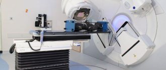
For such purposes, special radiosurgical inventions are used, the best of which is considered to be the gamma cutter, created by the famous Scandinavian neurosurgeon E. Lexill. The gamma cutter looks like a huge helmet that houses approximately 190 small gamma beam sources. Rays from absolutely all sources are directed at the same point.
The position of the patient's head in relation to this structure and the autocollimation of ray emission make it possible to obtain a zone of influence in the form of a clear geometric figure, which provides a chance to specifically destroy deep-lying tumors, virtually eliminating the chance of unsafe irradiation of all nearby organs.
In terms of accuracy, such an effect is equivalent to a surgical effect, which explains the name of such radiation treatment - “radiation surgery”. Similar results can be obtained by using precisely focused radiation from proton particles and electrons, as well as certain other types of elementary units of the structure of matter with high energy.
Rate this article:
1 votes, average:
5,00
out of 5
Ivan Drozdov 11/22/2016
A brain tumor, regardless of the nature of its origin and location, does not go away on its own. If there are clear prerequisites for growth and deterioration in well-being, it must be treated immediately. The treatment method for a brain tumor is chosen by an oncologist based on a preliminary examination and determination of the type, nature, size, and location of the tumor formation. It also takes into account the patient’s age, general health status and individual indicators for a particular treatment method.
Stereotactic procedures
Along with open manipulations on the brain that require craniotomy, a method called stereotactic (translated from Greek stereos - spatial, visual, and taxis - location) approach is also used. With this method, all manipulations are performed through a small milling mouth.
The purpose of stereotactic actions is that various devices are inserted into clearly defined parts of the brain (usually deep ones): electrodes for the purpose of destroying and stimulating medullary textures, cannulas for the purpose of cryogenic destruction, devices for biopsy or destruction of deep-lying tumors.
The mentioned devices are connected to the brain with the support of specialized stereotactic units attached to the patient’s brain. These units contain devices that allow you to volumetrically direct the device being introduced into the brain and predetermine the depth of its lowering. Stereotactic surgery is almost always the safest.
To establish the location of targets (subcortical ganglion nodes, thalamic centers, midbrain and other deep-lying brain systems, as well as deep-lying tumors, etc.), special stereotactic tables and summary comparative lists of radiographic results are used.
Current stereotactic devices make it possible to introduce the required devices into medullary textures with an accuracy of up to 1 millimeter during brain surgery.
Stereotactic procedures have found particularly widespread use in multifunctional neurosurgery (therapy of syndromes of increased motor activity, tremors, periodic pain, epileptic seizures, etc.).
The method of plastic orientation during the procedure on the cranium has in recent times become more probable, even without the use of stereotactic units. The negative consequences are minimal.
Brain tumor treatment
Depending on the factors described above, the oncologist can recommend to his patient one of the treatment methods or a complex combination of them:
Surgery . The method is justified and gives a positive prognosis if the tumor is detected in the early stages, is small in size and localized in an accessible location. In this case, it is highly likely that it can be completely removed and the patient has a chance to recover completely. If a brain tumor affects important parts of the brain and becomes large in size, it is partially removed. In both cases, the operation significantly alleviates the condition of the cancer patient by reducing the pressure of the tumor on the receptors and eliminating unpleasant symptoms. At the same time, surgical intervention in the brain structures can cause a number of complications - bleeding or inflammatory processes. Radiation therapy . Irradiation of the location of a brain tumor with a directed flow of radiation rays is used as an independent or complex therapy. In the first case, the technique is used to remove tumors of various etiologies that are located in places inaccessible for surgery or are concentrated in several parts of the brain. In the second case, radiation therapy is used after surgery, when the tumor is not completely removed. After irradiation, the patient may experience side symptoms - weakness, attacks of painful nausea, headache. Chemotherapy . The destruction of cancer cells occurs under the influence of medicinal chemicals. Chemotherapy is carried out before, after or instead of surgery, drugs are administered intravenously or taken in tablet form. The disadvantage of this method is the negative impact of chemicals not only on cancer cells, but also on all vital systems. As a result of acute intoxication of the body, the patient may experience hair loss, attacks of nausea and vomiting, and weakness.
Types of neurosurgical interventions
Depending on the purpose, brain procedures can be relatively divided into specific and palliative interventions.
The task of specific actions is to remove painful formations (bruises, ulcers, neoplasms), restore standard relationships of human anatomy (restoration) in the case of damage to the skull and congenital developmental defects acquired under the influence of external factors, etc. The concept of “radical impact” is used with a caveat. It determines the purpose of the procedure, but its result does not always correspond to the established problem (for example, with a tumor in the brain, it is often not possible to achieve its complete removal). Surgery may not remove a brain tumor at all, but it may improve the patient’s general well-being.
Palliative procedures are not intended to protect the patient from the disease itself, but are aimed at mitigating the condition of the victim. An example of a palliative procedure is the formation of new lines of cerebral fluid loss in case of incurable neoplasms that impair the patency of the lines and the circulation of the cerebrospinal fluid.
Depending on the time of the procedure, neurosurgical procedures are divided into planned and urgent. Emergency procedures are usually performed when clinically necessary. The need for urgent procedures appears in case of traumatic hemorrhages, with a sharp disruption of the patency of the cerebrospinal fluid lines, when the patient develops signs of deformation of the brain body and compression of its nodal zones in the foramen magnum or tentorium.