If an examination reveals a benign tumor in the structures of the brain, there is no need to panic. According to statistics, after the operation, most patients return to normal life, and with all rehabilitation procedures, a full recovery is possible. Why does pathology develop, what are its symptoms and what treatment methods exist?
Peculiarities
A benign brain tumor is an intracranial neoplasm that can be treated. ICD-10 (International Classification of Diseases) code – D33.0. The exact name of the disease is a benign neoplasm of the brain above the tentorium.
The neoplasm appears due to chaotic cell division. Unlike a normal cell, a diseased cell is underdeveloped and does not function.
Benign formations:
- develop to a specific stage;
- are formed within one organ;
- do not metastasize.
The reasons for the appearance of various neoplasms are the same. Benign and malignant tumors manifest themselves in the same way.
To identify the type, special examinations are carried out:
- MRI;
- CT scan;
- Submission of biomaterial for research (biopsy).
Causes
No doctor can answer why a brain tumor occurred. But there are factors that provoke development. It is impossible to predict which person will develop a brain tumor in the future.
Heredity
Human genes are modified due to the negative impact of various factors. Changes happen for the better and for the worse. An oncogene can manifest itself even after decades. Therefore, it is not necessary to expect negative consequences in a person under adverse influence. They may occur in the future in children, grandchildren or great-grandchildren.
Chemical exposure and radiation
Living in areas with high levels of radiation provokes the appearance of tumors. If a person’s work involves chemicals (mercury, lead, oil), such risks are likely.
Viral and bacterial infections
Infectious diseases can affect the human body, especially the brain and central nervous system. Against the background of weakened immunity, exposure to viruses or bacteria causes the development of a tumor.
What can cause brain cancer?
The appearance of cancer in the cranial cavity can be caused by many factors. In the case of a hereditary predisposition to oncology, the following reasons can provoke its onset and further development:
- constant exposure to electromagnetic fields (an example is the mobile phone, without which modern man cannot imagine his existence);
- infrared radiation;
- abundant evaporation of gases from a complex chemical compound of polyvinyl chloride (most plastic products in demand in everyday life are made with its help);
- poor nutrition;
- content of genetically modified organisms in the food consumed;
- alcohol abuse.
Knowing how many dangers lurk in the everyday life of any person, the above-mentioned potential provocateurs of the disease should be treated more carefully and not neglect the simplest safety rules.
People who once received a huge amount of radiation automatically fall into the risk group. A malignant form of education can develop in such people with a high percentage of probability. Not so long ago, in the treatment of dermatomycosis in children, found on the scalp, they resorted to radiation therapy. Thus, while saving children from fungal infections, they were endowed with a huge risk of cancer.
Carriers of papillomatosis viruses are also recognized as a risk group for tumor development. In particular, this applies to people affected by the 16th and 18th type of virus. Patients, as a rule, are not even aware that they are carriers of papillomatosis, and even more so have no idea how long they live with the virus in the blood. The optimal preventive and supportive measures in this case are regular consumption of large quantities of vegetables and hardening.
Classification
Primary and secondary neoplasms are distinguished. The difference is that the primary ones appear in the brain from the very beginning. They are localized in one place, preventing metastases. There are different types.
Secondary ones arise due to metastases from malignant neoplasms of another organ. The manifestation becomes a consequence of the development of other tumors. Secondary formations can be exclusively malignant.
Meningioma
A common type of neoplasm. Formed from the soft meninges. It is a clearly defined knot of a rounded shape or in the form of a horseshoe. Often adheres to the dura mater of the brain. There are also flat knots. The size of meningioma reaches several centimeters. The tumor tissue is gray-yellow.
Often meningioma is a benign formation. To determine the species, histological examination is used.
Meningiomas are removed surgically. Tumors do not cause relapses. If the tumor is in a hard-to-reach area, use a cyber and gamma knife. Chemotherapy and radiation are not performed.
Neuroma
Also called schwannoma. The diagnosis is common among children. Neuromas make up 8% of tumors that primarily developed in the brain.
Formed from nerve sheaths. It looks like a capsule with light nodules enclosed in it. Symptoms of schwannoma development include:
- hearing loss;
- dizziness;
- trigeminal neuralgia.
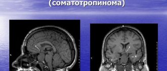
Pituitary adenoma
Formed from glandular tissue. Benign tumor. The pathology causes excessive production of hormones. When there is an excess of thyrotropin in the body, the functioning of the thyroid gland is disrupted. Excessive amounts of prolactin cause infertility. Due to an excess of somatotropic hormone, gigantism develops.
Astrocytoma
A benign tumor that forms in the brain from astrocytes - star-shaped neuroglial cells. The diameter of the formation sometimes reaches 10 cm. Children are more susceptible to astrocytoma. The tumor has a pale pink color and is close in density to the brain substance.
Oligodendrocytoma (oligodendroglioma)
Glial tumor, that is, developing in neuroglia. It has a gray-pink color. There are cysts in its structure. Develops slowly in the white matter. Capable of growing to large sizes. Men are more often affected (ratio 3/2).
Ependymoma
Education of the central nervous system. Formed from cells of the ventricles of the brain.
Ependymoma on the back of the head
It can be on their surface or inside. It can occur equally among children and adults. Has cystic and necrotic formations. Ependymoma can metastasize. Tumor cells spread along the cerebrospinal fluid outflow tract.
Craniopharyngioma
Present from birth. Develops from embryonic cells of the pituitary tract. Usually benign, but can become malignant. Rarely seen. Causes blurred vision, headaches, hormonal imbalances, and destruction of the nerves of the skull.
A benign formation becomes malignant due to negative influence. To avoid this, it is important to start treatment on time. Observation is excluded; malignancy will occur at any moment.
Stages of tumor growth
- Visiting a doctor if the initial symptoms of the disease occur: pain in the head, nausea and vomiting, change in gait, etc. This will allow you to perform a surgical operation and remove the tumor partially or completely.
- Monitoring when visiting a doctor is less positive. The affected cell structure begins to actively divide and put pressure on nearby tissues. At this stage, surgery may no longer be appropriate. The patient's age and personal characteristics of his body also matter. After 65 years of age, a patient who has undergone surgery, chemotherapy and radiation treatment has a greatly reduced chance of survival.
- A neoplasm at the third stage of development is very often inoperable. The patient can live with a tumor at this stage for no more than 2 years. The development of education proceeds quickly, the patient loses vitality in a short time and is rarely able to fight the disease.
- The likelihood of recovery at this stage is very low. Here, much depends on the support of relatives and friends, and the patient’s desire to live. On average, at this stage, with metastases that have already affected other organs in the body, life is calculated in several months, very rarely in years. Only every fifth person out of a hundred can overcome this line.
With timely detection, timely treatment and competently performed surgery, the patient can live for many more decades. However, unfortunately, even the use of modern treatment methods does not guarantee a 100% cure.
Despite this, the patient should not despair. The terrible diagnosis, of course, will turn his life 180°, but this is not a reason to stop fighting for life. In the history of medicine there are many examples of how illness receded before a person’s powerful will to live.
Symptoms of tumor development
It is often difficult to suspect the presence of a neoplasm. And given the slow development, a person does not feel its presence after years. Rapidly growing formations make themselves felt earlier. A person does not immediately think about a tumor. Often ailments are written off as signs of another disease.
The following should be taken seriously:
- Continuous headaches. Felt at a specific point or part of the head. For example, in the temporal, occipital, frontal region, depending on the location of the tumor. As it grows, the symptom intensifies due to compression of other areas. Painkillers do not have the desired effect.
- Uncoordination of movements. It occurs due to constant acute headache or under the influence of a tumor on the vestibular apparatus located in the cerebellum.
- Nausea or vomiting in the absence of gastrointestinal problems. May occur due to dizziness.
- Deterioration of memory and mental activity, sudden changes in mood.
- The appearance of seizures.
- Also, depending on the location of the tumor, speech is impaired, hearing is reduced, limbs become numb, and vision deteriorates.
If you have such symptoms, you should undergo a medical examination.
Medical examinations are carried out regularly to allow timely detection of developing tumors.
Diagnostic methods
To make a diagnosis, it is necessary to conduct several surveys and studies:
- During the interview with the patient, the existing symptoms are identified and the duration of poor health is determined. Tests are ordered if necessary. But they cannot detect a tumor. You can learn about the inflammatory process or other pathologies associated with the development of the tumor.
- Magnetic resonance imaging can determine the presence of a tumor. During this examination, images of the brain are taken from all sides. This way you can see the location of the tumor and its size.
- Computed tomography, electrical activity of the brain, angiography, craniography - these examinations provide detailed information about the tumor and allow you to determine the right direction of treatment.
- A biopsy (removal of tumor cells from the patient) determines whether the tumor is benign or not.
Treatment
Cancer therapy and benign tumor treatment have the same methods. The greatest effectiveness can be achieved by surgically removing the tumor. You can do without surgery. If the patient does not feel any discomfort and there is no threat to health, then the issue of surgical intervention is decided based on the person’s condition and the presence of contraindications to surgery. Chemotherapy is not required to treat a benign tumor.
X-ray examination of the pituitary gland
Possible methods of operation:
- Trepanation of the skull. The formation is excised after opening the skull.
- Exposure to ultrasound. The operation takes place without drilling the bones.
- Endoscopic intervention allows you to get by with small punctures.
- Radiosurgery does not require direct intervention into the organ. Removal is carried out with a gamma and cyber knife. Only tumor tissue is affected. Healthy cells are not affected. One of the most effective methods.
Treatment also involves radiation therapy. To get rid of the negative consequences, medications are prescribed: painkillers, corticosteroids, decongestants.
Possible complications and consequences
Negative consequences:
- An enlarged tumor compressing other areas of the brain. This may result in decreased hearing or vision. Speech defects appear. After the tumor is removed, the problems disappear. In rare cases, symptoms remain.
- The appearance of seizures is also associated with brain damage.
- Bleeding. Appear during or after surgery.
- Brain swelling.
- Cysts sometimes form at the site of a distant lesion.
- Thrombosis is the formation of blood clots (thrombi) inside blood vessels. The free flow of blood is disrupted.
- Infectious diseases. For example, meningitis or encephalitis.
- Consequences of surgery: headache, nausea, dizziness.
Benign neoplasms are perfectly treatable.
Forecast for the future
The development of a tumor places a serious burden on human health. But adults and very young patients can cope with it. Old age makes treatment difficult. This is due to the presence of other ailments and weak immunity in older people. Complications are more likely to occur after surgery.
Forecast and consequences
The negative consequences of the disease are expressed in irreversible changes in neural connections.
Even with timely treatment, some symptoms still remain, since the centers that regulate the functioning of one or another reflex have been affected (removed).
This also affects the functionality of internal organs. Some changes can be stopped or controlled with medication, others are smoothed out over time thanks to the brain itself, which learns to find “workarounds.”
Spontaneous convulsions are often noted among the consequences; there is always a risk of developing intracranial bleeding and chronic increase in ICP. In any case, the patient will take medications throughout his life and undergo regular neurological examinations.
The prognosis of the disease in comparison with malignant tumors is favorable. In the postoperative period, 90% of adult patients and 70% of children show clear improvements. Elderly patients tolerate treatment somewhat worse and this is explained by the age-related characteristics of their body, however, 65% of patients aged 60+ after recovery do not experience any particular difficulties in everyday life.
It is known that a malignant brain tumor is of oncological origin. The most common areas of damage are the meninges, brainstem and cerebellum. Read about the symptoms of brain tumors in children in the next topic.