| Service | Price, rub) | How is it carried out? |
| RHEOENCEPHALOGRAPHY (REG) ADULTS | RUB 800.00 | Video |
| RHEOENCEPHALOGRAPHY (REG) FOR CHILDREN | RUB 800.00 | Video |
Rheoencephalography is a narrowly focused diagnostic procedure, the name of which consists of Greek words meaning “flow”, “brain” and “I write, I depict”.
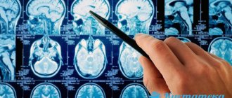
The REG procedure shows the real state of the brain vessels and even shows an image of the vascular system in general, indicating injuries or problems. REG of the head is carried out by exposing the brain to an electrical discharge, for which a medical cap with electrodes is put on the person’s head. During the reaction of tissues, arteries and vessels to an electrical discharge, all readings are recorded by a computer, and subsequent decoding of the card will reveal the presence of a disease or pathology.
The examination is painless and does not cause discomfort for patients, who need to relax and not worry, because this may distort the readings of the device.
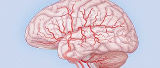
REG of blood vessels allows you to obtain the most accurate information about their condition, blockages or breaks, as well as choose an effective method of treatment or prevention.
Types of REG
Among the main types of REG, under certain conditions, the following are prescribed:
- Rheopulmonography (RPG).
Prescribed for bronchopulmonary pathologies. The essence of the method is to record the electrical resistance of the lung tissue and makes it possible to clarify the expected extent of resection. - Rheoencephalography (REG).
A technique that allows you to determine the degree of damage or abnormalities in the central nervous system, vascular system and cerebral blood supply. - Rheovasography (RVG).
One of the practical ways to diagnose blood circulation in the patient’s arms and legs. - Rheocardiography (RKG).
The study is aimed at studying the work of the heart and allows us to determine the degree of its stroke volume. - Integral rheography (IR).
A procedure that makes it possible to accurately measure the baseline impedance of the patient's entire body. - Polyrheocardiography (PRCG).
Diagnostics aimed at studying blood flow in the pulmonary and systemic circles to determine hypertension, hypotension or heart failure in the earliest stages. - Rheoophthalmography (ROG).
A technique for diagnosing and studying blood circulation in the tissues of the eye, with its reaction to the action of an electric discharge. - Rheohepatography (RGG).
A graphical method for studying blood circulation in the liver, based on recording frequency oscillations of the tissues of this organ in response to a high-frequency electrical discharge. - Rheorenography (RRG).
A special non-invasive method for studying blood flow in the kidneys, which makes it possible to identify or prevent renal failure and other pathologies associated with the functioning of this organ. - Reuterography (RUG).
A non-invasive way to study blood circulation in the uterus and pelvic organs. Allows you to detect vascular disorders in the early stages of pregnancy and other gynecological diseases. - Rheoprostatography (RPrG).
An effective way to study blood flow in the prostate gland, allowing timely detection of various diseases in the early stages and making a decision on the choice of quality treatment.
What is the difference between reg and eeg
REG and EEG are methods used to assess the state of the brain.
What is each examination, in what cases is it used, and how does REG differ from EEG? This is discussed below. articles
Rheoencephalography (REG) is a medical procedure aimed at studying the blood vessels of the brain.
Electroencephalography (EEG) is a method used when it is necessary to evaluate brain activity.
Comparison
Both types of examination are non-invasive. Patients do not have to experience pain. There is also no reason to be afraid of negative consequences.
In both cases, electrodes are attached to the head in a special way, and all data is recorded using appropriate instruments.
The images captured on the tape in the form of curves are then deciphered by a specialist. Information can also be displayed on a computer monitor.
The procedure allows you to find out how elastic and strong their walls are, whether there are lesions and cholesterol growths on these elements, and whether there is a predisposition to thrombosis. Blood circulation is also assessed.
It is recorded how the vessels are filled and whether the venous outflow is timely. The data obtained become guidelines for identifying the causes of diseases and conducting early diagnosis.
When can REG be prescribed? If your head often hurts or feels dizzy, you periodically experience tinnitus and high blood pressure. As a result, the development of hypertension, dystonia, atherosclerosis and other diseases is often detected.
Moreover, rheoencephalography helps not only to identify pathology, but also to check how effective the treatment method chosen in a particular situation will be.
This procedure is also indicated for monitoring a person’s condition after surgery or serious injury.
What is the difference between REG and EEG? The fact is that the latter of them is aimed at recording brain activity. Electroencephalography is a highly informative method based on capturing outgoing biocurrents.
Such an examination shows any pathological changes in brain activity. It becomes useful when it is necessary to find the cause of fainting and convulsive conditions, sleep disturbances, and developmental delays.
Such research is also carried out to monitor the course of brain diseases and adjust therapeutic actions. In certain cases, an EEG is prescribed to decide the issue of professional suitability.
This applies to those activities where a quick reaction is needed and there is any increased risk.
Source:
How does EEG differ from REG?
» Methods » Electroencephalography: differences between the procedure and REG
Electroencephalography (hereinafter EEG) is an analysis of the brain by recording the biological flows of a human organ. This method is simple and quite safe, and has a great impact on the ability to identify brain problems in the early stages.
EEG is the only way to examine a patient who has experienced attacks of loss of consciousness. Electroencephalography does not cause any harm to people. It has no contraindications or side effects; it is prescribed to people of all ages.
EEG carries a large flow of information that shows the functional state of the cerebral cortex. With its help, pathology is revealed that is hidden from the eyes of doctors and did not manifest itself until a certain time.
Thanks to EEG, there is a chance to monitor the development of the disease over a certain period of time, which allows for correction of the treatment procedure. The study shows how medications affect the course of the disease and the overall level of health.
At this stage, an overdose of certain medications or their insufficient effect on brain activity is detected.
The importance of electroencephalography in the study of epilepsy
Electroencephalography plays a key role in the study of epilepsy in patients. Every year, about 100 thousand new cases of this disease are registered around the world. Epilepsy often comes along with cerebral palsy.
The performed electroencephalography allows neurophysiologists to speak with complete confidence about the presence of “epilepsy” in the patient. Also, thanks to EEG, pathological activity in the brain is detected at an early stage, which creates difficulties for its normal functioning.
Electroencephalography technique
An EEG does not cause harm or pain to the patient. To conduct the examination, the patient is placed in a comfortable chair.
Then a number of electrodes are attached to the person using a helmet in the head area, which are connected to the device by wires.
When analyzing the functioning of the brain, the device amplifies the biological impulses coming from the sensors many times and records the results obtained on computer technology.
No specific preparation is required to undergo an EEG. You just need to follow a few simple rules:
- The patient should not experience a strong headache while undergoing the test. There is a high chance that this will lead to incorrect EEG results.
- Before undergoing electroencephalography, the patient should wash his hair. This will create the best conditions for contact of the electrodes with the surface of the head. Due to this, the research results will be more accurate.
- Before visiting the doctor's office, remove jewelry and let your hair down.
- You should not stop taking medications while the brain is being examined, as this will knock the body out of its usual state and provoke an attack.
The results obtained should be interpreted taking into account the patient’s age, existing diseases, and the names of medications that were previously prescribed by doctors.
Benefits of Electroencephalography
When using EEG, doctors perform the following actions:
- evaluate epileptic seizures according to their classification;
- identify areas of the brain where seizures occur;
- track the dynamics of the effects of drugs on the activity of the human brain;
- give answers about a person’s professional suitability, because the presence of epilepsy will become an obstacle for a person to get a job that involves driving vehicles and requires perseverance and concentration.
Electroencephalography is prescribed to patients who have a predisposition to epilepsy, diseases of the cardiovascular system, persistent headaches, and disorders of the nervous system.
To get the desired effect from the study, the patient is placed in a chamber. To identify hidden epilepsy, brighten the room and increase the noise effect inside the room. The patient is put into a sleep state and then woken up after some time.
Frequently turning on and off the light affects the human nervous system, thus trying to awaken latent epilepsy, if any. The duration of one procedure is at least 45 minutes. In especially severe cases, this procedure reaches 120 minutes.
On the eve of the EEG, get a good night's sleep and gain strength for the diagnosis. Before starting the procedure, the person should be at rest for about 20 minutes. In this case, high-quality results are obtained.
EEG causes a stressful situation in a person. Therefore, if a child is the object being studied, so that he is not afraid of her, a conversation is first held with him, during which the upcoming procedure will be described to him.
Rheoencephalography
Rheoencephalography (hereinafter referred to as REG) is a diagnosis of cerebral vessels. Just like EEG, it is absolutely safe for patients and does not cause them any harm. This method monitors the state of blood circulation in the head area. During the analysis, the resistance values of the electrical current tissue are recorded.
Reasons why rheoencephalography should be used include:
- constant severe pain in the head;
- presence of tinnitus;
- memory loss; regular fainting.
The main reason is the presence of cranial grass in the patient.
Functionality of rheoencephalography
What can REG do that makes it increasingly popular in diagnosing brain diseases?
Thanks to REG, doctors receive a large amount of information about the intensity of blood filling in the patient’s brain, the elasticity of human blood vessels, and affected vessels in the head.
When using rheoencephalography, doctors have a chance to detect vascular dystonia at an early stage. This can be done due to the fact that an unstable image of vascular tone appears in the pictures. Acute vascular lesions are detected.
REG is different in that it focuses on the vessels of the brain. In addition, the REG procedure is much shorter in its time scale and does not exceed 10 minutes.
Reg and eeg - is there a difference?
These two research methods are common when examining the human brain. At the same time, to give a more accurate assessment of brain pathology, EEG is used. It is especially good when the patient has epilepsy. Often, EEG is the main method of conducting research. The remaining methods are accompanying and are, as a rule, formal in nature.
During loss of consciousness, frequent headaches and severe fatigue over a short period of time, it is best to use EEG, as a method that has proven itself to be the best.
To make the right choice of research method, you need to proceed from what type of pathological process you strongly suspect. There are often cases when EEG and REG complement each other with maximum efficiency. In these cases, both methods are used in a complex state. This will allow you to make an accurate diagnosis as quickly as possible.
Contraindications
Brain studies are contraindicated during periods when a woman is preparing to become a mother. If the condition of the skin is poor, it is not advisable to perform REG and EEG before starting the electrodes.
The client's complete refusal to undergo testing also does not improve the therapeutic intervention. A recent disease of the skin and hair in those areas of the head where electrodes are supposed to be attached to a person also does not contribute to the effective treatment of the patient.
EEG and REG help medical workers prevent brain disease in a timely manner. Timely identified pathology will help to avoid serious problems with the human brain and begin timely treatment.
Correctly initiated treatment will be a lifeline for people suffering from these diseases.
Conclusion
Today, private and public medical institutions carry out similar studies. Based on the information received, doctors make an accurate diagnosis and begin the treatment process for a particular person.
Before undergoing a brain examination with electroencephalography or rheoencephalography, consult with your doctor. It depends on his opinion which of these methods is suitable for the patient.
Source:
Electroencephalography (EEG)
Electroencephalography (EEG) is an accessible and safe method of studying the brain by recording brain biocurrents. Neurons - the main elements of the central nervous system (and the brain, including) - are capable of generating and conducting electrical impulses that are recorded by an electroencephalograph.
This method is of great importance for the early detection of injuries, tumors, vascular and inflammatory diseases of the brain, and epilepsy. In addition, this is the only neurological outpatient study that is performed during attacks of loss of consciousness.
In our clinic, electroencephalography is performed by highly qualified neurologists-neurophysiologists with extensive practical experience in working with patients of various age groups.
EEG is absolutely harmless, has no contraindications, and therefore is used for patients of any age, both children and the elderly.
Electroencephalography is a highly informative study that reflects the functional state of the cortex, subcortical structures of the brain, as well as complex cortical-subcortical interactions, including the hidden pathology of diseases that have not yet manifested against the background of the complete clinical health of the subject.
EEG allows you to monitor the course of the disease over time, adjust treatment if necessary, and evaluate the effect of drug therapy (overdose or withdrawal of antipsychotics, tranquilizers, barbiturates, antidepressants) on brain activity. Unlike CT and MRI studies, EEG reveals structural and functional (reversible) changes that persist for a long time in the brain, for example, after a mild traumatic brain injury.
The role of EEG in the diagnosis of epilepsy
EEG is the most important diagnostic method for epilepsy. Every year, from 20 to 120,000 new cases of epilepsy are registered annually (on average - 70 - 100,000). In the CIS countries alone, about 2.5 million people suffer from this disease.
Source: https://otlichaem.ru/priroda/chem-otlichaetsya-reg-ot-eeg.html
Indications for rheoencephalography
A neurosurgeon or neurologist, after a thorough examination of the patient and discussion of his complaints, may decide to do an REG in Khabarovsk, if there are the following indications:
- Regular headaches, cramps and migraines;
- Suspicion of cerebral ischemia or stroke;
- Frequent dizziness;
- Concussion or serious traumatic brain injury;
- Venous congestion in the brain;
- Noises in the ears;
- Osteochondrosis;
- Vegetovascular dystonia.
What is REG (rheoencephalography) of the brain: normal indicators and interpretation of results
REG is a method of studying cerebral circulation, based on the use of a rheograph, a special recording device. The measure is based on determining vascular tone and the level of blood supply in a certain area. Based on parameters such as:
Deciphering the rheoencephalogram
To correctly interpret the data obtained from taking REG of the vessels of the head, the diagnostician must know information about the exact age of the person who underwent the procedure. This is due to the difference in the nature of blood circulation and vascular tone in subjects of different age categories. That is, what is considered the norm for the elderly and senile will be a clear deviation from the norm for young people or children.
The rheoencephalogram looks like a wave-like curve, and each of its fragments has a corresponding name. The ascending part is called anacrota, the descending part is called catacrota, the bend between them is called incisura and the small tooth after it is called dicrota. When decoding, the specialist evaluates the following characteristics:
- regularity of wave oscillations;
- appearance of anacrota and catacrota;
- wave peak features;
- the location of the initizura and dikrota, the depth of the latter;
- the presence and nature of additional bends.
- regularity of wave oscillations;
- appearance of anacrota and catacrota;
- wave peak features;
- the location of the initizura and dikrota, the depth of the latter;
- the presence and nature of additional bends.
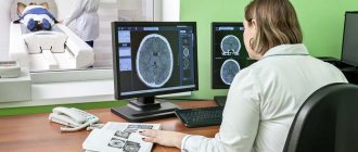
How to do REG
The technique is relatively simple. For both the doctor and the patient.
- The person arrives at the appointed time.
- Before starting, specialists measure blood pressure. It is necessary. It is important that it is in the usual range for a particular patient.
- Paired electrodes are placed on the head. One stimulates the brain in a certain area, the other, the opposite, reads the results. Such a system can also be closed, in the form of a special cap.
- The next thing that specialists do is actually stimulate the brain tissue.
- Based on the diagnostic results, the device automatically prints out a rheoegcephalogram. It looks like a sheet with a line showing the work of different parts of the brain.
Recognize on your own what won't work. Therefore, the patient should consult a specialist. Sometimes functional diagnostic doctors themselves describe the results and issue a conclusion; this is simpler and clearer.
At the end of the procedure he returns home. There are no restrictions after the examination.
Studying the brain in this way can be done as often as desired. Because there is no harmful radiation exposure or other problems.
Rheoencephalography is a rather narrowly focused technique.
Indications for use
- when complaining of neurological symptoms (pain and dizziness, tinnitus, cases of loss of consciousness);
- when sleep is disturbed and memory deteriorates, as well as cognitive abilities;
- if vision and hearing loss progresses, and the causes cannot be determined;
- with increased weather sensitivity;
- to assess the consequences of head injuries and stroke;
- in case of health problems, the cause of which may be a violation of vascular tone;
- for diseases of the cervical spine;
- to identify possible damage to veins, arteries and capillaries in patients with diabetes.
Among other things, REG is prescribed for preventive purposes to people who have a congenital predisposition to brain disorders.
In order for the results to be as informative and reliable as possible, it is necessary:
Preparation for REG for children and adults
Both in the case of adult patients and children, the REG procedure does not imply special or complex preparation.
It is enough just to follow a few simple rules that will allow you to achieve the most effective diagnostic result. In the case of adult patients, the most important thing is to maintain emotional stability and calm during the procedure. If the tone changes, it will immediately affect the blood circulation in the brain, and the readings on the rheograph will be distorted.
If serious pathologies are suspected, REG is prescribed as a highly effective and accurate diagnosis.
The exception is suspicion of venous or arterial defects, for which it is recommended to perform MR angiography, most often performed under general anesthesia. If a pediatric neurologist or neurosurgeon is referred for research, it is quite difficult to ensure that the child is even temporarily in a calm and balanced state. And if the procedure drags on, the child will definitely begin to fidget, move or open his eyes. In the first years of a baby’s life, the REG procedure is done when the child is in a sleepy state, 2 hours after feeding. And for older children, parents need to ensure compliance with several basic rules:
- Isolate the baby from the computer and TV for 24 hours;
- On the day of REG, walk with your child in the fresh air as long as possible so that he can throw out as much energy as possible;
- Mentally prepare the baby for the procedure by presenting it as a game;
- Give your child a favorite toy and offer to take a short nap with it;
- Convince the child to comply with all the doctor’s requests;
- Do not scold your child under any circumstances.
If the child is hyperactive and simply cannot cooperate with the doctor, even under parental supervision, then this type of diagnosis may have to be abandoned, because the results will still be incorrect.
How is REG performed for children and adults?
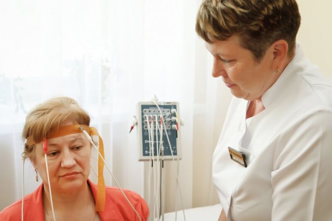
The duration of the REG examination for adults or children is the same and is approximately 10 minutes. To achieve optimal and correct results, a special medical device is used - a rheograph with 4 channels. The number of channels is proportional to the volume of area that needs to be examined. Consistent implementation of the procedure implies:
- The patient sits comfortably in a chair or on a soft medical couch;
- Metal electrodes are lubricated with a special medical gel, which prevents irritation of the scalp;
- The electrodes are applied to the places that need to be examined and fixed with a rubber band;
- The patient's eyes are closed so as not to react to various stimuli, such as sources of natural or artificial light;
- The record from the rheograph is recorded on a computer or paper.
If the procedure is prescribed for a child, then parents may even need to try to gently put the baby to sleep for a while, in order to avoid his mobility or fear at the sight of electrodes and other medical equipment. If the child is older, then during the diagnosis, the important role of the parents will be to support and reassure him, explaining that he will not experience any pain or discomfort.
Functional tests for REG
The severity of the reaction of arteries and veins to various irritations during REG will help determine the performance of functional tests. The patient may be offered certain medications during the procedure, he may be asked to make various movements, get up from a lying position, breathe deeply or, conversely, hold his breath.
For example, tilting the head when examining the vertebral arteries in a healthy person will not affect blood flow, but in case of pathology, the device will record changes.
REG of the head allows you to obtain a lot of data necessary for the doctor in one procedure. It is the result of functional tests that makes it possible to understand whether the patient simply has a disorder of vascular function, or whether we are talking about tissue changes.
What problems does rheoencephalography reveal?
EEG REG is prescribed after a strong blow to the head or problem blood circulation, the symptoms of which can also be determined by a qualified specialist. REG diagnostics helps to see:
- The actual condition of the blood vessels in general;
- Quality of blood supply to the brain;
- The work of each individual vessel;
- The speed of blood outflow and inflow.
Before choosing a medical institution where to have an REG done in Khabarovsk, it is important for parents to understand that the problems that the procedure reveals in children may differ from pathologies in adults. Among the childhood problems that diagnostics reveals are:
- The state in which the walls of blood vessels are now located;
- Activity of blood inflows and outflows;
- Viscosity and thickness of blood;
- Blood circulation speed.
Where can you take an REG in Khabarovsk with confidence in quality?
Most patients prefer to have rheoencephalography of cerebral vessels done in Khabarovsk at a medical clinic. The main distinctive features of this clinic that attract patients are:
- Minimum prices for EEG diagnostics in the region;
- Qualified and tactful doctors and medical personnel who can not only explain in detail what an REG examination is, but also prepare patients for the procedure;
- Spacious offices equipped with modern equipment;
- Reception of leading specialists, including a doctor of the highest category Suslova N.F.
In medical care, you can be sure that the patient will receive the highest quality service at an affordable price.