Ultrasound of the great vessels: essence and goals
MAG ultrasound is a combined type study that combines classic ultrasound with the use of the Doppler effect. This will allow you to check the condition of the blood vessels, as well as the movement of blood through them in the brain.
An ultrasound type angiogram is performed using sound waves that penetrate deep into the human body. In this case, the patient does not feel anything. Waves that the human ear cannot hear are emitted and then picked up by a specially designed sensor. When information is reflected from the walls, the computer converts it into a two-dimensional image in black and white. Doppler will image red blood cells (color image) as they move through the vessel. If you superimpose both results on each other, you will get an ultrasound image.
The objectives of conducting an examination of main arteries:
- detection of a congenital anomaly in the circle of arteries that are responsible for supplying blood to the brain
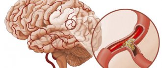
- determination of stenotic processes and angiospastic type conditions, as well as occlusion (all these processes affect the access of oxygen to tissues)
- identifying the effect of chronic diseases on blood circulation in the brain
- study of pathologies of blood vessels in the circle of Willis
- determination of vascular tone and their reactivity
- aneurysm detection
- regulation and quality control of therapeutic measures aimed at improving blood circulation in the brain
After all the data is received, the doctor will be able to draw conclusions about the need for treatment from another specialist, prescribing additional examinations or treatment procedures. Because the data is available immediately, it is easy to determine the impact of the disease and its treatment on blood flow.
Indications for the study
This examination of blood vessels is considered not dangerous and does not cause pain or discomfort. However, in order to appoint it, you need grounds, which are as follows:
- headaches of any type, including those caused by encephalopathy, migraine
- neck pain
- vestibular disorders
- problems with vision (blackouts) and hearing (extraneous noise)
- factors that cause vascular disorders (for example, this list includes diabetes, obesity, smoking, dystonia, etc.)
- surgical intervention in the myocardium;
- operations in the neck area
- traumatic brain injuries (serious bruises and severe concussions;
- obvious symptoms of stroke
- complications after compression and obliteration of blood vessels due to osteochondrosis of the cervical spine or oncological processes in the cervical area
- congenital anomalies in the spine (more precisely, in the cervical region)
Methodology
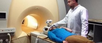
Ultrasound of the great vessels makes it possible to determine the state of blood flow in vessels of any size. This research technique is considered non-invasive, so there is no need to violate the integrity of the structures (as in laparoscopy). Thanks to this, ultrasound examination is the most convenient procedure for both the doctor and the patient. In addition, there is no need to carry out any complex manipulations. The procedure has no contraindications.
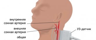
No lengthy or complex preparation is required. If a person is taking medications, then you can continue to do so as usual, but you need to warn your doctor. The patient should sit on the couch, lying on his back, and remove clothing from his neck. People usually wonder why the neck needs to be exposed when examining the vessels of the head. This is explained by the fact that arteries and veins pass through the neck, which is why they are called main.
The doctor will drive a device coated with a special gel that improves sound conduction. In addition, it is easier to move the sensor over the skin. Thanks to the location of the scanner in different planes, it is possible to evaluate the spinal arteries, carotid arteries, as well as spinal and jugular veins.
Typically the procedure takes 30-60 minutes. This study can be done at any age, even infants. This is because radiation does not affect the patient's body. The study is currently being conducted (without delays), so the results will be known immediately.
Contraindications and possible risks
There are no absolute contraindications prohibiting Doppler examination of the vessels of the head and neck. Restrictions include the serious condition of the patient and other factors that do not allow the patient to take a lying position (exacerbation of bronchial asthma, chronic heart failure).
Dopplerography is completely safe for the body and does not carry radiation exposure. This technique has no age restrictions. It is performed on infants, the elderly, pregnant and lactating women. During pregnancy, a gynecologist may refer you for an ultrasound scan if you suspect a circulatory disorder in the placenta.
Multiple ultrasound examinations are allowed. Due to the high accuracy and harmlessness of the technique, it is used to monitor ongoing treatment. It is also prescribed for the prevention of serious diseases. Some difficulties may arise during the procedure:
- The condition of small vessels is much more difficult to assess than large ones.
- It happens that the bones of the skull interfere with a full study of the condition of all cerebral vessels.
- Diagnostic results often depend on the quality of the instruments and the qualifications of the doctor.
- Doppler ultrasound often replaces angiography.
Patients who do not know what vascular Dopplerography is can ask the neurologist who prescribed this procedure about it in advance. Since there are no special preparations or contraindications, every patient can undergo examination in all medical institutions with functional diagnostic departments. The procedure is usually booked in advance. The patient must have a referral from a neurologist and an outpatient card.
Basic diagnostic methods
Vascular ultrasound is not the only option for studying the state of blood flow in the cervical and cephalic vessels. It is a comprehensive ultrasound-type method, which is focused on the most complete diagnosis of cerebral vessels. Ultrasound in the neck and head area is:
- USDG
- Triplex scanning
- Duplex study
You need to choose a technique depending on the purpose of the study, pathology and some other nuances. A duplex type of study will help check not only the blood flow, but also the walls of the blood vessels. The echolocation principle is used. As a result, a two-dimensional image will be obtained. The gap in them can be studied from different angles.
The triplex type of research is auxiliary to the duplex type. A color image is obtained showing blood circulation. Using this graph you can check the direction, speed and other indicators.
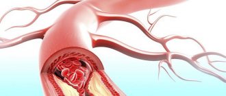
Both methods are considered visualization, because their combination helps to accurately identify the course of blood vessels, their structure, diameter, the presence of wall problems, various anomalies and atherosclerotic plaques.
Ultrasound scanning of the BCS is a fairly informative and accurate method that allows you to identify abnormalities in the neck and head. Ultrasound scanning of the BCA is the same as Ultrasound scanning of the BCA. In general, the BCS and BCA are arteries of the brachiocephalic type (trunk). The method is used to study the large arteries that branch from the aorta and carry blood to the brain. There are three arteries of the brachiocephalic type: the carotid, under the clavicle and in the spine.
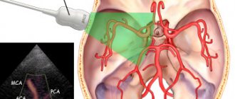
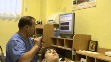
Transcranial Doppler ultrasound
Runa Aaslid introduced TCUS in 1982 to study blood flow in the basal intracerebral arteries. The method was originally used to detect vasospasm after subarachnoid hemorrhage. However, its use has expanded significantly over the past three decades.
TCUS has become a non-invasive and cost-effective tool for assessing the patency of cerebral arteries, detecting stenosis and thromboembolism. Currently, transcranial venous and arterial imaging is the only available diagnostic tool that provides real-time information about cerebral hemodynamics.
Transcranial ultrasound of vessels
The most important applications of transcranial Doppler ultrasound are:
- assessment of the condition of cerebral vessels;
- monitoring blood flow speed;
- diagnostic examination confirming brain death.
Important! Three-dimensional Dopplerography is a special duplex scanning mode. This type of study is not an independent diagnostic method. During it, the device operates simultaneously in 3 modes: B-mode, Doppler ultrasound and color Doppler mapping mode.
What the study shows
MAG ultrasound helps to study the sounds that the vessels reflect. During the study, the device records the frequencies of the ultrasonic signal, which changes when reflected from moving bodies. The device is based on the Doppler effect. This is a location-type device that helps examine signals that are sent to a person to probe an area. Doppler parameters may vary depending on different types of anomaly. Their presence in the area being surveyed may indicate the following:
- With occlusion, the level of blood circulation decreases to zero;
- Pathologies regarding the structure will have different versions of the Dopplerogram in the area that has disturbances;
- If the space of the vessel narrows, the blood flow will accelerate.
This procedure helps determine how tortuous the vessel is, what lumen it has, and whether there are any narrowings. You can also determine the integrity of the vessel, the condition of its walls, and the level of functionality of the valve system in the veins. If there are compactions or protrusions, they will also be noticeable. All defects will be immediately visible. It will be possible to make an assessment of blood flow parameters: turbulence, speed and its presence in general.
Data decryption
The results of the study are based on a comparative assessment of the artery with certain standards in several parameters:
- Wall thickness.
- Diameter.
- The nature of the symmetry of blood flow.
- Resistance indices.
- Peak systolic velocity.
Veins are assessed by the diameter of the vessel, the nature of blood exchange, and the condition of the venous walls.
If the results correspond to the norm:
- The gap between the vessels will be free and clearly visible.
- The thickness of healthy artery walls should be within 1 centimeter.
- In those areas where there is no vascular branching, there should be no turbulent blood flow.
- There should also be no arteriovenous malformations.
If the vessels are compressed among themselves, additional research is required.
When an abnormality is noticed in any area, the diagnostician indicates this with a alphanumeric abbreviation. The neurologist, knowing all the symbols, easily interprets the information received and prescribes treatment.
Data decryption
Ultrasound angiography is considered a very accurate, but at the same time a “blind” method, because Complete visualization of blood vessels will not be possible. The current in main-type vessels can be divided into parabolic and turbulent. In the first case, the streams will not mix, because they move at different levels of speed. Turbulent is formed due to the fact that some red blood cells move chaotically. As a result, the Dopplerogram will display two important points: the totality of red blood cells and those areas where they have different speeds. These flows can be assessed from a qualitative and quantitative perspective (speed, viscosity, mobility, reactivity).
Sometimes the data that the doctor received while the patient was calm is not enough. Then you need to hold your breath, do an inhalation test, compress the carotid artery and take nitroglycerin.
Such a study will help identify various abnormalities in the brain. The procedure is considered quite accurate. She uses both the ultrasound method and the Doppler effect. In addition, such an angiogram is completely painless and does not require serious preliminary preparation.