Ultrasound of cerebral vessels is a common diagnostic method that allows one to determine pathologies using sound frequencies. The study is based on the fact that sound radiation is absolutely harmless to the human body. Ultrasound penetrates the tissues and bones of the skull. It is reflected at the interface between solid and liquid media, and its signals are recorded. It is worth noting that the very substance of the brain and its structures are not visualized during such a study. Neurosonography is performed only in emergency cases.
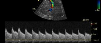
Ultrasound examination of the vascular network is based on the Doppler effect. A similar effect is achieved due to the fact that waves reflected from an object in motion return with a changed frequency. During this procedure, a non-two-dimensional image of the vessel is displayed on the monitor screen; using a certain graph, you can track the flow of blood in it.
Features of ultrasound examination of blood vessels
The name of the procedure itself suggests that this examination is carried out using ultrasound using special equipment. The bottom line is that a beam of ultrasonic waves is sent from a special tube into the body tissue, and then the same tube catches the reflected waves. By the nature of the reflection, you can determine the condition of the tissues and blood vessels of the body.
One of the popular additional functions of ultrasound is Dopplerography. It makes it possible to find out what the state of blood flow is in the vessels. The specialist activates this function on the device, after which the vessels on the screen turn blue or red. In the first case, this means that the blood flow moves in the direction from the sensor, and in the second - towards it. No shading means there is no blood flow.
Almost always, the doctor will prescribe an ultrasound not only of the vessels of the head, but also of the neck. Why is that? In an adult, the skull has fairly dense bones, so it is practically impossible to see all the vessels. Therefore, the vessels in the neck leading to the brain are examined, as well as through the neck and, if possible, through the thinnest bones, the head is examined.
What is ultrasound scanning of the vessels of the neck and head?
Doppler ultrasound is an instrumental research method using ultrasound.
Ultrasound waves are able to penetrate body tissues and be reflected from structures of varying densities, which is recorded by a special sensor. The signals from the sensor are processed by a computer and the doctor sees an image of the organs and internal environments on the monitor.
Dopplerography is an additional ultrasound diagnostic function that allows you to evaluate the nature and speed of blood flow in the arteries and veins.
If blood moves towards the sensor, the computer colors it red in the image. If in the opposite direction, then blue.
Who is the diagnosis indicated for?
Which patients are best to undergo an ultrasound of the cerebral vessels without delay? There are a number of indications for this. In some of them, Doppler sonography is simply necessary, while sometimes there is no urgent need. So, let's look at the reasons why the attending physician may prescribe an ultrasound examination of the cerebral vessels.
- severe, sharp headaches;
- frequent dizziness, especially if you suddenly change the position of your head;
- blurred vision, spots before the eyes;
- regular loss of consciousness;
- memory impairment, inability to concentrate, fatigue during mental work;
- causeless tinnitus;
- weakness in the arms and legs, as well as their unexpected numbness;
- diabetes mellitus and high blood cholesterol;
- atherosclerosis, stroke or heart attack;
- cervical osteochondrosis, when the neck often hurts, and a crunching sound is heard when turning;
- increased blood pressure, as well as pressure differences in each arm;
- obesity;
- long-term tobacco addiction;
- neck and head injury;
- vegetative-vascular dystonia;
- coronary disease and heart disease;
- systemic vasculitis;
- pulsation in the soft tissues of the neck;
- conventional sleeping pills are ineffective for insomnia;
- heavy head on waking in the morning.
The above list is not exhaustive; the doctor may send you for any other suspicion of circulatory disorders. In addition, nowadays it is not even necessary to go to the doctor for a referral, since many private ultrasound rooms do not require one at all. You can do an ultrasound of the cerebral vessels yourself if you find some of the listed symptoms, or you are over 55 years old, and your closest relatives have had similar problems. Just remember that only a specialist can decipher the results as accurately as possible and prescribe the correct treatment.
When is transcranial Doppler ultrasound of the brain necessary?
Duplex scanning, as vascular ultrasound is also called, is prescribed by a doctor if a cerebrovascular accident is suspected. The following symptoms indicate this pathology:
- bursting pain in the head;
- dizziness, especially when changing body position and throwing back the head;
- periodic darkening and flickering of spots before the eyes;
- fainting;
- progressive impairment of memory, attention, thinking;
- noise in ears;
- paroxysmal numbness, weakness in the limbs.
Some systemic diseases occur with vascular damage and poor circulation. Therefore, to assess their progression and the effectiveness of treatment, the doctor prescribes an ultrasound examination of the blood vessels. It is shown when:
- atherosclerosis;
- diabetes mellitus;
- after a cerebral stroke;
- increased levels of cholesterol in the blood (hypercholesterolemia);
- cervical osteochondrosis;
- systemic vasculitis;
- heart defects;
- neurocirculatory dystonia;
- obesity;
- long history of tobacco smoking;
- arterial hypertension;
- head and neck injuries;
- coronary heart disease.
All people over 55 years of age should undergo vascular ultrasound once a year if their immediate family members have had a heart attack, stroke, coronary heart disease, atherosclerosis or hypertension. This indicates a hereditary predisposition and a high risk of developing such conditions.
Contraindications
The study does not violate the integrity of tissues, is painless and does not have a negative effect on the body. Therefore, there are no absolute contraindications to its implementation.
Difficulties arise only if a person for some reason cannot take the position necessary for research.
Examination of children
In almost any case, the pediatrician will send your newborn baby for an ultrasound of the brain. Most often, the prescription is prescribed at the age of one to two months. At this age, the procedure that will be performed is called neurosonography. It is possible as long as the baby’s fontanel on the skull is not overgrown, and is usually performed on children under one year of age.
If the child is older and the fontanel is already overgrown, the procedure is carried out in the same way as for adults. It is prescribed when a child complains of frequent headaches, fatigue, decreased attention, or developmental delays.
In what case will a child be prescribed an ultrasound examination of the cerebral vessels:
- premature and pathological birth;
- the shape of the head and facial skeleton differs from standard indicators;
- the child is not gaining weight well;
- oxygen starvation during pregnancy and childbirth;
- various neurological diseases;
- diseases of any internal organs.
How to make a diagnosis
The stages of Doppler ultrasound look like this:
- The patient removes jewelry from the neck and head area.
- Lies down on the couch.
- The doctor places a special cushion under the head.
- Gel is applied to the Doppler.
- The sleep specialist conducts a study of the vessel, the result of which is visible on the screen.
There are no differences in the procedure for children and adults. The examination up to one year is called neurosonography and is done through the parietal fontanel.
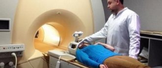
To conduct the study, the person is placed on his back on a soft couch. A cushion is placed under the neck, the head lies without a pillow. The doctor applies a special gel to the sensor and skin - this is necessary for the passage of ultrasonic waves into the internal environment of the body.
The vessels of the neck are examined by pressing the sensor to its lateral surface. At this moment you cannot move your head or talk. During the procedure, the doctor will press the sensor several times to assess the elasticity of the blood vessels.
The vessels of the head are examined through the thinnest areas of the cranial bones: the orbit, temporal bone, occipital bone and foramen magnum. The sensor is placed on the closed eye, above the auricle and behind it. After this, the patient is seated and the back of the head and the place where the neck connects to the head are examined.
In this way, the doctor examines all the vessels that bring blood to the brain and carry it back to the heart.
The procedure takes about half an hour and does not cause any discomfort. During this procedure, the diagnostician may ask you to hold your breath, breathe frequently, and turn your head. This is necessary for the best image accuracy and assessment of the functional state of the vessels.
On the day of the test, you should not take medications that affect blood pressure. It is advisable to refrain from drinking strong coffee, nicotine and alcohol - all these substances change the state of the vascular bed and can distort the results of the study.
What can be revealed
So, what does a doctor expect to see when ordering an ultrasound examination of the cerebral vessels? First of all, it will be assessed where the vessels are narrowed or dilated, as well as in what direction and at what speed the blood flow moves. In addition, an experienced specialist will be able to notice the presence of thrombotic masses and vascular obstruction. You can also find thickening of the walls of blood vessels and a deterioration in their elasticity.
You can also identify the following indicators:
- the degree of patency of each vein or artery;
- compliance with the speed and trajectory of blood flow relative to standard indicators;
- to what extent the lumen of the vessel is closed by various formations;
- condition of the tissues around the vessel.
In a newborn baby, the following indicators are also examined:
- size of the hemispheres and ventricles of the brain;
- subarachnoid space;
- the presence of growths and formations.
In case of any deviations from the norm or detection of brain pathologies, you must consult a doctor. Depending on the intended diagnosis, this may be a cardiologist, neurologist or neurosurgeon.
What results should be normal?
To determine whether the cerebral vessels are indeed susceptible to any pathological process, the results obtained should be compared with the norm. During the study, the specialist evaluates several important parameters. And only if these parameters are normal, then we can say that there are no pathologies. The main parameters are shown in the table.
| Vascular tortuosity | There should be no kinks |
| Vessel diameter | The narrowing and dilation of blood vessels indicates a violation of their functionality |
| Arterial lumen | In normal condition, the lumen of the artery does not contain any plaques |
| Blood flow speed | The assessment of blood flow velocity is carried out in two stages to eliminate any inaccuracies. Since this parameter is one of the most important |
Carrying out an ultrasound procedure
An ultrasound examination of the brain is not a complicated procedure and is no different from ultrasound examinations of other parts of the body. You need to contact a special diagnostic center, and in some cases, a district clinic.
For adults, ultrasound of the head and neck is performed in the following form:
- the patient lies down on the couch;
- first, the soft tissues of the neck are examined;
- then, through the thinnest bones of the skull, for example, the eye sockets, temples, or under the lower jaw, an examination is carried out from the side of the face;
- then the patient takes a sitting position, and the examination is carried out from the back of the head.
For a newborn baby, an ultrasound of the head is performed through an unovergrown fontanel. This allows you to more objectively and accurately assess the condition of the baby’s body.
Ultrasound is performed using a special sensor in the form of a tube onto which a special gel is applied. The gel can also be applied to the skin. The specialist presses the tube to the body and turns it to get the highest quality image on the screen. During the procedure, try to move less and not talk.
How is an ultrasound performed and what does it show?
The main advantages of ultrasound examination of the head and neck vessels are that there are no contraindications to this type of examination. No special preparation is required. The procedure itself does not cause any discomfort. It is prescribed even to children with characteristic symptoms such as headache, nausea, and tearfulness. Unlike x-rays and x-rays, ultrasound does not ionize the body.
To perform an ultrasound of the vessels of the head and neck, the patient is placed on a couch. The doctor applies a special gel to the area being examined to improve contact of the sensor. Before the examination, the sensor is applied to the eyelids or in the area of the temporal bones of the skull.
The whole procedure takes no more than 30 minutes. The duration depends on the condition of the veins and arteries of the brain. Decoding is carried out simultaneously with the examination. The result of the ultrasound will be photos and graphs, from which the specialist will be able to assess the condition of all the great vessels of the brain. Based on the conclusion, the question of treatment methods is decided: therapy, surgery, bypass surgery.
- Cocktails with rum - recipes with photos. How to make rum-based cocktails at home
- How to restore intestinal microflora after taking antibiotics
- Feeling anxious for no reason
Indications for ultrasound of the blood vessels of the head and neck are as follows:
- headache;
- severe dizziness;
- frequent fainting;
- pathologies of blood vessels or brain - hypertension, stroke, heart attack, diabetes mellitus, smoking;
- high cholesterol, atherosclerosis;
- planned surgical interventions;
- tumor-like formations of the neck.
Based on the results of the ultrasound, the doctor will be able to assess the condition of the artery walls, the presence or absence of plaques and blood clots. In addition, the method will help to identify life-threatening brain diseases in time: malformations, aneurysms, anastomosis.
Decoding the results
In most cases, the specialist who conducts the ultrasound examination will provide general information and a basic interpretation of the results. He can also advise which specialist you should contact. In some medical centers you can get a doctor's consultation right on the spot.
Under no circumstances should you try to decipher the results of the study or prescribe treatment yourself. Be sure to consult a doctor, even if you were examined on your own initiative.
Contraindications
There are no contraindications as such. True, there are some limitations associated with the technical features of the procedure. For example, angiography cannot be replaced by ultrasound to fully conduct a study of the condition of blood vessels. It is also very difficult to examine small vessels. Some vessels are almost impossible to see at all due to the thick bones of the skull. In addition, the condition of the vessels due to atherosclerosis may interfere with the passage of the ultrasound beam.
But, be that as it may, everything depends on the quality of the equipment and the professionalism of the doctor. A true professional will be able to see everything he needs, even on not the most modern equipment.


