CT scan of the brain vessels is part of the general examination of the head. The diagnostic method is based on the absorption of X-ray radiation by tissues.
To examine the vessels, a contrast agent is used, which helps to clearly see all blood connections. As a result, many pathologies are detected at early stages.
How and when tomography of the nasal sinuses is done, read this article -
Cerebral angiography
To study the condition of the vessels of the brain and skull, a modern method is used - cerebral angiography. This is an X-ray instrumental examination method, the essence of which is the introduction of a contrast agent into the vascular bed of the brain, followed by radiography.
Angiography is a method of studying blood vessels, which is carried out to determine their condition and functioning. It is performed using radiography, during which a special substance is used. When there are suspicions of cerebral vascular pathologies or their diagnosis is necessary, angiography of the cerebral vessels (cerebral angiography) is performed.
Indications for use
Cerebral angiography is performed only for special indications, which include:
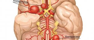
- circulatory disorders in the vessels of the brain of the hemorrhagic type (predisposition to the occurrence of intracranial hemorrhage) - aneurysms (pathological dilations) of the arteries of the brain, diverticula (protrusions) of the walls of intracranial vessels, malformations and angiomas (tumor formations);
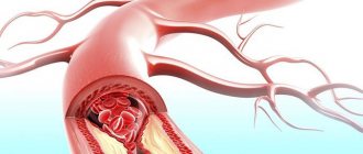
- pathological blood circulation in the vessels of the head is of the ischemic type - in this case there are obstacles to normal blood flow - blood clots, atherosclerotic plaques (cerebral atherosclerosis), deformations (changes in shape) of the arteries of the brain;
- brain tumors - benign or malignant tumors that lead to an increase in the vascular pattern at the site of their localization;
- lack of information content when using other instrumental methods of the brain, in the presence of symptoms of disease or circulatory disorders.
Types of cerebral angiography
According to the established classification, this diagnostic technique is divided into 2 types:
- Selective - targeted, local. When performing selective cerebral angiography, an iodine-containing contrast agent is injected into an arterial vessel that supplies one of the brain regions.
- Survey is an extended research method. A contrast agent is injected into the area of the main artery, which is responsible for the general cerebral blood supply and nutrition. Using this technique, a specialist can carefully examine all cerebral vessels.
The optimal type and method of implementation is determined by a specialist individually, taking into account the characteristics and severity of the clinical case.
Contraindications to the procedure
As with any other procedure, there are contraindications for cerebral angiography. They are associated both with the procedure itself and with the contrast agent that is injected into the bloodstream. Iodine compounds are used as the administered substance. The amount of the substance depends on the volume of the examination; it can be 5-10 ml.
Examination during pregnancy and lactation
This diagnostic technique is strictly prohibited for pregnant women and women during breastfeeding. This limitation is due to the lack of a full range of information regarding the effects of x-rays on the fetus and the health of the newborn.
How is the examination carried out?
Important Terms
- Aseptic conditions for the procedure,
- Availability of a team of doctors: radiologist, anesthesiologist, cardiac resuscitator.
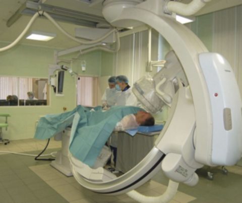
The patient examination process itself takes approximately half an hour to an hour. The procedure is considered invasive because a puncture is made to access the artery, where a special catheter is inserted. Therefore, cerebral angiography is often combined with other interventions in the body that occur through access through large blood vessels, for example, with the removal of an aneurysm.
To avoid complications associated with infection through the catheterization site, the skin is treated with an antiseptic solution. Next, local anesthesia is performed. Puncture (puncture) of the vessel is carried out with a special needle. A flexible catheter is inserted through this site to supply contrast. As a rule, the puncture is done in places through which it is easy to “get” to the necessary vessels.
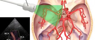
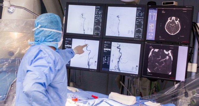
A contrast agent is injected into the blood through a special catheter. After contrast, a series of X-ray images of the cerebral vessels are taken. These images show the different phases of blood circulation: capillary, arterial and venous.
Modern medical equipment makes it possible to take layer-by-layer images to subsequently form a three-dimensional image using special computer programs.
When the filming is completed, the patient's catheter is removed and the bleeding stops. Next, the received information is decrypted. A vascular surgeon and a radiologist are involved in deciphering and establishing or clarifying the diagnosis.
Methodology for cerebral angiography
The entire procedure of this study can be divided into several stages:
- preparing the site for puncture - treating the skin with an antiseptic solution to destroy microorganisms and local anesthesia;
- direct puncture of the vessel with a special needle and insertion of a flexible catheter;
- introduction of a contrast agent into the vascular bed of the brain through a catheter;
- performing a series of X-ray images of blood vessels; during CT cerebral angiography, a series of layer-by-layer images is taken;
- interpretation and interpretation of the received images with a conclusion - is carried out by a radiologist together with a vascular surgeon.
Considering the paramount importance of proper cerebral circulation, adequate and correct diagnosis of vascular problems is the basis for their further successful treatment.
Classic angiography
When carrying out this technique, a puncture is made, after which a contrast compound is injected into the area of the patient’s carotid artery. The procedure is carried out under the influence of local anesthetics. At subsequent stages, X-rays are taken in two projections with a break of several seconds to assess the different phases of cerebral blood flow.
CT angiography
An innovative and reliable method of diagnostic research. This method involves the introduction of a contrast agent into the area of the cubital vein. Then the doctor takes a series of X-ray images of the brain in various projections with further computer visualization of the vascular bed and reconstruction of the 3D image.
The main advantages of this technique (compared to the method described above) include lower radiation exposure to the patient’s body.
MR angiography
The technique involves conducting research using magnetic fields. For these purposes, it is not always necessary to use contrast agents, due to which the technique has fewer contraindications. During diagnostics, specialists evaluate the phases of blood circulation and the condition of blood vessels, observing changes in their tissues.
This technique allows you to most accurately examine the vascular pattern and determine even the most minor deviations in the arterial structure. This method is considered less dangerous, but is not used in people with obesity or claustrophobia.
Advantages of the procedure
- Possibility to create a three-dimensional image,
- Visualization of blood vessels, which allows the doctor to detect blood clots, hematomas, aneurysms,
- The possibility of an individual approach to the patient, and therefore an accurate diagnosis.
The modes and programs of this type of examination can be very wide, so you can always choose the best option in each specific case.
The absence of inaccuracies in diagnosis makes it possible to timely and accurately identify problem areas in the blood vessels and understand the cause of diseases or disorders in the functioning of the blood vessels of the brain. Adequate diagnosis is the main condition for proper treatment.
Approximate cost of angiography of cerebral vessels
The price depends on the method, the respectability of the clinic, the qualifications of specialists and the quality of the equipment used. The cost of a classic angiographic study is 4000-5000 rubles. KT will cost patients 6,500-18,000 rubles; MP - varies from 5,000 to 30,000 rubles.
Result evaluation
A specialist will be able to decipher the results obtained during angiography on the basis of X-ray images, which allow one to assess the arterial bend, the presence or absence of vascular narrowings and lumens.
In the presence of a tumor neoplasm, straightening and displacement of blood vessels localized near the pathological focus are noted.
Atherosclerotic disorders are diagnosed when there is uneven narrowing of the vascular lumen.
Possible complications
Possible complications of angiography:
- bleeding;
- embolism;
- convulsive syndrome;
- vascular spasms;
- disruption of cerebral circulatory processes;
- damage to the walls of cerebral vessels;
- manifestation of neurological symptoms;
- inflammatory processes caused by the penetration of a contrast compound into tissues adjacent to blood vessels;
- renal failure occurring in acute form;
- manifestation of allergic reactions.
If the patient has no absolute contraindications and proper preparation for diagnosis, the risks of the complications listed above are reduced to minimal levels.
Sources: https://vashnevrolog.ru/metody-diagnostiki/kogda-zachem-i-kak-provoditsya-cerebralnaya-angiografiya.html https://idiagnost.ru/issledovaniya/rentgen/chto-takoe-tserebralnaya-angiografiya https: //prososud.ru/profilaktika/angiografiya-golovnogo-mozgaa.html
Source: https://uzibook.ru/angiografiya/tserebralnaya-angiografiya.html
Indications
When abnormal processes occur in the human body, symptoms always appear that indicate that the person’s health is deteriorating. There may be several reasons for such changes. In such cases, MR angiography of cerebral vessels is prescribed.
Indications for the procedure are the following symptoms:
- Headache and dizziness that constantly do not go away. There is no way to get rid of them using pills or other methods.
- Noise in ears.
- Constant nausea and vomiting attacks in the absence of obvious reasons.
- Head injuries.
- Diabetes.
- Suspicion of a tumor.
- Diseases of the heart and circulatory system.
- Complete or partial loss of hearing or vision.
MR angiography can visualize aneurysms, which is not always possible with tomography. The same applies to the expansion or narrowing of the lumen of blood vessels, anastomosis, dissection, malformation and other pathologies.
Angiography is used in the following branches of medicine:
- Oncology. The technique helps to visualize various tumors, formations and metastases.
- Phlebology. It is possible to determine the places where the vessel is blocked or narrowed, various pathologies, blood clots, congenital or acquired changes, and atherosclerosis develop.
- Vascular surgery. Angiography is used during surgical intervention in blood vessels in order to accurately determine the location of dislocation and various disorders.
- Neurology. Makes it possible to visualize hematomas, aneurysms, tumors in the brain, and is used for bleeding and hemorrhagic stroke.
- Pulmonology. Required for diagnosing various pathologies in the lungs, as well as to find the source of bleeding.
Angiography can be either general or selective. For general examination, all vessels are examined. With selective - only the circulatory system of individual parts of the body. In this case, individual vessels are contrasted.
As a rule, MRI angiography is prescribed in cases where there is suspicion of:
- damage to the kidney arteries;
- aneurysms;
- aortic damage;
- defects of veins and arteries;
- the presence of blood clots in the duct of an artery of the lungs (called thromboembolism);
- deep vein thrombosis (the procedure will also help determine the nature of the changes).
Angiography of the vessels of the neck and brain is performed in the presence of the following indications:
- aneurysms;
- tumors in the brain;
- angiomas;
- heart attack;
- hemorrhagic stroke;
- vascular malformation;
- hematomas;
- injuries.
Indications for the procedure on the vessels of the lower extremities are:
- atherosclerosis;
- obliterating type endarteritis;
- thrombosis;
- thrombophlebitis;
- thromboembolism;
- various damage to blood vessels due to injury;
- aneurysms of dissecting type;
- control of surgical intervention.
Angiography of the coronary vessels is prescribed for:
- heart attack;
- ischemic heart disease;
- atherosclerosis on such vessels;
- pathology of blood vessels, which is a congenital change.
Not all people are allowed to undergo angiography of the head region, because this type of examination can lead to side effects and complications. Before prescribing the procedure, the doctor must make sure that the person has no contraindications. They can be various diseases, as well as special conditions of the body.
It is not allowed to do CT angiography of cerebral vessels in the presence of mental disorders, extensive tumors and cysts located on the vessels. The procedure is not performed for people who have an allergic reaction to contrast agents. If there are metal implants, a different type of examination is selected, because due to foreign objects in the body, angiography is not performed.
Also a contraindication is the presence of liver, heart and kidney failure. Women during pregnancy should avoid this procedure. During lactation, a contrast agent should also not be used so as not to harm the baby.
When a person is obese, he is not allowed to undergo the procedure because he will not fit into the equipment. If blood clotting is poor, the patient should be given a test using a contrast agent with caution.
The main indications are:
- aneurysm;
- atherosclerosis;
- thrombosis;
- tumor processes;
- suspicion of malformation;
- determining the presence of narrowing or expansion of the lumen of blood vessels;
- controlling the location of surgical clips.
Angiography is prescribed only in cases where other diagnostic methods are unsuccessful.
Angiography cannot be performed if:
- intolerance to iodine-containing drugs;
- pregnancy;
- mental illness;
- thrombophlebitis;
- acute infectious and inflammatory process;
- decompensated failure of the kidneys, heart, liver.
Contrast or magnetic resonance angiography may be prescribed for people with complaints of:
- frequent headaches that occur for no particular reason;
- pressing, bursting pain in the neck;
- attacks of dizziness;
- noise in the head, flickering of spots before the eyes;
- progressive deterioration of memory and attention;
- personality changes;
- loss of consciousness, frequent fainting;
- nausea, vomiting not associated with gastrointestinal diseases.

Do you often have a headache?
Get examined In addition, this diagnostic method is recommended if cerebrovascular diseases are suspected based on the results of clinical, laboratory and instrumental examination. Angioscanning of the vessels of the head and neck is also indicated for monitoring the treatment of chronic diseases.
Despite its high efficiency and safety, there are conditions in which manipulation cannot be performed.
The following contraindications to diagnostic testing are identified:
- decompensated failure of internal organs (heart, kidney, liver);
- acute psychosis and other mental illnesses;
- vascular tumors, cysts;
- laboratory-confirmed blood clotting disorders;
- pregnancy and breastfeeding.
The price of the procedure starts from 1600 rubles.
Cerebral angiography of cerebral vessels: indications and reviews
Modern medicine is developing incredibly quickly. Now you won’t surprise anyone with ultrasound and X-ray examinations. But even these surveys are becoming more and more advanced from year to year. Angiography is one such method that allows you to see the size, shape, and contours of the vessel.
How can you see the blood vessels of the brain?
Cerebral angiography is an X-ray method for visualizing cerebral vessels, which consists of staining the vascular bed with previously injected contrast. This is a highly effective and modern diagnostic method that allows you to make an accurate diagnosis.
The method of visualizing blood vessels using a contrast agent has been known to medicine for about a century. Back in 1927, a neurologist from Portugal began using this method, and it came to Russia in 1954. Despite such a long use, cerebral vascular angiography has changed significantly over this time, becoming more sophisticated.
The essence of the method
In order for the radiologist to see the vessels of the brain, an injection of an iodine-based X-ray contrast agent (Triiodtrust, Ultravist) is performed into one of the cerebral arteries.
Injection is possible either into a vessel in the brain or through a catheter through an artery on the periphery, for example, the femoral one.
Without this procedure, cerebral angiography of the cerebral vessels will be ineffective, since the arteries will be poorly visible on the image.
Next, two x-rays are taken, in frontal and lateral projection. After which the radiologist writes his report.
Indications
A doctor's referral is required to examine the brain using cerebral angiography. This diagnostic method is not carried out only at the request of the patient.
The main indications are:
- suspicion of the presence of a cerebral aneurysm (sac-like bulging of the artery wall);
- determining the degree of narrowing of the lumen of the vessel by atherosclerotic plaques (narrowing of more than 75% significantly aggravates blood circulation in the brain and is an indication for surgical intervention);
- control of the location of clips pre-installed on the vessels;
- diagnosis of arteriovenous malformation (pathological connections between arteries and veins; usually congenital);
- suspicion of the presence of tumors, while the angiogram visualizes a change in the normal vascular pattern at the site of the tumor;
- visualization of the arteries of the brain during volumetric processes in it (tumors, cysts) in order to establish the location of the vessels relative to each other;
- suspicion of the presence of a brain angioma (a benign tumor formed by the vascular wall);
- lack of information when using other neuroimaging methods (CT, MRI), but in the presence of patient complaints and symptoms of the disease.
Contraindications
Carrying out both indirect and direct cerebral angiography has a number of contraindications:
- Allergy to iodine and iodine-containing substances. In this condition, the contrast can be replaced with gadolinium. If there is an allergy to other contrast components, it is necessary to completely abandon this examination method.
- Kidney and liver failure in the stage of decompensation. These conditions lead to impaired removal of contrast from the body.
- Severe chronic diseases.
- Acute inflammatory diseases, as the symptoms of infection may worsen.
- Age up to two years, as radiation impairs the growth and development of the child.
- The period of pregnancy and breastfeeding, since X-ray radiation has a detrimental effect on the fetus.
- Mental illnesses during exacerbation.
- Bleeding disorders (hemophilia, thrombocytopenic purpura), which increases the possibility of bleeding after contrast administration.
Preparing for the examination
Since the examination method is X-ray with the introduction of a contrast agent, you need to carefully prepare for cerebral angiography. Preparation includes the following steps:
- A maximum of 5 days before the examination, take a general blood and urine test (to determine the condition of the kidneys and exclude the presence of infectious diseases), a coagulogram (to determine the coagulation function of the blood).
- Do electrocardiography and phonocardiography (to exclude heart diseases).
- Do not drink alcohol for at least two weeks before the examination.
- Do not take medications that affect blood clotting for at least a week before angiography.
- 1-2 days before the examination, perform an allergy test with contrast, which is carried out by administering 0.1 ml of the drug to the patient and further observing the reaction on the skin. If redness, rash, and itching do not appear on the skin, then the test is negative and angiography is possible.
- Do not eat anything 8 hours before the examination and do not drink anything during the last 4 hours.
- It is possible to take tranquilizers or herbal sedatives if there is significant anxiety. However, it should be remembered that these medications can only be taken as prescribed by a doctor!
- If necessary, shave the contrast injection site.
- Remove all jewelry and other metal objects before angiography.
- Immediately before undergoing the examination, medical personnel must explain to the patient the methodology, goals and possible risks of this examination method.
Methodology
Before conducting the examination, the doctor must obtain written consent from the patient. After placing a catheter in a peripheral vein, necessary for immediate administration of drugs, the patient is premedicated.
He is administered painkillers and a tranquilizer to achieve maximum patient comfort and relieve pain.
The patient is connected to special devices to monitor his vital functions (oxygen concentration in the blood, pressure, heart rate).
Next, the skin is treated with an antiseptic to prevent infection, and contrast is injected into the carotid or vertebral arteries during direct angiography, and into the femoral artery during indirect angiography.
If indirect angiography is performed, a catheter is also inserted into the femoral artery, which is pushed through the vessels into the desired artery in the brain. This procedure is completely painless, since the inner vascular wall has no receptors.
The movement of the catheter is monitored using fluoroscopy. Most often, indirect angiography is performed.
When the catheter has reached the required location, a contrast volume of 9-10 ml is injected into it, having previously been warmed to body temperature. Sometimes, a few minutes after the administration of contrast, the patient is bothered by a feeling of heat and the appearance of an unpleasant metallic taste in the mouth. But these feelings quickly pass.
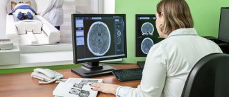
After the contrast is administered, two x-rays of the brain are taken - in the lateral and direct projections. The images are assessed by a radiologist. If there are still uncertainties, it is possible to reintroduce contrast and take two more photographs.
At the end, the catheter is removed, a sterile bandage is applied to the insertion site, and the patient is monitored for 24 hours.
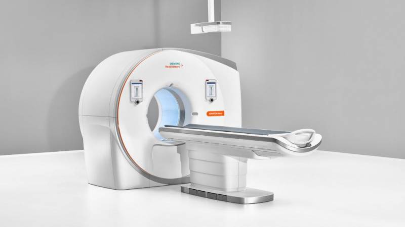
Features of MR angiography
MR angiography is even more informative than CT. It allows you to see soft tissues that are difficult to visualize on CT. It is carried out using a magnetic resonance imaging scanner and is not an x-ray method, unlike other angiography methods. This avoids exposure to radiation.
Another advantage is good visualization even without the use of contrast, which is why MR angiography without contrast can be used for allergy sufferers.
The main contraindication for use is the presence of any metal objects in the body (artificial pacemakers, prostheses, implants, metal clips on blood vessels).
Perhaps selective cerebral angiography of the brain has already become commonplace and routine for doctors. It may be inferior in effectiveness to CT and MRI angiography. However, being more affordable and not requiring special high-tech equipment, it is still actively used 100 years later for diagnosing brain diseases.
Source: https://FB.ru/article/396624/tserebralnaya-angiografiya-sosudov-golovnogo-mozga-pokazaniya-i-otzyivyi
Possible contraindications
Despite the relative safety and information content of the manipulation, it is not always permitted . The main contraindications will be the following:

- period of pregnancy and breastfeeding;
- allergic reaction to iodine and drugs that contain it;
- coma, when the patient is unable to respond to the doctor’s instructions;
- mental illnesses accompanied by emotional agitation, when the patient’s behavior cannot be controlled;
- thyroid diseases, such as hyperthyroidism;
- severe form of renal failure;
- blood pathologies with impaired blood clotting and a tendency to bleed;
- the presence in the patient’s body of titanium implants, piercings or tattoos made with paint containing metal particles.
Diagnostics are not carried out without the consent of the patient. A relative contraindication would be any pathology in the acute stage. In this case, the specialist postpones the study until the patient’s condition improves.
Complications usually do not occur, but sometimes the patient may experience nausea caused by the contrast agent, headache, weakness, and drowsiness. As a rule, symptoms disappear within a few hours. If the condition does not improve, symptomatic therapy is used.
Cerebral angiography of cerebral vessels: what is it?
Cerebral angiography is a hardware diagnostic examination aimed at studying the state of cerebral vessels and circulatory processes. This technique makes it possible to promptly identify cerebrovascular disorders and prevent the further development of pathologies with extremely dangerous consequences.
What do angiography indicators mean?
The amount of radiation that will penetrate the veins and other brain tissue is determined by their density. It is expressed in various color shades.
The bone in the image will be white, and the cerebrospinal fluid will practically not appear on the resulting images. Other brain substances have different colors and densities. Using them, doctors evaluate the internal structure.
The doctor will provide a detailed transcript of the received images.
Preparation for the procedure
Before the study, the patient should not eat for 10 hours and not drink for 4 hours. He needs to remove all metal objects. If surgical intervention is required to administer contrast, the following is prescribed:
- iodine allergy test;
- urine and blood tests;
- ECG;
- kidney function testing;
- consultations with an anesthesiologist and therapist.
Before performing an MRI or CT scan of the brain with angiography, you must adhere to a diet for several days. Eliminate from your diet carbonated drinks, refined sweets and sweet fruits, dishes made from legumes and other products that cause increased gas formation in the gastrointestinal tract.
Preparing for the study
No special preparation is required for the procedure, but there are rules that must be followed. Doctors recommend not eating heavy foods on the eve of diagnosis to eliminate the possibility of indigestion and other problems. In addition, it is necessary to stop drinking alcohol in any doses 7-10 days before the procedure.
If the patient takes any medications on a regular basis, it is worth consulting with a doctor. Some may need to be discontinued before the study or the dosage reduced.
You should not take a mobile phone or other devices with you into the office where the equipment is located, especially when using a tomograph. On the eve of the procedure, the doctor conducts a conversation with the patient, explains the essence of the procedure and its importance for establishing the correct diagnosis. The patient should not experience fear or uncertainty, since the manipulation is absolutely safe.

A mandatory stage of preparation is conducting a test for sensitivity to contrast agents. Gadolinium-based agents are usually used. The patient is given a minimal dose intradermally, and the doctor observes the reaction. If there are no negative symptoms for several hours, the required dose of the drug is allowed to be administered. In case of allergies or other manifestations, a specialist examines the brain vessels in a different way.
Diagnosis using selective angiography
Angiography is a method of fluoroscopic examination of body systems, namely blood and lymphatic vessels, veins and capillaries. There are general and selective (selective) angiography, using one or another type, guided by the goals set by the doctor. General angiography examines all the main vessels of the area, a selective procedure examines individual vessels.
Diagnostics involves the introduction of radiocontrast agents (organic iodine compounds) into the vessels under study using catheterization or puncture.
The phases of angiography are distinguished by the stages of passage of the radiopaque substance through the arterial system:
- Arterial.
- Capillary.
- Venous.
The rate of disappearance of the radiopaque substance characterizes the hemodynamics in the organ under study.
This method successfully diagnoses various abnormalities in the functioning of the vascular system, as well as various tumors, parasitic and other organ lesions.
With its help, the topographical features and functional states of blood vessels, the paths of the roundabout circulation, and the speed of blood flow are studied.
Types of angiography depending on diagnostic purposes
Depending on the disease and the goals and objectives set during diagnosis, angiography can be classified into:
- Cerebral selective angiography is an angiographic study of cerebral vessels. In general and selective angiography of cerebral vessels, immediately after the invasion of the organic compound of iodine, several angiograms are made, which display the capillary, arterial and venous phases of blood flow. For selective cerebral angiography, both indirect and direct (carotid and vertebral) angiography are used. The most common type of examination is the carotid one, in which a radiopaque contrast agent is injected into the carotid artery. During a vertebral examination, iodine is injected into the vertebral artery. Indirect diagnosis involves the injection of contrast through a catheter into large vessels, for example, on the thigh.
- Arteriography - allows you to diagnose blockage or narrowing of the lumen in the artery, disturbances in the blood flow, as well as neoplasms near the examined blood vessels.
- Lymphography - studies the state of the lymphatic system. The study is in demand in oncology for the diagnosis of malignant tumors of the ovaries and uterus. The procedure is necessary to assess the stage of the disease and the effectiveness of chemotherapy.
- Venography is successfully used to diagnose problems in the venous circulation of the extremities. Angiograms show both deep and superficial veins, their length and location. Research using this method is effective for varicose veins and various degrees of thrombosis, even diagnosing heart pathologies and heart failure.
- Cardiac angiography - used to study the chambers and arteries of the heart muscle, identifies vascular pathologies (blockages, thrombosis, pathological narrowing and dilatation), assesses the pressure in the chambers of the heart and arteries, and diagnoses the contractile function of the myocardium. This type of study provides a more informative picture of heart disease than venography.
- Fluorescein angiography is an angiography method for examining the ocular vessels and fundus. Used in the diagnosis of glaucoma, dystrophic and inflammatory processes in the retina and membrane of the eye, diabetic retinopathy. In this study, fluorescein sodium salt 10% is used as a radiopaque composition; administration occurs through a vein on the elbow.
Preparation and performance of selective angiography
Before examining a patient using angiography, the doctor must familiarize himself with the test results (general urine and blood tests, biochemistry), conduct a medical examination, and obtain intelligible answers to the following questions:
- Do you have a history of allergies?
- Chronic and past infectious diseases.
- What medications are being used at the time of the study.
- If the patient is a woman, make sure there is no pregnancy.
It is important! Some time (the doctor will determine it exactly) before the angiography procedure, you will need to adjust your diet, and possibly stop some of the medications you are taking. This is necessary to minimize errors in the study results.
A catheter with a contrast agent is inserted under local anesthesia. Sometimes, before or during the procedure, if indicated, a sedative is administered to help the body relax. The procedure itself is practically painless. However, there may be a feeling of some discomfort during the insertion of the catheter and a feeling of nausea when the X-ray contrast agent is administered.
The examination can take from 10 minutes to several hours, it all depends on the scale of the study. After the angiography, the doctor will suggest that you stay in the hospital for some time to monitor your condition, thereby reducing the risk of possible complications.
It is important! Accurate information about whether the patient is allergic to X-ray contrast agents, as well as medications and seafood will help reduce the risk of complications.
Upon discharge, the doctor:
- will write down recommendations for the care and treatment of the catheter insertion site;
- will recommend drinking a lot of water to remove the contrast agent from the body;
- writes instructions for taking medications;
- will note the need to reduce physical activity for 24 hours after the procedure.
Even if the angiography passed without visible consequences, the patient should be attentive to his health and, if one of the symptoms appears, immediately consult a doctor:
- temperature increase;
- pain, swelling, or discharge at the catheter insertion site;
- changes in the functioning of the gastrointestinal tract;
- pain in the chest or organ being examined;
- numbness or pallor of the hands and feet;
- labored breathing;
- weakness of facial muscles, problems with speech or vision.
Source: https://VashNevrolog.ru/metody-diagnostiki/diagnostika-metodom-selektivnoj-angiografii.html
What does the patient experience during the procedure?
When the needle is inserted into the vessel, the patient may experience minor pain.
If the procedure is performed with sedation, the patient feels sleepy and relaxed.
The patient may feel slight pressure as the catheter is inserted.
As the contrast agent passes through the patient's body, they may experience a warm sensation that quickly dissipates.
The patient must remain motionless during the examination.
After the procedure is completed, the catheter is removed from the vessel.
After the procedure, the patient remains in the recovery room for several hours for observation.
The patient should immediately inform the doctor if after the procedure he develops symptoms such as:
- weakness or numbness in the muscles of the facial limbs
- speech disorders
- vision problems
- signs of infection at the catheter site
- dizziness
- chest pain
- difficulty breathing
- rash
- pain in the limb where the catheter was inserted; difficulty
What is cerebral angiography?
Cerebral angiography is a hardware diagnostic examination aimed at studying the state of cerebral vessels and circulatory processes. This technique makes it possible to promptly identify cerebrovascular disorders and prevent the further development of pathologies with extremely dangerous consequences.
What is angiography?
Angiographic examination of blood vessels is an instrumental technique for studying the human circulatory system. Such methods suggest damage to cerebral vessels and vascular beds. The procedure is invasive because During the diagnostic process, a contrast agent is injected into the patient's vessel area.
Angiography of cerebral vessels helps to detect the following potentially dangerous disorders in a timely manner:
- narrowing and dilation of the walls of blood vessels;
- thrombus formation;
- tumor development;
- bleeding.
The technique facilitates the process of making a correct diagnosis for a patient and developing the most effective, safe therapeutic course.
Indications and general contraindications
Indications for cerebral angiography of cerebral vessels are:
- hematomas and internal hemorrhages;
- tumor neoplasms of both benign and malignant origin;
- circulatory disorders occurring in severe or acute form;
- cerebral aneurysm;
- arterial embolism;
- stenosis, cerebral type atherosclerosis;
- epileptic syndrome;
- recent stroke;
- thrombosis of cerebral arteries;
- intracranial hemorrhage;
- arteriovenous type malformation;
- ruptures, traumatic injuries of cerebral arteries;
- inflammatory lesions in the area of cerebral vessels;
- atherosclerosis;
- hemorrhagic damage to the central nervous system;
- pathologies in the structure of veins and arterial vessels, both congenital and acquired;
- varicose veins;
- heart attack
In addition, cerebral angiography is done when a patient has the following clinical symptoms indicating the presence of severe vascular pathologies:
- increased intracranial pressure;
- impaired hearing function, presence of tinnitus;
- frequent fainting;
- periodic dizziness;
- attacks of headache;
- migraine attacks;
- manifestation of local neurological signs accompanying damage to any cerebral lobes;
- constant feeling of nausea.
This procedure is carried out not only for the purpose of diagnosing and monitoring the patient’s condition, but also during the period of preparation for surgery to assess the patency of the vascular bed.
Such diagnostic testing is not always carried out. It is not recommended in the following cases:
- a history of allergies, hypersensitivity and individual intolerance to iodine-containing products;
- mental disorders and disorders;
- coma;
- kidney failure;
- violation of liver functions, occurring in a complicated form;
- general severe, weakened condition of the patient;
- acute or severe form of heart failure;
- decreased blood clotting, thrombocytopenia, hemophilia;
- acute form of myocardial infarction;
- processes of an inflammatory, infectious nature, occurring in an acute form;
- severe anemia.
This technique is also contraindicated for children under two years of age due to the lack of sufficient information about the effect of X-ray radiation on the child’s body.
Possible complications
Adverse reactions and complications during cerebral angiography of cerebral vessels do not occur often, up to 3% of cases. However, such reactions may occur, and the patient must be informed about them. Among the main possible complications are the following conditions:
MR angiography: how the study works
The technique involves conducting research using magnetic fields. For these purposes, it is not always necessary to use contrast agents, due to which the technique has fewer contraindications. During diagnostics, specialists evaluate the phases of blood circulation and the condition of blood vessels, observing changes in their tissues.
This technique allows you to most accurately examine the vascular pattern and determine even the most minor deviations in the arterial structure. This method is considered less dangerous, but is not used in people with obesity or claustrophobia.
How does CT angiography work?
An innovative and reliable method of diagnostic research. This method involves the introduction of a contrast agent into the area of the cubital vein. Then the doctor takes a series of X-ray images of the brain in various projections with further computer visualization of the vascular bed and reconstruction of the 3D image.
The main advantages of this technique (compared to the method described above) include lower radiation exposure to the patient’s body.
Result evaluation
A specialist will be able to decipher the results obtained during angiography on the basis of X-ray images, which allow one to assess the arterial bend, the presence or absence of vascular narrowings and lumens.
In the presence of a tumor neoplasm, straightening and displacement of blood vessels localized near the pathological focus are noted.
Atherosclerotic disorders are diagnosed when there is uneven narrowing of the vascular lumen.
When is it appointed - video
Angiography can be prescribed for such types of damage and vascular malformations as:
- aneurysm;
- atherosclerosis;
- defects and damage to internal organs;
- malformation;
- thrombosis;
- tumor processes.
However, this procedure also has its contraindications.
Angiography is not performed if the following problems exist:
- allergy to iodine-containing drugs;
- mental disorders;
- acute inflammatory processes and infectious diseases;
- heart, kidney, liver problems;
- thrombophlebitis;
- serious condition of the patient.
There are a number of indications for which cerebral angiography is prescribed. The doctor can send the patient for this procedure after hearing a certain list of complaints. This study is necessary in order to accurately assess the condition of the head and identify deviations.
Indications:
- Constant headache for no apparent reason.
- Dizziness and tinnitus.
- Pain or stiffness of movement in the cervical spine.
- Nausea not associated with eating poor quality food.
- Low blood pressure that is not normal for a particular person.
- Fainting or loss of consciousness.
Angiography of cerebral vessels and MRI are necessary in order to confirm or refute the presence of abnormalities. Thanks to the procedure, it is possible to identify narrowing of the lumen, blockage of a vessel with a blood clot or cholesterol plaque, spasm, inflammation of the walls, aneurysms, as well as damage leading to hemorrhage. In addition to these pathologies, angiography of cerebral vessels makes it possible to diagnose tumors in the early stages.
Advantages of the procedure
- Possibility to create a three-dimensional image,
- Visualization of blood vessels, which allows the doctor to detect blood clots, hematomas, aneurysms,
- The possibility of an individual approach to the patient, and therefore an accurate diagnosis.
The modes and programs of this type of examination can be very wide, so you can always choose the best option in each specific case. The absence of inaccuracies in diagnosis makes it possible to timely and accurately identify problem areas in the blood vessels and understand the cause of diseases or disorders in the functioning of the blood vessels of the brain. Adequate diagnosis is the main condition for proper treatment.