Ultrasound of the vessels of the brain and neck is a diagnostic procedure that allows you to assess the condition and function of the vessels responsible for blood supply in the head area: the main arteries of the brain, carotid arteries, vertebral and subclavian arteries and veins of the neck. Significant advantages of ultrasound of neck vessels are harmlessness for the patient (the study is not associated with exposure to radiation), painlessness (the procedure does not involve instrumental intervention and is non-traumatic), high information content, reasonable price, and the possibility of repeating the study many times if it is necessary to clarify the diagnosis.
Ultrasound of the vessels of the brain and neck is indicated for patients with acute or chronic cerebrovascular accident; those who have undergone open or closed traumatic brain injury or neurosurgical intervention; patients with various types of pathology of the cervical spine (congenital disorders of posture, injuries of the cervical spine and intervertebral discs, osteochondrosis, etc.).
There are several types of ultrasound examination of blood vessels. The modern diagnostic base includes traditional ultrasound examination (direct ultrasound) and duplex scanning of blood vessels (doppler ultrasound). Traditional ultrasound allows you to determine and evaluate the geometry of blood vessels, their thickness and patency, and the diameter of the vessel lumen. Doppler ultrasound (USDG) makes it possible to assess the speed of blood flow in real time and identify various vascular patency disorders (the presence of vasoconstrictions, blood clots, atherosclerotic plaques).
Ultrasound of the vessels of the brain and neck is indicated for the following group of patients:
- patients with acute or chronic cerebrovascular accident;
- patients who have suffered an open or closed craniocerebral injury;
- patients who have undergone neurosurgical intervention.
- patients with various types of pathology of the cervical spine (congenital disorders of posture, injuries of the cervical spine and intervertebral discs, osteochondrosis);
- patients with identified chronic diseases (hypertension, diabetes mellitus, atherosclerosis, etc.);
- patients with neoplasms (tumors) of various origins in the head and neck area.
An ultrasound scan of the vessels of the brain and neck is indicated if the patient has the following symptoms and conditions:
- persistent headaches of unknown etiology;
- systematic dizziness, darkening of the eyes;
- repeated episodes of loss of consciousness;
- noise in the head and ears;
- sudden weakness, numbness and tingling in the limbs;
- sudden disturbances in visual and speech function.
The study shows high efficiency in the early diagnosis of cerebral circulatory disorders, which allows for timely identification of the risk of stroke. The price of vascular ultrasound is quite acceptable and allows, among other things, routine preventive examination of patients with a hereditary predisposition to cardiovascular diseases and associated risk factors (age over 40 years, smoking, being overweight, high blood pressure, high cholesterol levels). blood, sedentary lifestyle, systematic stress).
Preparation for ultrasound of the blood vessels of the brain and neck
On the day of the study, the patient must stop taking all medications, with the exception of those that cannot be stopped even for a short period. It is necessary to discuss the possibility of interrupting medications with your doctor. Also, 2 hours before the procedure, you should stop drinking tea and coffee and refrain from smoking (caffeine and nicotine have a direct effect on the tone of blood vessels and can distort the results of the study). Before the examination, it is necessary to remove all jewelry from the head and neck.
At the Center, ultrasound of the blood vessels of the brain and neck is performed by appointment. After the procedure, the patient receives a photo and a detailed description made by a diagnostician.
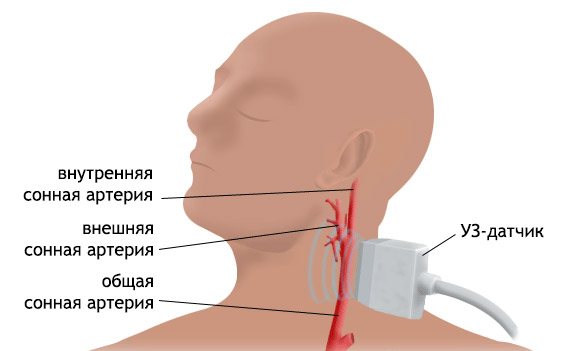
Extracranial duplex scanning of neck vessels
Two groups of main blood vessels are responsible for the blood supply to the brain - extracranial (located outside the skull) and intracranial (located directly in the skull and bone canals). A danger to the normal blood supply to the brain is posed by mechanical injuries to the neck and head, vascular diseases that lead to changes in the vascular wall, blockage of the vessel and disturbances in its patency. Pathologies are often irreversible, but they can be tracked in the early stages and development can be slowed down. One of the main diagnostic methods for examining blood vessels and blood flow is duplex scanning of the cervical vessels.
What will the examination show?
Duplex scanning combines two types of examination - conventional ultrasound and Doppler ultrasound. It allows you to evaluate:
- The structure of blood vessels.
- Violations of venous outflow and its possible causes.
- Blood flow parameters and the nature of their disorders.
- Vascular pathologies - spasms, blood clots, cholesterol plaques, inflammation, compression of blood vessels by cervical vertebrae or other structures, aneurysms and others.
- What are the reserve capabilities of the brain's blood circulation if there is a disturbance in one of the vessels.
- The first signs of pathologies, when there are no changes in well-being yet.
Extracranial duplex scanning of neck vessels is an ultrasound examination of the cervical and brachial veins and arteries. If complaints characteristic of cerebrovascular disorders occur, this area is examined first. The cervical spine is mobile, and the muscular frame here is relatively weak, so there is a high risk of vascular compression due to injuries of the cervical spine, osteochondrosis and other diseases. Extracranial arteries include the carotid artery and parts of the subclavian and vertebral arteries.
Indications
The main symptoms of circulatory disorders, for which the doctor will prescribe a duplex scan of the veins of the neck:
- Frequent headaches.
- Dizziness that occurs when changing body position or turning, throwing back the head.
- Gait disturbances – unsteadiness, involuntary deviation from moving in a straight line.
- Changes in facial expressions and symmetry of facial movements.
- Visual distortions – darkening before the eyes, “spots”, blurred vision.
- Memory impairment, difficulty concentrating.
- Noise or ringing in the ears, deterioration of hearing acuity.
- Numbness in the upper extremities, muscle weakness, deterioration of sensitivity.
- Noticeable strong pulsation on the neck of the blood vessel.
- The difference in blood pressure in the arms is more than 15 mm. rt. Art.
- Sleep disorders.
Duplex scanning of neck veins is recommended even without such symptoms. Direct indications are systemic diseases that impair blood circulation. These are diabetes mellitus, atherosclerosis, high cholesterol, osteochondrosis, heart disease (coronary artery disease, defects), excess weight, hypertension and others. It is also worth examining blood vessels for preventive purposes every year after forty years. This is especially necessary for those who lead a sedentary lifestyle, smoke, and have a history of strokes and cardiovascular diseases in close relatives.
How it goes
No special preparation is required to assess the condition of extracranial vessels. On the day of the examination, it is advisable not to smoke or drink alcoholic or caffeinated drinks. You must warn your doctor if you are taking any medications. Diagnosis takes place in the supine position. There may be a small cushion under the neck. In some cases, the study is carried out with functional tests: turning the head, briefly pressing the arteries, holding the breath.
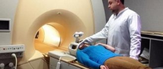
After the ultrasound, a conclusion is issued, which is deciphered by the attending physician. In some cases, other types of ultrasound or vascular examination using MRI may be prescribed.
Examination in Moscow
We invite you to the Kutuzovsky Children's Center for the diagnosis of vascular pathologies. The clinic has all the necessary modern equipment to obtain the most accurate and detailed data.
Contraindications
There are no absolute contraindications to Doppler ultrasound.
Relative contraindications include the patient’s serious condition and other factors that prevent the body from taking a horizontal position.
This technique is completely safe for the body and is highly informative at the initial stages of the pathological process . Thanks to its use, it is possible to make a correct diagnosis in a timely manner and begin therapy.
The study does not cause side effects and is not associated with radiation exposure. In addition, this study does not require the use of drugs. It can be performed on all people, including small children.
Varieties of Research Approaches
There are a number of ultrasound examinations of the head:
- Dopplerography of the main arteries . In this case, the object of analysis is blood flow in the vessels, its intensity and speed. Based on these data, the doctor can judge the presence of pathological formations and processes occurring in the walls of blood vessels.
- Duplex scanning . With this study, the doctor is able to obtain a two-dimensional image of the blood vessels. This allows you to assess the condition of the lumen and walls of the cerebral vessels.
- Triplex scanning . It is an addition to duplex scanning. When it is performed, a color image is displayed on the screen, which will allow a more detailed study of the condition of the vessel. Thanks to its implementation, the doctor can obtain information about the direction and speed of blood flow.
All these methods are highly informative and accurate, making it possible to diagnose brain disorders.
Using the data obtained, it is possible to visualize the vascular system of the head and record all existing deviations.
A severe blow to the integrity of the brain, a hemorrhagic stroke can cause disability and even death. More details in the article. In what cases can dyscirculatory encephalopathy of the 3rd degree develop - treatment and preventive measures to prevent a dangerous disease.
Advantages of Doppler ultrasound method
Dopplerography of the vessels of the neck, brain and other organs is one of the most popular diagnostic methods today. Doctors choose it for the following benefits.
- Safety. The use of this diagnostic method is not associated with any side effects. The patient is not exposed to radiation, does not experience painful or even simply unpleasant sensations. He is not given any contrast agents. The procedure can be used even on children and the elderly. The safety of the procedure allows it to be used to monitor the effectiveness of treatment.
- Wide scope of application. Due to the fact that using the Doppler effect it is possible to study the network of vessels in detail, this diagnostic method is used to identify vascular pathologies of various types. The method makes it possible to identify early, barely manifested preclinical symptoms of diseases of the circulatory system, vascular damage, and disturbances in blood flow, making it of great importance for early diagnosis.
- Speed of execution. The procedure for examining blood vessels using ultrasound takes only fifteen minutes. A similar MRI study lasts at least half an hour.
- Low cost. Ultrasound costs the patient much less than angiography, MRI or CT with high information content.
On the importance of diagnosing the great vessels
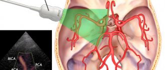
It is known that the so-called brachiocephalic arteries (BCA) of the brachiocephalic vascular trunk (on the right) depart from the aorta. It consists of the subclavian as well as the carotid arteries. On the left side, when departing from the aorta, they do not form the brachiocephalic vascular joint (trunk). The vessels of the vertebral region also depart from the aorta and the arteries of the subclavian trunk, which, connecting with each other, form the so-called basilar artery. It, together with the carotid artery (on both sides), are the main channels of blood supply to the brain, thereby forming the Circle of Willis.
Scanning of the brachiocephalic arteries
The vascular apparatus of this strategically important node is located in such a way that if the functioning of any of its sections is disrupted, other vessels will provide nutrition to the brain. Disruption of blood flow in any part of the cerebral arteries of the brain can lead to a stroke. In addition, brachiocephalic (BCA) vessels are at high risk of developing atherosclerosis.
Ultrasound duplex scanning of the brachiocephalic arteries (BCA) allows us to identify disturbances in the flow of the bloodstream and pathological changes in blood vessels in the early stages of development. The treating specialist can also determine the condition of the vascular tissue, the degree of atherosclerotic changes, as well as identify stenosis and assess its severity.
Progress of the procedure
To conduct the study, a person needs to lie on a special couch face up, turn his head to the left or right and tilt it back slightly. A little acoustic gel is applied to the neck - it is along this gel that the sensor moves.
The sonologist places the sensor in those places on the neck where large and medium-sized veins and arteries are projected.
To obtain a holistic picture of the state of the blood vessels, functional tests are performed with holding the breath and changing body position. Thanks to this, it is possible to assess the regulation of vascular tone.
Also, different modes can be used when conducting research. Thus, when performing diagnostics in two-dimensional or B-mode, vessels passing outside the cranium are studied.
The carotid and vertebral arteries, jugular veins, etc. are analyzed. Typically, indicators such as the condition of the vascular walls, the patency of veins and arteries, the correctness of the structure, and the condition of the tissues in the vascular area are assessed.
When performing duplex color scanning, it is possible to obtain information regarding the vessels in the cranial cavity and those located in the neck and in the brain area. Such a study is called extracranial.
Carrying out this procedure allows you to obtain data on the speed of blood movement through the artery, the zone of turbulent blood flow, and the uniformity of filling of veins and arteries.
The study also makes it possible to study the condition of the lumen, assess the condition of the venous vessels and the vein wall.
The results obtained at rest and during functional tests are compared with the norm. If pathology is detected in certain places, the sonologist designates it with an abbreviation consisting of letters and numbers.
Based on this information, the neurologist interprets the results and prescribes treatment.
Method for obtaining information during duplex scanning
Duplex scanning (from “duplex” - double) is a method of visualizing internal organs and structures using an ultrasound sensor with Doppler examination. The image acquisition mechanism is based on the device emitting high-frequency waves (more than 40,000 Hz; for reference, the human ear hears sounds in the range from 16 to 20,000 Hz).
Duplex scanning involves simultaneous examination in two modes:
- B-mode (from “brightness” - brightness). Penetrating through the skin and soft tissues, depending on the density of the latter, the emitted signal is reflected to varying degrees and hits the recording screen of the sensor. The signal is then converted into a two-dimensional image. Internal organs, depending on their structure, are displayed in different shades of gray.
Duplex scanning is an improved diagnostic method that combines the already familiar ultrasound and Doppler ultrasound
- DC (Doppler mapping) is a method of obtaining a color image of the movement of blood through the vessels. In this case, blood cells (erythrocytes) become the structure from which the ultrasonic wave is reflected. The method allows you to evaluate the speed and direction of blood flow.
Modern devices allow you to get moving images when connecting the M-mode. In this case, the monitor screen displays the state of the organ in real time.
About the need for preparation
Dopplerography does not require any special preparation.
Immediately before the procedure, you need to remove all jewelry from your neck.
This study can be carried out repeatedly, since it does not affect the body. Due to the complete safety of Doppler ultrasound, it is often prescribed continuously for the purpose of monitoring and monitoring treatment.
It can also be used to prevent serious diseases.
Carrying out an ultrasound examination of cerebral vessels allows the doctor to detect a vulnerable area of the vascular bed and choose effective treatment tactics.
Thanks to the research, it is possible to choose the right medications.
Indications for duplex angioscanning
Duplex scanning of blood vessels may be prescribed for:
- the need to monitor the correctness of a number of surgical operations performed on the vascular apparatus (abdominal aorta, carotid, subclavian and a number of other arteries);
- screening diagnostics, which allows to identify the asymptomatic course of a number of diseases;
- thrombosis of the intestinal vascular apparatus (abdominal aorta);
- detection of diseases of the vascular apparatus of the neck and brain;
- the presence of venous thrombophlebitis and phlebothrombosis;
- aortic aneurysm (abdominal);
- varicose veins and vasculitis;
- damage to atherosclerosis of the visceral parts of the aorta;
- angiopathy in diabetes and endarteritis.
Advice: if you notice a sharp deterioration in vision, the presence of noise and ringing in the ears, changes in blood pressure, as well as impaired coordination of movements, you must consult a doctor to identify diseases of the vascular system of the brain and the need to prescribe therapeutic treatment or surgery on the vessels of the neck .
Evaluation and interpretation of survey results
Duplex examination of the cervical arteries allows us to reconstruct their position in the tissues, anatomical structure, their diameter, features of the adjacent interstitium, and the tortuosity of their collateral branches. In addition, duplex scanning of the brachiocephalic arteries shows the characteristics and changes in blood flow. When displaying a picture from the sensor on the monitor, you can visualize blood flow indicators in color. Spectra of its speeds, turbulence and regurgitation are visible.
Images from the duplex scanning system monitor
Based on these elements, diagnoses are formed about the presence or absence of stenoses, obliterations, occlusions, blood clots, aneurysms and plaques. All values obtained during scanning are recorded by a functional diagnostics specialist, who, at the end of the study, formulates them in the form of a conclusion indicating specific problems.