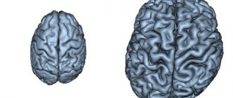
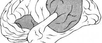
The conditional division of people into techies and humanists is based on the peculiarities of the development of the individual’s brain. The discovery of interhemispheric brain asymmetry provides an answer to the questions: why one person solves problems with ease, while another creates masterpieces in art.
The issues of the functioning and interaction of the brain hemispheres have not been fully studied, but already now theories of interhemispheric asymmetry make it possible to explain some features of the perception of information, the learning of children, or the rehabilitation of people with damage to parts of the brain.
General information about interhemispheric asymmetry of the brain
The term “interhemispheric asymmetry” is used as a division of the functions of brain processes (perception, analysis, production of information) between the left and right halves of the brain.
Research also speaks of anatomical differences in the size and morphology of the halves of this organ. In 1836, M. Dax published observations of patients with partial lesions in the left region. In particular, such patients had a speech disorder (aphasia), a symptom that was not present in people with brain damage in the similar (mirror) area of the right side.
Observations made it possible to draw a conclusion about lateration - the distribution of mental processes between the left and right hemispheres.
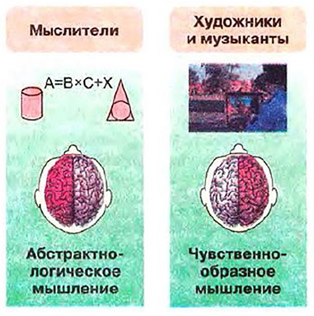
Conventionally, each half is “responsible” for its own range of thought processes: the left - for logic, calculation, the right - for imaginative thinking, intuition.
To study brain lateration the following was used:
- Observation of patients with damage to areas of the brain due to trauma, tumor, parasitic cyst.
- Dissection of the corpus callosum, which serves as a conductor between parts of the brain.
- Specific studies measuring electrical impulses of a particular part of the brain when solving a specific problem.
It was found that the hemisphere controls the sensory of the opposite part of the body: the right - the left side, the left - the right. Thus, the action of the right hand is controlled by the left, and vice versa.
This fact allows us to conduct psychological tests that allow us to determine the share of participation of each hemisphere (dominance) in a particular individual. So, in 90% of people the left is dominant, since most people are right-handed. For a more objective picture, tests are used:
- Sensory - asked to perform an action, for example, untie a knot or fold your arms across your chest. During performance, the subject manipulates his right hand more confidently.
- Visual - the test determines the dominant eye.
- Auditory - a test to determine the perception of sounds.
The totality of the results of such a study allows us to classify an individual into one of five groups:
- right-handed;
- right-handed;
- left-handed;
- left-handed;
- ambidextrous.
In the latter, dominance - the property of interhemispheric symmetry - is absent. Such people can use both hands equally, but this phenomenon is rare.
Basic concepts of functional brain asymmetry
Functional asymmetry of the cerebral hemispheres is the morphophysiological characteristics of cerebral structures that determine the dominance of the hemisphere when processing information of a certain type. It manifests itself in the difference in functional loads performed by the symmetrical parts of the hemispheres.
Important! For each person, the severity of hemisphere dominance and the location of specific functions between them are very individual.
At the same time, a lack of stability in the asymmetry of the human cerebral hemispheres has been established. With unilateral lesions, the opposite hemisphere, forming new connections between the projection fields, is able to take over the performance of lost functions. Functional asymmetry is considered as the ability of a person’s cerebral adaptation to changing conditions in normal and pathological conditions.
Interesting! Paleontological studies have discovered the existence of morphological asymmetry in prokaryotes during the Archean period.
How was brain asymmetry formed?
There are different opinions as a result of which brain asymmetry was formed. Is this quality innate or acquired? Right-handedness - a sign that the left side of the brain is dominant - was inherent in human cave ancestors.
Scientists make this conclusion based on an analysis of rock paintings. It is believed that ancient people made images of hands using the stencil method, tracing the palm with the dominant hand. Since the drawing of the left hand is found in 90 percent of all images of the limb, it is concluded that the ancient “artists” were for the most part right-handed, and therefore left-hemisphere.
On the other hand, many researchers say that newborns do not have asymmetry, and in the first 3 years of life, right-hemisphere dominance predominates in children - after all, at first they communicate with emotions and non-verbal methods. And only with the development of speech does the left-hemisphere type begin to predominate.
A number of researchers believe that asymmetry is present from birth. It appeared as a result of natural selection and is passed on to offspring. But due to the lack of thinking in infants, this postulate has the status of a theory.
Scientists have a unanimous opinion only on the issue of the occurrence of asymmetry as a consequence of development (ontogenesis).
It has been noticed that the more intellectually developed a person is, the greater the asymmetry.
In uneducated primitive individuals the anatomical and functional difference between left and right is insignificant.
Remarkable! Interhemispheric asymmetry is a property not only of humans, but also of animals. Differentiated functioning and anatomical differences were found in dolphins, rats, and monkeys.
Properties of brain asymmetry
Neurophysiologists believe that asymmetry is formed before adulthood. During this period, a person has a high learning ability, and in the learning process a dominant type of thinking develops.
The left (logical analysis) and the right hemisphere (operating with images) can “work” on one problem, each with its own “tools”. Such interaction between the parties provides high quality data processing and synthesis, which is characteristic of an intelligent person, a person with high brain development.
It is known that despite the differentiation of responsibilities between the hemispheres, when part of the brain is damaged, the function of the damaged area is taken over by the remaining healthy cells and areas. That is, narrow “specialization” can change if necessary.
A particularly high degree of reorganization occurs at a young age (period 10–15 years).
Thus, interhemispheric asymmetry has the following properties:
- Dominance.
- Switchability.
- Plasticity.
These properties are used in neuropedagogy. The development of figurative or logical thinking is possible not only with the help of intellectual tasks and imagination exercises. Physical activities involving fine motor skills for the left or right hand force the corresponding hemisphere to work (and develop).
In the light of new knowledge about lateration, modern pedagogy does not recommend retraining left-handed children to write with their right hand. Right-hemisphere children are more vulnerable and sensitive, and being forced to write with a subdominant hand worsens the learning outcome - psychological stress due to the dissatisfaction of a parent or teacher is added to the discomfort of writing with a passive hand.
Lyrical digression: An illustration of the plasticity and switchability of brain asymmetry are facts from the biography of some famous scientists (A. Einstein, N. Bohr). It seems that among representatives of the exact sciences, left-hemisphere (logical) thinking should prevail. However, in childhood these people were of the right-hemisphere type, as evidenced by their poor success at school. However, the dominance of the right (the ability to think and operate with images) did not prevent them from becoming great scientists and developing analytical abilities (the left hemisphere).
Neurophysiologists explain this by the fact that right-hemisphere people use images for intellectual activity. The right, unlike the left, perceives information as a whole, and not one by one. This ability to work in parallel increases the speed of mental processes, including analysis and synthesis of information.
After the work is completed, it is transferred to the left hemisphere for encoding into signs: words, numbers, formulas.
It is known that only about 5% are left-handed. However, among great or famous people their percentage increases to almost half. This suggests that asymmetry is not a defect, but an acquisition of humanity as a result of evolution.
Functions of the left hemisphere
The left hemisphere works with symbols and processes information sequentially, analyzing each element. With its help, they remember dates, words, numbers, names. The left hemisphere is responsible for:
- language;
- speech;
- letter;
- mathematical analysis;
- logical constructions;
- right half of the body.
The left side is characterized by inductive processing of information. Despite the responsibility for speech, a person understands words and phrases in the literal sense on the left side of the brain; humorous associations or figurative expressions are not understood by the left hemisphere.
Functions of the right hemisphere
The right side of the brain perceives nonverbal information. Images, metaphors, sounds and gestures are perceived as a complete picture. Let's list the areas of responsibility:
- spatial orientation;
- perception of painting, music, any kind of creativity;
- religious or mystical beliefs;
- left half of the body.
The right hemisphere not only perceives, but also synthesizes and transmits information in images. Such signals in the form of vague anxiety or a decision that came from nowhere are usually called intuition. In fact, this is all the same information, only received (processed, transmitted) using images.
Interhemispheric brain asymmetry and interhemispheric interaction
Interhemispheric asymmetry is a fundamental property of the human brain, reflecting a complex integral system of interaction between its many formations. In recent decades, the study of this problem has been one of the most popular topics of scientific research both in neuropsychology and in related branches of science - neurophysiology, psychophysiology, sports psychology, etc.
It is necessary to distinguish between hemispheric asymmetry and functional specialization of the hemispheres (Leutin V.P., Nikolaeva E.I., 2005). Interhemispheric asymmetry is considered as a temporary dominance of the activity of structures of one hemisphere, associated with the type of tasks presented. The functional specialization of the hemispheres refers to the preference of each of them for processing information of a certain type.
The first works in the field of studying interhemispheric asymmetry concerned observations of patients with local brain lesions. Analyzing the differences in symptoms between patients with left-sided and right-sided lesions, experts sought to link them with the characteristics of the normal activity of each hemisphere. Currently, the problem of interhemispheric asymmetry of the brain in relation to higher mental functions is being studied as a problem of the functional specificity of the hemispheres. The main task of researchers is to determine the contribution that each hemisphere makes to the implementation of a particular mental function.
The basis of interhemispheric asymmetry is the anatomical inequality of the brain hemispheres. Below are the main differences in the structure of the right and left hemispheres identified in right-handed people (Blinkov S.M., Glezer I.I., 1964; Witelson SF, 1985; Zhavoronkova L.A., 2009):
- it is shown that the left temporal lobe is one third larger than the right in both the majority of adults and children.
- cortical areas involved in the implementation of speech signals have a greater representation in the left hemisphere. This is especially typical for the posterior part of the postcentral gyri, which provides kinesthetic afferentation of the articulatory apparatus, as well as for the premotor area.
- the left occipital pole is longer and often extends beyond the midline compared to the right, while the right hemisphere is wider than the left in the central and frontal regions.
- the posterior association area is large in the right hemisphere.
- Primary projection zones are represented more in the left hemisphere, while association zones are more pronounced in the right hemisphere.
- gray matter is proportionately larger in relation to white matter in the left hemisphere, while in the right hemisphere this ratio is reversed.
- the internal carotid artery is larger on the left and has higher blood pressure.
Left-handed people are characterized by less anatomical asymmetry of the brain. Thus, it was found that in 71% of left-handers the location of the Sylvian fissure is symmetrical in both hemispheres (Zhavoronkova L.A., 2009). Some studies have also shown that in left-handed people the size of the corpus callosum is larger than in right-handed people (Witelson SF, 1985). According to MRI data, they also have a smaller diameter, but a larger number of fibers connecting different parts of the cortex. At the same time, the largest sizes of the corpus callosum were found in those left-handed people for whom a discrepancy between the leading hand and the center of speech was identified (Maffat SD, Hampson E., Lee DH, 1998).
The identified features create the preconditions for unequal functional capabilities of the hemispheres. In the left hemisphere, short-axon connections predominate, especially in the primary projection areas of the cortex, which ensures the dominance of this hemisphere in relation to unimodal processes. Thus, a study of infants showed a lower threshold for audio stimulation for the right ear and clearer responses to tactile stimuli addressed to the right side of the body, as well as a pronounced tendency to prefer the perception of visual stimuli to the right visual field. The right hemisphere has the structural prerequisites for processing complex information, exhibiting a more pronounced ability to activate the entire cortex as a whole. Interhemispheric differences observed during the performance of cognitive functions are ensured not only due to differences in the cortical systems of the right and left hemispheres, but also in subcortical structures. Thus, it has been shown that in the right hemisphere the support of mental processes is carried out with the active participation of diencephalic formations, which have closer connections with it, while in the left hemisphere connections with the stem structures are more pronounced.
Thus, in the picture of neuroanatomical asymmetry of the brain of right-handed people, two basic features can be distinguished:
- a more pronounced representation of sensory and motor areas in the left hemisphere, while the right hemisphere is characterized by a more prominent representation of associative areas;
- in the left hemisphere there is a predominance of vertical connections, and in the right - horizontal ones.
Psychophysiological basis of brain asymmetry.
Psychophysiological asymmetry is realized in the difference in physiological and psychological parameters due to the unique functioning of each hemisphere. It can be divided into motor, sensory, cognitive and emotional-motivational (Leutin V.P., Nikolaeva E.I., 2005). The differentiated role of the hemispheres is manifested in the use of various cognitive strategies, differently adapted to the assessment of certain stimuli. According to numerous literature data, the left hemisphere plays a leading role in solving verbal problems, ensuring conscious psychomotor activity and memory, while the right hemisphere dominates in solving spatial tasks, perceiving the world and oneself in this world, remembering events in the form of sensory images.
The asymmetry of the biopotentials of the hemispheres is characteristic of the norm and is especially clearly manifested in conditions of voluntary mental activity. The connection between the type and degree of asymmetry of biopotentials and the individual profile of the lateral organization of mental functions is shown. In healthy subjects, different patterns of interhemispheric asymmetry are observed in terms of alpha and beta rhythms when performing different types of activities. Thus, when moving from nonverbal to verbal tasks, there is a decrease in the right hemisphere dominance of the activation reaction or a change from right hemisphere dominance to left hemisphere dominance. Evoked potentials (EPs) in response to visuospatial stimuli in the posterior parts of the right hemisphere are ahead in time of EPs in the symmetrical parts of the left hemisphere.
Using the example of a study of the perception of words by healthy subjects, it was shown that the perception of words is determined by the ratio of many factors that determine the participation of each hemisphere in the processing of words (Nikolaenko N.N., 2013). These include meaningfulness or meaninglessness, concreteness or abstractness, non-producibility or derivativeness, early or late appearance in the language. The first members of these oppositions assume the predominant participation of the right hemisphere in word recognition, the second - the left. For example, the right hemisphere plays a leading role in the perception of nouns denoting specific objects. At the same time, the dominant participation of the left hemisphere is revealed when presented with slang words, which are vulgar synonyms of normative verbs: “tjapnut” in the meaning of “drink”, “drip” in the sense of “write a denunciation”. These words are late acquisitions of a person in his vocabulary.
In infants aged 1 week to 10 months, presentation of speech sounds produced greater ERP amplitude in the left hemisphere, whereas presentation of non-speech sounds (e.g., a piano chord) produced greater ERP amplitude in the right hemisphere (Molfese DL, 1975).
These results were compared with data obtained in children from 4 to 11 years of age and in adults. It turned out that the amplitude of the asymmetry was greatest in infants and decreased with age.
Numerous commissures, especially the corpus callosum, play a special role in the interaction of the hemispheres. Studies using fMRI have shown that in right-handed people, interhemispheric interaction with the participation of the corpus callosum is realized with elements of mutual inhibition. The newborn's corpus callosum appears disproportionately small compared to the adult brain. During the first two years of a child’s life, it continues to grow rapidly, increasing approximately 2 times and reaching the size characteristic of young people. The corpus callosum matures gradually by 5-10 years of age, which is accompanied by the death of neurons that were unable to form connections with neurons that have a similar function on the opposite side of the brain. Within the corpus callosum there is a high specialization of functions depending on which areas are connected by fibers. So, in the anterior part of the corpus callosum, somatosensory information is exchanged, and in the posterior part - visual information. The corpus callosum reaches its maximum size at approximately 25 years of age, as demonstrated by studies performed using nuclear magnetic resonance (Pujol J., Deus J., Losilla JM, Capdevila A., 1999).
By exchanging information between the hemispheres, the corpus callosum synchronizes their work, and also creates conditions under which there is no competition or repetition of the same actions. Such types of interaction between the hemispheres are described as cooperation (distributing the load between the hemispheres), comparison (comparing information received by different hemispheres), summation during perceptual transfers, interhemispheric transfer, interference, excitation and inhibition (Wyke MA, 1983).
Currently, the most common are three models of functional brain asymmetry (Nikolaenko N.N., 2013).
The first of them is called the direct access model (Zaidel E., Clarke JM, Suyenobu B., 1990). According to this concept, functional asymmetry is caused only by different ways of processing information. Both hemispheres take part in processing all information, but their inherent differences lead to an asymmetrical response to incoming information. The role of the corpus callosum is limited to the mechanical transmission of information from one hemisphere to the other. This model is considered obsolete.
The second model is called callosal transmission (that is, carried out through the corpus callosum - the corpus callosum) and suggests that not all cortical modules are represented in both hemispheres of the brain (Umilta C., Rizzolatti G., Anzola GP, Luppino G., Porro C., 1985 ). If information cannot be processed in one hemisphere, then it is transmitted through the corpus callosum to the other. In this model, the corpus callosum plays a leading role in distributing the flow of information between the hemispheres, and the time delay of callosal transmission (as well as the partial loss of information during this transmission) leads to the appearance of functional asymmetry.
The third model places increased emphasis on callosal inhibition and suggests that functional asymmetry arises primarily as a result of inhibitory interhemispheric interactions through the corpus callosum. Callosal connections between symmetrical cortical areas suppress identical information processing in the contralateral hemisphere. As a result, the strength of inhibition from one hemisphere to the other is proportional to the strength of activity of each hemisphere involved in solving the problem. That is, the more a hemisphere is involved in solving a particular task, the stronger its inhibitory effect on the other hemisphere, which leads to even greater differences in the activity of the hemispheres. For example, activation of the speech zones of the left hemisphere leads to ipsilateral inhibition of neurons located directly next to the source of excitation, and to inhibition of symmetrical parts of the right hemisphere through the corpus callosum. At the same time, in the right hemisphere, areas located near the source of inhibition are activated. It is assumed that it is these neurons that are included in the analysis of the context of the situation (Leutin V.P., Nikolaeva E.I., 2005). Thus, the corpus callosum not only ensures the coordinated functioning of the hemispheres, but also reveals the consistency of incoming information (Ottoson D., 1987).
People born without a corpus callosum (with so-called agenesis of the corpus callosum) often do not show severe cognitive impairment, although intellectually they are at the lower limit of normal. At the same time, they have impairments when performing tasks that require interhemispheric transfer of motor and visuospatial skills, and when performing some tasks that require the integration of visual and tactile information from the right and left sides of the body (Lassonde M., Sauerwein HC, Lepore F., 1995, 2003). According to other studies, children with agenesis of the corpus callosum with normal IQ do not have significant impairments in oral speech and reading. However, they have difficulty performing tasks related to the generation and perception of rhyme (for example, when subjects were asked to say as many words as possible in one minute that rhymed with a given word). There is also a deficit in constructive praxis, which involves coordinating a series of movements, such as putting puzzle pieces together to form an abstract drawing (Temple C., Ilsley J., 1994).
As the brain matures, the systems and processes necessary for speech, visuospatial and motor functions are formed and maintained in one hemisphere while being suppressed in the other (Geschwind N., Galaburda AM, 1987). At the same time, it has been shown that the left-handed population is characterized by a fairly independent formation of motor, speech and perceptual zones in the right and left hemispheres during ontogenesis (Semenovich A.V., 1991).
The formation of interhemispheric asymmetry has age-related characteristics and occurs differently in different parts of the brain (Polyakov V.M., Kolesnikova L.I., 2005). Normally, there is a certain sequence of inclusion of various brain structures in the overall integrative activity of the brain. The functions provided by the posterior brain structures, especially the right hemisphere, are formed earlier than the functions provided by the anterior frontal lobes. Functions associated with the work of the right hemisphere of the brain are formed earlier, and those associated with the work of the left hemisphere - later. It is this pattern that underlies compensatory or so-called “pathological left-handedness,” in which during intrauterine development the right hemisphere takes over the functions of the left if the fetal brain is affected by certain harmful factors. For example, among children with a hemiparetic form of cerebral palsy, right-sided hemiparesis (caused by damage to the left hemisphere) is more common than left-sided hemiparesis (Shipitsyna L.M., Mamaichuk I.I., 2001).
A lot of evidence has accumulated regarding the restructuring of interhemispheric relationships that accompany mental illness. Thus, in depressive states there is a shift in the balance of interhemispheric activation towards the right hemisphere, and in manic states and paranoid schizophrenia - towards the left (Nikolaenko N.N., Egorov A.Yu., Ivanov O.V., 1999).
Clinical data . One of the first experimental studies were neurosurgical operations on transection of the corpus callosum, carried out by R. Sperry and M. Gazzaniga in order to improve the condition of patients with epilepsy (Sperry R., 1968, 1973). This work was continued by doctors D. Bogen and P. Vogel, who performed 10 such operations from 1961 to 1972 (Sperry R., Gazzaniga M., Bogen J., 1964). Based on the results of observations of the behavior of patients who underwent commissurotomy, it was concluded that the left hemisphere is functionally associated with the use of verbal symbols, logic and analysis, and the right hemisphere is functionally associated with the perception of visual, spatial and kinesthetic stimuli, as well as with the perception of music. Researchers have discovered a number of symptoms that have been combined into the so-called “split brain syndrome”:
- Anomia is the inability to name objects presented to the left field of vision (that is, to the right hemisphere) or offered for palpation with the left hand. At the same time, a flash of light in the left field of vision was noticed by patients, which indicated that the transmission of visual information through the chiasma was intact. In addition, a longitudinal study of this category of patients showed that 14 years after the operation the subject could name 25%, and after 15 years - 60% of objects presented for recognition in the left hand (Gazzaniga M., 1996).
- inability to read or write a word presented in the left visual field. Despite these difficulties, the patient could choose from a number of objects the one that corresponded to the stimulus word or belonged to the same semantic field (for example, a pear instead of an apple, a teapot instead of a cup, etc.).
- discopia and dysgraphia - a right-handed patient loses the ability to write with his left hand and draw with his right, while before the operation he could carry out these types of activities with both hands, although with varying efficiency.
- violation of reciprocal (joint) coordination of arms and legs, as a result of which the patient lost the ability to ride a bicycle, climb a wall bars, or type.
In 1981, R. Sperry, together with D. Hubel and T. Wiesel, received the Nobel Prize in Physiology or Medicine “for their discoveries concerning the functional specialization of the cerebral hemispheres.”
Comparing the clinical manifestations of damage to the sensorimotor cortex of the left and right hemispheres, J. Semmes noticed that the lesion in the left hemisphere leads to more pronounced, but at the same time more clearly defined motor and sensory deficits. Damage to the symmetrical parts of the right hemisphere in patients was accompanied by more “blurred” neurological symptoms (Seemes J., 1968).
In later studies, it was shown that patients who had their left hemisphere removed in infancy for medical reasons, in adulthood show difficulties both in understanding syntactic structures and in distinguishing speech sounds (Nikolaenko N.N., 2013). However, no other differences were found in speech production between patients with the left hemisphere removed and patients with the right hemisphere removed, as well as with the norm. Consequently, on the one hand, we can talk about a high degree of plasticity of the nervous tissue in childhood, as a result of which the remaining hemisphere is able to take on the functions of the remote one. On the other hand, this plasticity has certain limits, which is why the right hemisphere in patients with the left hemisphere removed is better at controlling speech production at the semantic level than at controlling the processing of speech sounds or the generation of syntactic structures.
In studies in which suppression of the right hemisphere was used (after the end of right-sided unilateral seizures (UP) as a result of electric shock), accompanied by increased activity of the left, patients often refuse to draw, citing the fact that they lack imagination. Often, instead of depicting an object, they wrote its name (Nikolaenko N.N., Egorov A.Yu., 1998). In addition, depression of the right hemisphere is characterized by an increase in speech activity, the use of complex grammatical phrases, conceptual vocabulary, an abundance of rarely used words, as well as an improvement in mood against the background of a decrease in the intonation expressiveness of the voice. On the contrary, depression of the left hemisphere is characterized by a decrease in speech activity, the use of simplified grammatical phrases, subject vocabulary, a predominance of nouns, a deterioration in mood, tense facial expressions and an increase in the intonation expressiveness of the voice.
In this regard, it is of interest to study speech disorders that occur in Japanese who speak two types of writing: kana, which consists of writing words using letters corresponding to sounds, and kanji, which is writing words in the form of hieroglyphs. After a left-sided stroke, Japanese people lose the ability to read kana, but can perceive kanji (Hatta T., 1977).
Right-handed subjects showed different emotional disturbances depending on the side of the lesion. When the left hemisphere is damaged, signs of anxiety, tearfulness, and negativism are revealed, while when the right hemisphere is damaged, there is an underestimation of the severity of one’s condition, even to the point of completely ignoring the existing defect (anosognosia) and euphoria.
A study of memory impairment in patients with local brain lesions showed lateral differences depending on varying memory tasks, conditions of mnestic activity and the experimental material itself. For example, O.A. Krotkova notes that when listening to a short fable story and receiving instructions to retell it as closely as possible to the text, a patient with damage to the left hemisphere, as a rule, reproduced the general plot of the story, but missed individual episodes. At the same time, a patient with damage to the right hemisphere could include fragments in his story that were not contained in the text and distort the very essence of the fable. In a spontaneous conversation, to the specialist’s question: “What did you do yesterday?” — a patient with damage to the left hemisphere answered something like this: “I don’t remember exactly, there were some medical procedures.” The patient with damage to the right hemisphere confidently described the events of the past day, and in his description, along with the correct facts, there were both long-standing events and those that were completely absent in the patient’s life (Krotkova O.A., 2010, 2014).
Of particular interest is the study of aphasia in left-handed patients. Aphasia in combination with left-sided hemiparesis occurs in left-handed people with damage to the right hemisphere of the brain and amounts to 40.7% if the patient has dominance of the left ear in the auditory-speech sphere. At the same time, aphasia in combination with right-sided hemiparesis occurs in left-handed people with damage to the left hemisphere, if it is “right-eared,” and accounts for 38.2% (Dobrokhotova T.A., Bragina N.N., 1994). At the same time, almost all experts note a higher frequency of expressive speech disorders compared to expressive speech, a high mobility of speech defects with a more rapid regression compared to right-handed patients.
Agnosia in left-handed patients is characterized by a higher frequency of occurrence compared to right-handed patients, and is less dependent on the side of the brain lesion. For example, facial agnosia in left-handed patients can manifest itself with damage to the left hemisphere of the brain, combined with severe amnestic aphasia, the phenomenon of alienation of the meaning of words, difficulties in maintaining speech, as well as a slowdown in the speed of perception of object images (Dobrokhotova T.A., Bragina N.N. ., 1994). In general, left-handed people are characterized by the absence of a clear correlation between the side of the brain lesion and the onset of certain HMF disorders.
Neurochemical basis of interhemispheric asymmetry
Biochemical asymmetry manifests itself in different amounts of mediators, enzymes and other biologically active substances in the left and right structures of the brain. Researchers have found increased levels of dopamine in the basal ganglia of the left hemisphere of the human brain, in particular in the globus pallidus, compared to the right. Along with this, it has been shown that in the postnatal period, the asymmetry of dopamine levels in various brain structures increases as it matures (Zhavoronkova L.A., 2009). An increased content of acetylcholine was found in the frontal regions of the left hemisphere (Vartanyan G.A., Klementyev B.I., 1991). Studies of the distribution of serotonergic metabolites in the human brain have shown that their predominance is often noted in the right hemisphere, which is especially noticeable in the mediofrontal regions (Polyakov V.M., 1986). Other studies have shown a predominance of norepinephrine concentrations in the right hemisphere. Some authors hypothesize that norepinephrine and serotonin, characteristic of sympathetic structures and the right hemisphere, activate glycolysis processes in nerve cells, which increases the excitability of nerve centers. Part of the released energy of chemical bonds is used to implement right-hemisphere functions, while the other is lost, increasing entropy (irreversible dissipation of energy). In the left hemisphere, parasympathetic influences determine such features of mediator systems as a decrease in the level of afferent noise, greater discreteness and organization of nervous processes, thus reducing entropy and energy loss (Rebrova N.P., Chernysheva M.P., 2004).
Clinical observations indicate that the same pharmacological agents, for example, anesthetics, can cause a different therapeutic effect in left-handed people than in right-handed people, which may be due to the characteristics of the neurochemical asymmetry of their brain. Different amounts of testosterone were found in people with different manual asymmetries, as well as a more frequent occurrence of diseases of an immunological nature in left-handed people (2.5 times) compared to right-handed people. This made it possible to put forward an immune theory of the origin of left-handedness, which is considered by specialists along with genetic, cultural and pathological ones.
In higher animals, a closer connection is found between the right hemisphere and afferent flows from internal organs and neurohumoral changes. Clinical studies prove the greater sensitivity of the functions of the right hemisphere to interventions in brain metabolism: the effects of alcohol, drugs, chemical intoxications in hazardous industries (Kostandov E.A., 1990; Baulina M.E., 2002). Taking nootropic drugs by patients who have suffered severe traumatic brain injury is accompanied by unidirectional (nonspecific) dynamics of left hemisphere symptoms, while the symptoms of damage to the right hemisphere change selectively, revealing the heterogeneity of the neurochemical processes underlying it (Krotkova O.A., Naidin V. .L., 1992). More and more new experimental facts are emerging about a more extensive neural network characterized by a greater number of synaptic contacts in the left hemisphere compared to the right (Novoselova N.Yu., Sapronov N.S., 2012).
Interhemispheric asymmetry in women and men
The classic humorous division into ordinary and feminine logic has grounds from the point of view of neurophysiology. It is believed that in men the left hemisphere is better developed, that is, their logic is inductive, based on the analysis of detailed information.
In women, the right hemisphere works better, perceiving information in the form of images and sensations. This is why the explanations of right-hemisphere women to left-hemisphere men seem incomprehensible or absurd. The thing is that each hemisphere perceives and generates information in its own way, in other words, men and women speak different languages.
However, among new theories there is an opposite opinion: men, as less valuable productive material (for the reproduction of offspring), are much more likely to have a right-hemisphere type of asymmetry. On such individuals, nature develops new qualities that contribute to more successful survival of the species.
