| Lateral groove, lateral fissure | |
| Lateral sulcus of the brain | |
| Part | Cerebral cortex |
| Catalogs | |
| |
| Media files on Wikimedia Commons | |
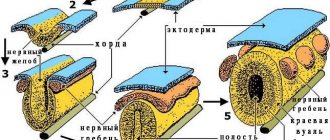
Lateral groove
(lat. sulcus lateralis), also known as
the lateral fissure
(lat. fissura lateralis),
lateral fissure
,
Sylvian fissure
- one of the largest fissures of the telencephalon, separates the frontal and parietal lobes from the temporal lobe. The insular lobe is located deep in the sulcus.
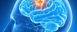
Meningiomas
Meningiomas can be considered the most common tumors localized in the intracranial space - they account for approximately 30% of the total number of all primary brain tumors. These tumors are formed from the cells of the arachnoid (arachnoid) membrane of the brain, and are mostly benign. The World Health Organization classifies meningiomas into three grades, depending on their malignancy: grade 1 – typical (completely benign); 2nd degree – atypical (conditionally benign); Grade 3 – anaplastic (malignant).
Medical statistics indicate that atypical and anaplastic meningiomas are quite rare - in 5 percent of cases, both occur.
Meningiomas most often develop in patients aged 40 to 70 years; in addition, they are much more common in women than in men. In children, such brain tumors are extremely rare - 1-1.5% of the total statistics.
Functions of the midbrain: Structure of the human brain
The structure of the brain can be imagined as a complex technical device. A great many cells are concentrated and functioning in it, making human life truly unique.
Or rather, they make our life possible in general. It is responsible for all functions of the human body, from the first breath, movement and ending with new skills.
It is always changing, improving, adapting to the constantly changing external environment.
The average brain, as its component, a very complex “device”, should go from the particular to the general. The structure is the first thing we start with. Also from the article the reader will be able to learn about what functions are inherent in the midbrain, i.e. what is he responsible for? In addition, the portal’s specialists are happy to share recommendations on how to maintain its functionality.
Structure and its features
When you touch on the structure and functionality of the midbrain, you cannot do without the use of terminology. All functions of this area are unique due to their complex structure.
The most important thing is that in this part of the brain there is the quadrigeminal and peduncles. Of course, they are not the only ones in the structure, but they are the main ones. The background includes nuclei, as well as matter (black and red).
Each of these elements is responsible for certain functions of the human body.
As mentioned above, the structure of the midbrain is complex. But despite this, its size is only 2 cm and is responsible for many reflection functions.
Its main components are the isthmus, roof and tire, which have a special structure. The isthmus is a combination of three elements:
- auditory loop;
- sail;
- legs.
The roof is a quadrigeminal structure, which, together with the lateral geniculate bodies, is responsible for the functionality of the visual apparatus.
The legs, together with the medial geniculate bodies, are responsible for the functioning of the hearing organs.
Thus, a person is able to see the reality around him, admire the beauty of objects and objects, distinguish sounds and enjoy the singing of birds. In addition, the structure of the lower hills is directly involved in the formation of an orientation reflex to sound, scientifically called the “start reflex.”
By the way, gray matter determines the thickness of each of the midbrain hills. The black substance is located next to the tire. There is also the Sylvian aqueduct, which is its continuation.
Presentation: “Structure and functional purpose of the midbrain”
The tegmentum itself contains nuclei that not only serve as unique links between the cerebellum and the human brain, but are mainly responsible for the coordination of the body and the manifestation of automatic movements.
The substantia nigra is an incredibly interesting element of the midbrain. With its help, a person can move his fingers very clearly, which helps him write, play musical instruments, especially the violin, and also draw. It turns out that the average brain is a real patron of those areas of creativity in which it is important to be able to hold a certain pose for a long time, spending a minimum amount of energy.
Functional meaning
Each part of the human brain is equally important, since together they create a completely unique system, incomparable to anything else.
Not a single technique, even the most modern, or even one that will appear in the future, will most likely be able to even repeat its functions, let alone surpass it. To date, only a minimal part of it has been studied.
But, with regard to its functions of controlling the human body, a lot of definite things can be said.
The middle part of the brain is responsible for many functions, among which the main ones are the following:
- sensory;
- conductor;
- possibility of movement;
- reflexes.
The centers of the subcortex, which are part of the structure of the midbrain, are aimed at influencing and maintaining the performance of the auditory and visual systems. It is here that the nuclei of the cranial nerves are located, ensuring the functioning of the eye muscles.
Of course, the other elements also work. For example, a muscle contracts due to the injection of a black substance and a red nucleus. However, this applies only to automatic actions, such as raising and lowering the arms and legs, as well as all other mechanical movements acquired through experience.
But maintaining muscle tone is the direct responsibility of the middle part of the brain of the human body.
But, despite this, each function is important for normal human life.
Every movement is under his control - coordination in time and space, the presence of muscle tone, as well as such habitual and often unnoticed actions as the ability to swallow food and water, move and even breathe air.
We don’t even notice how many reactions our brain uses when a person performs a seemingly simple movement - throwing a ball. Therefore, such a state as irritability is also a function of the middle part of the brain.
It is noteworthy that it is also responsible for reflex functionality, i.e. synchronous movement of the eyes, reaction to a sudden switch on of light or sound, turns, sensations of someone else's gaze on the back.
And it is not unimportant that there is also a pain center here. If you constantly make him excited, then over time the person will feel less physical pain.
Often the midbrain functions with the medulla oblongata and together they control almost every reflex action of the human body. Their functioning allows people to navigate in space, instantly respond to external stimuli, and also turn their body in the direction they are looking.
As you can see, the functions of the midbrain are difficult to overestimate, and this once again proves the unique structure of the human brain.
Symptoms of Sylvian fissure meningioma
The Sylvian fissure (sulcus) separates the temporal and frontoparietal lobes of the brain. This groove is one of the deepest in the brain; it runs along the lateral periphery of the hemisphere from top to bottom/anterior, dividing into three branches.
Meningioma of the Sylvian fissure has symptoms that are characteristic of almost most tumors of the frontal lobe of the brain:
- mental disorders (emotional instability, primitive behavior);
- personal changes;
- epileptic seizures;
- Broca's aphasia (speech disturbances/difficulties);
- impaired coordination of movements;
- olfactory disturbances;
- hyperkinesia (uncontrolled movements);
- convulsions.
Treatment of meningioma
The choice of the optimal treatment option depends on many factors, the main ones being:
- tumor size;
- the degree of its infiltration into neighboring tissues;
- proximity to vital brain centers;
- degree of malignancy, etc.
The most effective method of treating benign brain tumors is transcranial surgery, which allows the operating surgeon to gain full access to the site of the operation.
Stereotactic radiation therapy can be used as an additional treatment for Sylvian fissure meningioma. If, due to some circumstances, craniotomy is not possible, radiation therapy can be used as the main treatment method.
“NEIRODOC.RU is medical information that is maximally accessible for assimilation without special education and created on the basis of the experience of a practicing physician.”
If you are looking for information on the topic “cyst in the brain” or an answer to the question “what is a cyst in the brain?”, then this article is for you. A cyst in the brain, or more precisely, an arachnoid cerebrospinal fluid cyst, is a congenital formation that arises during development as a result of splitting of the arachnoid (arachnoid) membrane of the brain. The cyst is filled with cerebrospinal fluid (CSF), a physiological fluid that washes the brain and spinal cord. True congenital arachnoid cysts should be distinguished from cysts that appear after damage to the brain due to traumatic brain injury, stroke, infection, or surgery.
Arachnoid cyst ICD10 code G93.0 (cerebral cyst), Q04.6 (congenital cerebral cysts).
Sylvian aqueduct: structure and functions
The Sylvian aqueduct was known back in ancient times. Already in those days, scientists interested in studying human anatomy knew about the circulatory system and the heart, the digestive system. But the brain still holds the most mysteries. Both in ancient times and now, when medicine has the latest technologies.
Origin of the term
Hippocrates theorized that blowing your nose is harmful to the body, since this action causes the loss of part of the brain. In this regard, having learned and gained the opportunity to dissect corpses, scientists began to devote a lot of time to studying the brain.
The brain consists of many components: the shell, the enclosure, the globus pallidus, the nuclei, the tegmentum, the circle. The midbrain aqueduct is located among its formations.
Earlier in the seventeenth century there was a famous scientist named Francis Silvius. He was just doing brain research.
It is he who is credited with the discovery and description of such a department as the Sylvian aqueduct, which was later named in his honor.
Probably almost everyone knows about a disease called meningitis. If it is suspected, doctors select cerebrospinal fluid (CSF) for analysis through a puncture. There is only about half a glass of such liquid in the human body. But it is very important for maintaining normal pressure levels in the brain.
In simple terms, the spinal cord is immersed in cerebrospinal fluid. Inside it there is another channel with the same cerebrospinal fluid. The canal ends at the foramen magnum, where it expands into the lateral ventricles.
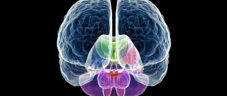
Midbrain
Very often questions arise about the brain. A table of its structural elements helps explain their functionality.
| Divisions of the brain | Functions |
| Medulla | Regulates heart rhythm, breathing, blood pressure |
| Bridge | Responsible for eye movement and facial expressions |
| Midbrain | Reflex movement of the head |
| Diencephalon | Controlling the functioning of internal organs |
| Cerebellum | Responsible for clear coordination |
It is the midbrain that is responsible for visual and auditory reflexes. And its central part regulates unconscious stereotypical movements: tilts and turns of the head and torso.
In terms of complexity, the midbrain is inferior to other parts of the brain. That's why it is studied the least.
The parts of the midbrain are the roof, the tire, and the legs. Inside there is a narrow channel called the cerebral aqueduct. It is designed to connect the ventricles of the diencephalon and rhombencephalon.
The midbrain is responsible in the human body for orientation reflexes, postures, vision, hearing, chewing and swallowing movements, and muscle tone.
Silviev water supply
As mentioned earlier, in the brain there is a canal that connects the third and fourth ventricles to each other. This is the Sylvian aqueduct, which is an integral part of the central canal. The cross-section of a water pipeline may look like a triangle, rhombus or ellipse. Its length does not exceed two centimeters.
Why was the Sylvian aqueduct invented and created by nature? Its function is trophic, that is, it is to deliver nutrients to brain cells. Without food they may die.
In addition, the brain is located around it. The table of his departments shows this clearly. These are the nuclei of the reticular formation, the oculomotor nerve. Thanks to the Sylvian aqueduct, cerebrospinal fluid circulates in the brain, creating pressure.
In total it contains just over one hundred milliliters.
Liquor is indispensable for creating the phenomenon of depreciation and balance. In addition, it serves to form a hydrostatic membrane and ensures a position of the nerve roots that reduces the tension of the blood vessels.
Liquor is also indispensable for providing tissues with nutrition. With its help, nutritional cells are delivered to them. And after the process of processing them, the liquor removes waste substances.
The cellular immune systems included in its composition protect against microbes.
The plumbing is extremely important for maintaining normal intracranial pressure. If the fluid is lost, the pressure will drop, which will immediately affect it in the form of unbearable headache, nausea, vomiting, and decreased vision. If there is a loss of cerebrospinal fluid, then an urgent magnetic resonance imaging (MRI) procedure is necessary.
Thus, the role of the aqueduct of Sylvius in the brain is irreplaceable. It is thanks to him that a person feels good, his intracranial pressure remains normal, and brain cells can feed normally and, therefore, function.
Source: https://fb.ru/article/290268/silviev-vodoprovod-stroenie-i-funktsii
Classification of arachnoid cerebrospinal fluid cyst.
- Sylvian fissure arachnoid cyst 49% (the fissure formed by the frontal and temporal lobes of the brain), is sometimes called a temporal lobe arachnoid cyst.
- Arachnoid cyst of the cerebellopontine angle 11%.
- Arachnoid cyst of the craniovertebral junction 10% (junction between the skull and the spine).
- Arachnoid cyst of the cerebellar vermis (retrocerebellar) 9%.
- Arachnoid cyst sellar and parasellar 9%.
- Arachnoid cyst of the interhemispheric fissure 5%.
- Arachnoid cyst of the convexital surface of the cerebral hemispheres 4%.
- Arachnoid cyst of the clivus 3%.
Notes[ | ]
- ↑ 12
Foundational Model of Anatomy - Jee G. Chi;
Elizabeth C. Dooling; Floyd H. Gilles. Gyral development of the human brain (English) // Annals of Neurology (English) Russian. : journal. — 1977. — January (vol. 1, no. 1). - P. 86-93. - doi:10.1002/ana.410010109. — PMID 560818. Archived December 16, 2012. - Carpenter, Malcolm.
Core text of neuroanatomy. - 3rd. — Williams & Wilkins. - P. 22. - ISBN 0683014552. - Furrows and convolutions of the brain, cerebral hemispheres // Great Medical Encyclopedia / ch. ed. B.V. Petrovsky. — 3rd ed. - M.: Soviet Encyclopedia, 1980. - T. 3. - P. 339.
- ↑ 1 2 3 Collice, M.;
Collice, R.; Riva, A. Who discovered the sylvian fissure? (undefined) // Neurosurgery. - 2008. - T. 63, No. 4. - P. 623-628. - doi:10.1227/01.NEU.0000327693.86093.3F. - PMID 18981875. - Zanchin, G.;
De Caro, R. The nervous system in colors: the tabulae pictae of GF d'Acquapendente (ca. 1533-1619). (English) // J Headache Pain: journal. - 2006. - October (vol. 7, no. 5). - P. 360-366. - doi:10.1007/s10194-006-0340-0. - PMID 17058037. - Riva, A.
GF d'Acquapendente tabulae pictae on the nervous system. (English) // J Headache Pain: journal. — 2007. — September (vol. 8, no. 4). - P. 253-254. - doi:10.1007/s10194-007-0408-5. - PMID 17906833. - Stark, Tanja
Crashing Out with Sylvian: David Bowie, Carl Jung and the Unconscious (English) (22 June 2015). - Penfield, Wilder and Marshall Faulk Jr., (1955) “The insula. Further observations on its function', Department of Neurology and Neurosurgery”, Brain, 78: 445-471. 1955; Arzy et al., 2006. Arzy Shahar, Margitta Seeck, Stephanie Ortigue, Laurent Spinelli L and Olaf Blanke. (2006) “Induction of an Illusory Shadow Person.” Nature, 443: 287.
- Meshberger, Frank Lynn (10 October 1990). "An Interpretation of Michelangelo's Creation of Adam Based on Neuroanatomy". JAMA. 264(14):1837–41. doi:10.1001/jama.1990.03450140059034. PMID 2205727. Retrieved September 24, 2012. Pdf. Excerpt on Mental Health & Illness.com. Retrieved 21 September 2010.
- Di Bella, Stefano.
The “Delivery” of Adam: A Medical Interpretation of Michelangelo // Mayo Clinic Proceedings (English) Russian. : journal. — 2020. — Vol. 90, no. 4. - P. 505-508. - doi:10.1016/j.mayocp.2015.02.007. - PMID 25841253.
Clinical signs of an arachnoid cyst.
Clinical manifestations of arachnoid cysts usually occur in early childhood. In adults, symptoms appear much less frequently. They depend on the location of the arachnoid cyst. Often cysts are asymptomatic, are an incidental finding during examination and do not require treatment.
Typical clinical manifestations of an arachnoid cyst:
- General cerebral symptoms due to increased intracranial pressure: headache, nausea, vomiting, drowsiness.
- Epileptic seizures.
- Protrusion of the skull bones (this happens rarely, I have never personally encountered it).
- Focal symptoms: monoparesis (weakness in the arm or leg), hemiparesis (weakness in the arm and leg on one side), sensory disturbances of the mono- and hemitype, speech disorders in the form of sensory (lack of understanding of spoken speech), motor (inability to speak) or mixed (sensory-motor) aphasia, loss of visual fields, paresis of cranial nerves.
- Sudden deterioration, which may be accompanied by depression of consciousness up to coma:
- Due to hemorrhage into the cyst;
- Due to cyst rupture.
Sylvian aqueduct of the brain
It is known that anatomy has been descriptive for quite a long time. Its tasks were completely satisfied not by a functional explanation of the work of certain organs, but simply by stating a set of certain facts, and confirming the relationship of these facts through a fairly large number of autopsies.
Thus, already ancient doctors knew about the function of blood and heart. It was not difficult to guess the role of the stomach and intestines, to connect it with digestion and its role in life. It was more difficult with breathing; scientists for a long time could not understand where arterial blood becomes venous, and only Marcello Malpighi, using a microscope, discovered the existence of a capillary network.
The largest number of terms that came from this era are hidden in the brain. He “got it” the most.
Consider, for example, Hippocrates’ statement that blowing your nose is harmful to life, since in doing so a person loses brain matter, and the brain, as everyone knows, serves to cool the “hot vital force.”
Therefore, after the advent of modern times, having received the opportunity to dissect corpses, anatomists began to methodically describe the structures of the brain.
The brain has a shell, a fence, and a pallidum. There you can find the caudate and lenticular nuclei, the tegmentum, and the quadrigeminal. The brain contains both the Circle of Willis and the septum pellucidum.
The flight of fancy is limitless, and therefore, to the question, “which organ has running water”? – you can safely answer that the water supply must be looked for among the formations of the brain. It is named Silviev, after the doctor and anatomist who first described it during the life of Johann Sebastian Bach, that is, not so early. What is it, and why is such a structure needed?
About the importance and circulation of cerebrospinal fluid
It is known that when a person is suspected of meningitis, doctors take cerebrospinal fluid, that is, cerebrospinal fluid, for analysis by performing a puncture. This fluid is contained in the central nervous system in small quantities - only half a glass or a small glass for an adult, but its functions are very important.
Thus, cerebrospinal fluid maintains a certain level of pressure, performs a nutritional and trophic function, and also mechanically protects the brain from shock, playing the role of a “shock-absorbing cushion.”
It circulates in the subarachnoid spaces and washes the spinal roots. The cerebrospinal fluid is located inside the blood-brain barrier, so many substances cannot penetrate there.
The cerebrospinal fluid begins from the basal cistern at the level of the base of the spinal cord . The spinal cord itself seems to “hang”, immersed in the cerebrospinal fluid, and inside it there is a central canal, which also contains cerebrospinal fluid.
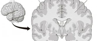
This central canal continues when the spinal cord enters the foramen magnum, but since the brain in its phylogenetic development slightly “rotated” and twisted, then inside the brain there are extended extensions of the central canal and its lateral branches, which are called the lateral ventricles .
In the image below, the lateral ventricles are shown in green, and the unpaired structures, which include the cerebral aqueduct, or aqueductus cerebri, are shown in purple. Let us remember that the word aqueductus is Latin.
All anatomical structures, according to the established rules of the BNA, or international anatomical classification, must be Latin. But they are often duplicated in Greek. So, “cerebrum” is the brain translated from Latin.
Hence the terms “cerebral”, “cerebrovascular” and so on. And translated from Greek “brain” is “encephalon”, that is, “what is inside the head”.
And terms such as “encephalitis” and “encephalopathy” are also applicable and exist on a par with the Latin names.
Unpaired liquor structures
The central structures are the unpaired ventricles of the brain, namely the IV and III ventricles, if you rise up from the spinal cord. The floor of the fourth ventricle is nothing more than the famous “diamond-shaped fossa”, in which the nuclei of the cranial nerves lie.
Gradually, the roof of the fourth ventricle narrows, turning into a narrow canal, which then opens into the cavity of the third ventricle, ventriculi tercii. This narrow cerebral canal, which connects the unpaired ventricles, is the aqueductus cerebri, or cerebral aqueduct.
It lies directly under the quadrigeminalis, and is about 2-2.5 cm long in an adult. If you draw its cross section, the aqueductus cerebri will look like a triangle, an ellipse, or a rhombic cross-section.
All of these are variants of the norm.
The diagram below shows the projection of the aqueduct, when viewed from above, and its relationship between the third and fourth ventricles.
More about plumbing
Since this central narrow duct leaves the region of the trunk and pons, running in the structures of the midbrain, sometimes, taking into account its topography, it is called the aqueduct of the midbrain, aqueductus cerebri mesencephali.
Further, after flowing into the third ventricle, the unpaired or middle part of the liquor ends, giving way to the lateral ventricles of the brain. On a sagittal section, the topography of the aqueduct is clearly visible: ventrally it passes over the cerebellar peduncles, leaving above the lamina tecti, the plate of the roof of the midbrain.
What is the role of the plumbing? It performs a trophic function for the surrounding brain structures, and the following formations are located around the Sylvian aqueduct:
- separately lying nuclei of the oculomotor and trochlear nerves, that is, the III and IV pairs of cranial nerves;
- A little more ventral in the thickness of the gray matter are the Yakubovich nuclei, which carry out the autonomic innervation of the eye: they compress and dilate the pupil, more precisely its sphincter, or the orbicularis muscle, depending on the amount of light entering the retina. The pupil itself is an opening and does not contain any structures.
Also, the role of the water supply is very important in maintaining normal intracranial pressure. When the aqueductus cerebri is compressed by a tumor, or a space-occupying formation of another nature, for example, a traumatic or parasitic cyst, intracranial hypertension syndrome may occur, which is manifested by bursting headaches, nausea, vomiting, and progressive decrease in vision.
Such clinical manifestations should be a reason for urgent MRI, fundus examination and consultation with a neurosurgeon.
Pogrebnoy Stanislav Leonidovich, neurologist
Rate this article:
Total: 124
4 124
Source: https://mozgius.ru/stroenie/silviev-vodoprovod.html
Diagnosis of arachnoid cyst.
Neuroimaging methods are usually sufficient to diagnose an arachnoid cyst. These are computed tomography (CT) and magnetic resonance imaging (MRI).
Additional diagnostic methods are contrast studies of the cerebrospinal fluid pathways, such as cisternography and ventriculography. They are required occasionally, for example, when examining median suprasellar cysts and when affecting the posterior cranial fossa for the purpose of differential diagnosis with Dandy-Walker anomaly.
Examination of the fundus by an ophthalmologist for hypertension syndrome (intracranial hypertension).
Electroencephalography (EEG) in case there was an epileptic seizure, to determine whether it was really caused by a cyst.
Treatment of arachnoid cyst.
As I said above, most congenital arachnoid liquor cysts are asymptomatic and do not require any treatment. Sometimes a neurosurgeon may recommend dynamic monitoring of the size of the cyst; for this, it will be necessary to periodically perform computed tomography or magnetic resonance imaging.
In rare cases, when an arachnoid cyst is accompanied by the symptoms described above and has a mass effect, surgical treatment is resorted to.
In some cases with a sharp deterioration, due to rupture of the arachnoid cyst or hemorrhage into it, urgent surgical treatment is resorted to.
There is no standard size for an arachnoid cyst. Indications for surgery are determined taking into account the location and symptoms of the arachnoid cyst, and not just its size. This can only be determined by a neurosurgeon during an in-person examination.
Absolute indications for surgery:
- intracranial hypertension syndrome caused by an arachnoid cyst or concomitant hydrocephalus;
- the appearance and increase of neurological deficit.
Relative indications for surgery:
- large “asymptomatic arachnoid cysts” that cause deformation of neighboring lobes of the brain;
- progressive increase in cyst size;
- deformation of the cerebrospinal fluid tract caused by the cyst, leading to disruption of the cerebrospinal fluid circulation.
Contraindications for surgery:
- decompensated state of vital functions (unstable hemodynamics, breathing), terminal coma (coma III);
- the presence of an active inflammatory process.
There are three possible options for surgical treatment of arachnoid cysts. Your treating neurosurgeon chooses the tactics, taking into account the size of the cyst, its location and your wishes. Not all three methods are suitable for all arachnoid cysts.
Evacuation of an arachnoid cyst through a burr hole in the skull using a navigation station. The advantage is simplicity and speed of implementation with minimal trauma to the patient. But there is a drawback - the high rate of cyst recurrence.
Open surgery, that is, craniotomy (cutting out a bone flap on the skull, which is placed in place at the end of the operation) with excision of the walls of the cyst and fenestration (drainage) into the basal cisterns (cerebrospinal fluid spaces at the base of the skull). This method has the advantage of allowing direct examination of the cystic cavity, avoiding a permanent shunt, and is more effective for the treatment of arachnoid cysts consisting of multiple cavities.
Shunt surgery with installation of a shunt from the cyst cavity into the abdominal cavity or superior vena cava near the right atrium through the common facial vein or internal jugular vein. Many foreign and domestic neurosurgeons consider shunting of an arachnoid cerebrospinal fluid cyst to be the best treatment method, but it is not suitable in all cases. The advantage is low mortality and low rates of cyst recurrence. The disadvantage is that the patient becomes dependent on the shunt, which is installed for life. If the shunt becomes blocked, it will have to be replaced.
Early postoperative complications - liquorrhea, marginal necrosis of the skin flap with dehiscence of the surgical wound, meningitis and other infectious complications, hemorrhage into the cyst cavity.
Outcomes of treatment of arachnoid cyst.
Even after successful surgery, part of the cyst may remain, the brain may not fully expand, and displacement of the midline structures of the brain may persist. It is also possible to develop hydrocephalus. As for focal neurological symptoms in the form of paresis and other things, the longer it exists, the less chance of its recovery.
The materials on the site are intended to familiarize you with the characteristics of the disease and do not replace an in-person consultation with a doctor. There may be contraindications to the use of any medications or medical procedures. You cannot self-medicate! If there is something wrong with your health, consult a doctor.
If you have questions or comments about the article, leave comments below on the page or participate in the forum. I will answer all your questions.
When using materials from the site, the active reference is obligatory.