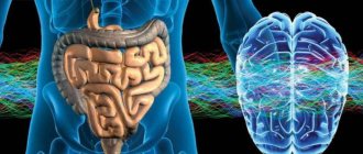
The parasympathetic nervous system is a component of the autonomic system. The latter is responsible for human growth, controls the energy received from the lungs and intestines, and regulates blood circulation. It is completely interconnected with heart rate.
The autonomic system works with impulses coming from the brain, due to which the tone of the vessels through which blood and lymph move is regulated. If a person has suffered a head injury, this functionality may be impaired.
Structure of the vegetative system
The vegetative system has a special structure. Its main components:
- hypothalamus;
- pituitary;
- "nervus vagus";
- nerve nuclei located in the occipital zone and responsible for head control;
- sacral nuclei, which are responsible for returning blood to the heart muscle;
- cardiac plexuses, which provide cardiac impulses;
- thoracic nerve plexuses;
- celiac nerve plexuses;
- lumbar nerve plexus.
The basis for the autonomic system are the nuclei of the cranial nerves (pituitary gland, vagus nerve, pineal gland and hypothalamus). The left organs are controlled through the left nuclei, and the right ones through the right ones. Such a system depends on the volume of blood in the body; its “quality” is influenced by the skull, spine, pelvis, and the condition of the throat, nasal and frontal sinuses, lungs and liver.
The visceral system has two sections: sympathetic and parasympathetic.
Parasympathetic nervous system
This subsection “turns on” its functions when a person is at rest.
Thanks to it, the pupils constrict, the heart begins to beat more slowly, blood pressure decreases, and accordingly, the blood vessels come to a relaxed state. Also, the functions of the parasympathetic nervous system include providing secretion from the glands, helping to contract the intestines and relaxing the sphincters. If the body begins to tense up, then the sympathetic department begins to work, which performs all the actions in reverse: dilates the pupils, makes the heart beat faster, contracts the sphincters, etc.
For the work of internal organs, the autonomic system has the solar, pericardial, pelvic and mesenteric plexuses. The control involves the nuclei of the hypothalamus, which are located in the limbic-reticular complex. At this time, a unification of both mental and somatic processes occurs. The parasympathetic part of the autonomic nervous system “helps” empty the gallbladder and bladder, and is also involved during bowel movements.
The center of such a system is the brain and spinal cord. In these areas of the body, efferent fibers of the anterior roots are formed, which are responsible for vasodilation, delayed sweat secretion, contraction and relaxation of the hair muscles located on the body and limbs.
The cranial area has its own centers, which are located in the mesencephalic and bulbar parts. The peripheral department has areas:
- Preganglionic fibers that pass through the 3rd, 5th, 9th and 10th pairs of cranial nerves.
- Terminal nodes that are located next to organs.
- Postganglionic fibers, which can be independent or part of a nerve complex.
Irritation of such fibers activates one or another organ. If the hypogastric plexus comes into action, then the organs of the corresponding area will be emptied, which also takes part in contractions of the uterus in women.
The pudendal nerves also deserve special attention, as they have a complex structure. The fact is that they have animal and autonomic fibers that enter the hypogastric plexus. They are connected to sympathetic fibers, which connect in the pelvic cavity, and there, after passing through the lower hypogastric plexus, they reach the organs located there.
One of the components of the parasympathetic system is the intramural nervous system. It is most localized in the digestive tract and has several plexuses:
- muscular-intestinal, which is located between the longitudinal and annular muscles of the digestive tube;
- submucosal, which, accordingly, is located in this area.
By the way, it is the last plexus that further grows into a network of glands and villi. Nerve fibers of the entire autonomic system approach them.
SYMPATHETIC AND PARASYMPATHETIC DIVISIONS AND THEIR DIFFERENCES
⇐ PreviousPage 3 of 5Next ⇒Based on anatomical and functional differences in the autonomic nervous system, two divisions were identified - sympathetic and parasympathetic.
The sympathetic department is trophic in its main functions. It provides increased oxidative processes, increased respiration, increased heart activity, i.e. adapts the body to conditions of intense activity. In this regard, the tone of the sympathetic nervous system predominates during the day, and at night - the parasympathetic one (“the kingdom of the vagus”). The parasympathetic department plays a protective role (constriction of the pupil, bronchi, decreased heart rate, emptying of the abdominal organs).
The sympathetic and parasympathetic divisions often act as antagonists (Table 1). However, this antagonism is relative. With a sharply changed functional state of the organ, they can act unidirectionally as synergists. In response to increased activity of the body, parasympathetic shifts occur, aimed at restoring energy potential and homeostasis. Thanks to the activity and synergy of both parts of the autonomic nervous system, long-term, adaptive activity of the body is possible.
Thus, between them there is not so much antagonism as interaction, which ensures the most subtle regulation of organ activity.
The sympathetic and parasympathetic divisions also differ in mediators - substances that transmit nerve impulses at synapses. The mediator in sympathetic nerve endings is sympathin (similar to norepinephrine). The mediator of parasympathetic nerve endings is a substance close to acetylcholine.
Along with the functional ones, there are a number of morphological differences in the sympathetic and parasympathetic divisions of the autonomic nervous system, namely:
1. The foci of parasympathetic fibers exiting the brain are separated from each other (mesencephalic, bulbar, sacral sections), sympathetic fibers exit from one, but more extended focus (thoracolumbar section).
2. The sympathetic nodes include nodes of the 1st and 2nd order, and the parasympathetic nodes include the 3rd order (terminal). In this connection, preganglionic sympathetic fibers are shorter, and postganglionic fibers are longer than parasympathetic.
3. The parasympathetic division has a more limited area of innervation, innervating only internal organs. The sympathetic department of the autonomic nervous system, in addition to internal organs, innervates all blood vessels, sweat, sebaceous glands and hair muscles of the skin, as well as skeletal muscles, providing it with trophic innervation.
SYMPATHETIC DIVISION OF THE AUTONOMIC NERVOUS SYSTEM
The sympathetic nervous system consists of central and peripheral divisions.
The central section is represented by the nuclei of the lateral horns of the gray matter of the spinal cord (nuclei intermediolaterales) of the following segments: C8, Th1-12, L1-3 (thoracolumbar region).
The peripheral division of the sympathetic nervous system consists of:
1) nodes of the first order, ganglia trunci sympathici;
2) internodal branches, rami interganglionares;
3) connecting branches are white and gray, rami communicantes albi et grisei;
4) nodes of the second order, ganglia intermediae, involved in the formation of plexuses;
5) visceral nerves, consisting of sympathetic and sensory fibers and heading to the organs, where they end in nerve endings;
6) sympathetic fibers running as part of the somatic nerves.
SYMPATHETIC TRUNK, truncus sympathicus, paired, located on both sides of the spine in the form of a chain of first-order nodes, ganglia trunci sympathici (Fig. 7).
| Rice. 7. Diagram of the structure of the sympathetic trunk (from Foss and Herlinger) 1 - cervical nodes; 2 - thoracic nodes; 3 - lumbar nodes; 4 - sacral nodes; 5 - g. impar. | In the longitudinal direction, the nodes are interconnected by branches, rami interganglionares. In the lumbar and sacral regions there are also transverse commissures that connect the nodes of the right and left sides. The sympathetic trunk extends from the base of the skull to the coccyx, where the right and left trunks are connected by one unpaired coccygeal node, gangllion impar. Topographically, the sympathetic trunk is divided into 4 sections: cervical, thoracic, lumbar and sacral. The sympathetic trunk in the cervical spine is covered with fascia, fascia prevertebralis. In the thoracic, lumbar and sacral regions, it is respectively covered by fasciae endothoracica, subperitonealis et fascia pelvis. The nodes of the sympathetic trunk are connected to the spinal nerves by white and gray communicating branches. |
The white communicating branches, rami communicantes albi, consist of preganglionic sympathetic fibers, which are axons of the cells of the intermediolateral nuclei of the lateral horns of the spinal cord. They are separated from the spinal nerve trunk and enter the nearest nodes of the sympathetic trunk, where part of the preganglionic sympathetic fibers are interrupted. The other part passes through the node in transit and through the internodal branches reaches more distant nodes of the sympathetic trunk or passes to nodes of the second order. Sensitive fibers, the dendrites of the cells of the spinal ganglion, also pass through the white connecting branches.
The white connecting branches go only to the thoracic and upper lumbar nodes. Preganglionic fibers enter the cervical nodes from below from the thoracic nodes of the sympathetic trunk through the rami interganglionares (Fig. 8), and into the lower lumbar and sacral nodes - from the upper lumbar nodes also through the internodal branches.
| Rice. 8. Sympathetic nervous system (according to S.P. Semenov). Sh8 - P3 - segments of the spinal cord with sympathetic nuclei; 1 - superior cervical sympathetic ganglion; 2 - middle cervical; 3 - lower cervical; 4 - stellate ganglion; 5 - solar plexus ganglia; 6 - large and 7 - small splanchnic nerves; 8- inferior mesenteric ganglia. |
From the nodes of the sympathetic trunk, part of the postganglionic fibers joins the spinal nerves - gray connecting branches, rami communicantes grisei, (there is no myelin sheath) and as part of the spinal nerves, sympathetic fibers are sent to the soma, where they end with nerve endings on the sebaceous and sweat glands, smooth muscles, levator hair of the skin, in the wall of peripheral vessels, as well as in skeletal muscles in order to ensure the regulation of its trophism and maintain tone. The gray connecting branches arise from all nodes of the sympathetic trunk and constitute the somatic part of the sympathetic nervous system.
In addition to the gray connecting branches, visceral branches depart from the nodes of the sympathetic trunk to innervate the internal organs - the visceral part of the sympathetic nervous system. It consists of: postganglionic fibers (cell processes of the sympathetic trunk), preganglionic fibers that passed through the first order nodes without interruption, as well as sensory fibers (cell processes of the spinal nodes).
It is important to note that preganglionic fibers in the ganglia of the sympathetic trunk branch repeatedly and form synapses on many cell bodies of effector neurons. The ratio of preganglionic fibers to postganglionic fibers can reach 1: 100. This leads to the phenomenon of animation (multiplication), i.e. to a sharp expansion of the excitation area (generalization of the effect). Due to this, a relatively small number of central sympathetic neurons provide innervation to all organs and tissues. For example, when an animal’s preganglionic sympathetic fibers passing through the anterior roots of the IY thoracic segment are irritated, constriction of the blood vessels of the scalp, neck, forelimb, dilatation of the coronary vessels, and constriction of the vessels of the kidneys and spleen can be observed.
The cervical sympathetic trunk most often consists of three nodes: superior, middle and inferior. The nodes of the cervical spine do not have white connecting branches. Preganglionic fibers come to them from the upper thoracic nodes through the internodal branches.
The upper cervical ganglion, ganglion cervicale superius, is fusiform, about 2 cm long, lies in front of the transverse processes of the II-III cervical vertebrae, on m. longus capitis. The following branches depart from it:
1. Gray connecting branches to the I-IY cervical spinal nerves;
2. The internal carotid nerve, n.caroticus internus, which in the form of two branches approaches the artery of the same name and, entwining it, forms the internal carotid plexus, plexus caroticus internus. The continuation of this plexus in the cranial cavity is the cavernous plexus, plexus cavernosus. Branches extend from the internal carotid plexus: nn. caroticotympanici, which together with the branches of the glossopharyngeal nerve form the plexus tympanicus; n. petrosus profundus, which connects to the parasympathetic nerve - n. petrosus major and forms n. canalis pterygoidei, entering the pterygopalatine ganglion. Without interruption in this node, sympathetic fibers follow to the vessels and glands of the mucous membrane of the nasal cavity and palate. From the cavernous plexus come plexuses for the branches of the internal carotid artery (plexus of the ophthalmic artery, anterior and middle cerebral arteries, plexus of the artery of the choroid plexus), as well as individual branches to the pituitary gland, trigeminal ganglion, oculomotor, trochlear and abducens nerves.
Following the course of the ophthalmic artery, sympathetic fibers are directed to the lacrimal gland, and also as part of the sympathetic root, radix sympathicus, enter the ciliary ganglion. In the node, the fibers are not interrupted, but are sent further as part of the short ciliary nerves, nervi ciliares breves, to the eyeball for innervation of the m.dilatator pupillae and the vessels of the eye. When the upper cervical node is affected, a narrowing of the pupil is observed on the side of the same name.
3. External carotid nerves, nn.carotici externi, which form a plexus around the artery of the same name - plexus caroticus externus. Due to the secondary plexuses along the branches of the external carotid artery, the salivary glands, dura mater and partially the pharynx, thyroid gland and larynx are innervated.
4. Laryngopharyngeal branches, rami laryngopharyngei, which, together with the branches of the vagus and glossopharyngeal nerves, form a nerve plexus in the wall of the pharynx, plexus pharyngeus, and some of the branches, together with n. laryngeus superior (from n. vagus) are directed to the larynx.
5. The superior cardiac nerve, n.cardiacus cervicalis superior, involved in the formation of the superficial (left) and deep (right) cardiac plexuses.
6. Ramus sinus carotici - goes to the bifurcation of the carotid artery, and the sensitive branch from n.glossopharyngeus also comes there.
7. Jugular nerve, n.jugularis, running along the internal jugular vein and disintegrating in the area of the jugular foramen into gray connecting branches to the lower node of the glossopharyngeal, nodes of the vagus and branches of the accessory and hypoglossal nerves.
The middle cervical node, ganglion cervicale medium, is located at the intersection of the inferior thyroid artery with the common carotid artery, at the level of the VI cervical vertebra. Sometimes he is missing. Its internodal branch to the lower cervical node is divided into two bundles, covering the subclavian artery in front and behind like a loop - ansa subclavia. Branches extend from it:
1. Gray connecting branches to the V, VI cervical spinal nerves.
2. Branches to the common carotid artery, forming the plexus caroticus.
3. Branches to the inferior thyroid artery - plexus thyroideus inferior.
4. Middle cardiac nerve, n. cardiacus cervicalis medius, entering the deep cardiac plexus.
The lower cervical ganglion, ganglion cervicale inferius, is located in the region of the initial section of the vertebral artery, at the level of the head of the 1st rib and often merges with the 1st thoracic node, forming the cervicothoracic node, ganglion cervicothoracicum (stellate, ganglion stellatum). Branches extend from it:
1. Gray connecting branches to the VII, VIII cervical and I thoracic spinal nerves.
2. Branches to the subclavian artery, forming the plexus subclavius along its branches.
3. Branches to the vertebral artery, forming the plexus vertebralis, due to which the membranes and vessels of the brain and spinal cord are innervated.
4. Inferior cardiac nerve, n. cardiacus cervicalis inferior, entering the deep cardiac plexus.
5. Branches to the phrenic nerve for innervation of the vessels of the abdominal cavity.
6. Branches to the trachea, bronchi, esophagus, where, together with the branches of the vagus nerve, they form plexuses.
The thoracic section of the sympathetic trunk consists of 10-12 nodes, ganglia thoracica, lying in front of the heads of the ribs. White connecting branches from the thoracic spinal nerves come to the nodes of the thoracic sympathetic trunk. The following branches depart from them:
1. Gray connecting branches to the thoracic spinal nerves.
Visceral branches depart from the upper 5-6 nodes to innervate the organs of the thoracic cavity, namely:
2. Thoracic cardiac nerves, nn. cardiaci thoracici, entering the deep cardiac plexus. All cardiac nerves extending from the nodes of the sympathetic trunk consist of sensory, postganglionic and partially preganglionic sympathetic fibers. The latter are interrupted in the nodes of the cardiac plexuses.
3. Branches to the aorta, forming the thoracic aortic plexus, plexus aorticus thoracicus, which is connected at the top with the cardiac plexus, and at the bottom with the celiac plexus.
4. Branches to the trachea and bronchi, participating together with the branches of the vagus nerve in the formation of plexus pulmonalis.
5. Branches to the esophagus directly from the nodes or from the aortic plexus, forming the plexus esophageus.
6. Branches depart from the V-IX thoracic nodes, forming the large splanchnic nerve, n. splanchnicus major.
7. From X-XI thoracic nodes - small splanchnic nerve, n. splanchnicus minor.
8. N departs from the XII thoracic node (if present). splanchnicus imus.
The splanchnic nerves pass between the crura of the diaphragm and enter the celiac plexus. They consist predominantly of preganglionic sympathetic and sensory fibers.
The lumbar section of the sympathetic trunk consists of 4-5 nodes, ganglia lumbalia, which lie on the anterior surface of the vertebral bodies (along the medial edge of the m. psoas major). A feature of these nodes is the presence of transverse fibers connecting the right and left nodes, which increases the extent of the spread of excitation.
Only the upper lumbar nodes have white connecting branches. Preganglionic fibers to the lower nodes come through the internodal branches from the upper lumbar. Branches extend from them:
1. Gray connecting branches to the lumbar spinal nerves.
2. Visceral nerves - splanchnic lumbar nerves, nn. splanchnici lumbales, consisting predominantly of preganglionic sympathetic and sensory fibers. The upper ones enter the celiac plexus, the lower ones enter the aortic and inferior mesenteric plexuses.
The sacral section of the sympathetic trunk is represented, as a rule, by four nodes, ganglia sacralia, located near the medial edge of the foramina sacralia pelvina, and one unpaired coccygeal node, ganglion impar. All nodes are connected by transverse commissures. They do not have white connecting branches. Preganglionic fibers come to them through the internodal branches from the upper lumbar nodes. Branches extend from them:
1. Gray connecting branches to the sacral and coccygeal spinal nerves.
2. Visceral branches - splanchnic sacral nerves, nn. splanchnici sacrales, consisting predominantly of preganglionic sympathetic and sensory fibers and entering the superior and inferior hypogastric plexuses.
PRESPINAL NODES AND VEGETATIVE
PLEXUS
Prevertebral nodes (ganglia intermedia) are part of the autonomic plexuses and are located in front of the spinal column. On the effector neurons of these nodes, preganglionic fibers end, passing through the nodes of the sympathetic trunk without interruption.
Autonomic plexuses are located mainly around blood vessels, or directly near organs. Topographically, the autonomic plexuses of the head and neck, chest, abdominal and pelvic cavities are distinguished.
In the head and neck area, the sympathetic plexuses are located mainly around vessels from the carotid artery system (many of them were mentioned above). They give fibers to the lacrimal gland, m. dilatator pupillae, to the salivary glands, thyroid, parathyroid glands. Next comes the laryngopharyngeal plexus, formed together with the branches of the vagus and glossopharyngeal nerves. Some fibers from the cervical plexuses innervate the trachea and esophagus.
In the chest cavity, the sympathetic plexuses are located around the descending aorta, in the region of the heart, at the hilum of the lung and along the bronchi, around the esophagus.
The most significant plexus of the thoracic cavity is the cardiac plexus, plexus cardiacus. It is formed by three pairs of cardiac nerves from the cervical nodes of the sympathetic trunk and branches of the vagus nerve. From these sympathetic and parasympathetic sources, two main nerve plexuses are formed: the superficial, plexus cardiacus superficialis, located between the concave side of the aortic arch and the division of the pulmonary trunk, and the deep, plexus cardiacus profundus, located behind the aortic arch - between it and the bifurcation of the trachea. A continuation of these plexuses are the plexuses along the coronary arteries - plexus coronarius dexter et sinister, as well as plexuses located in the wall of the heart. The most significant plexuses are located under the epicardium. There are 6 such plexuses that innervate the myocardium of the atria and ventricles, the septum between them, which are connected to the nodes of the conduction system of the heart and continue into the atrioventricular bundle (His).
The cardiac plexuses contain many vegetative (intramural) nodes, as well as afferent fibers - processes of the sensory nodes of the spinal nerves and the vagus nerve.
In the abdominal cavity, sympathetic plexuses surround the abdominal aorta and its branches (Fig. 9). Among them, the largest plexus is distinguished - the celiac plexus, as N.I. puts it. Pirogov - “brain of the abdominal cavity”.
| Fig.9. Diagram of the abdominal sympathetic plexuses (from Foss and Herlinger). 1- plexus coeliacus et gg. coeliaci; 2-g. aorticorenale et plexus renalis; 3-g. lumbale II; 4- truncus sympathicus; 5- plexus rectalis superior; 6- plexus aorticus abdominalis; 7- plexus mesentericus inferior; 8- plexus rectalis inferior; 9-nn. pelvini; 10- plexus hypogastricus inferior. |
Celiac plexus (solar), plexus coeliacus s. solaris, surrounds the beginning of the celiac trunk and the superior mesenteric artery. The plexus is bounded above by the diaphragm, on the sides by the adrenal glands, and below it extends to the level of the renal arteries. The following nodes take part in the formation of this plexus:
1. Right and left celiac ganglia, ganglia coeliaca, semilunar in shape.
2. Unpaired superior mesenteric ganglion, ganglion mesentericum superius.
3. Right and left aorto-renal nodes, ganglia aorticorenalia, located at the point of origin of the renal arteries from the aorta. These nodes receive preganglionic sympathetic fibers, which switch here, postganglionic sympathetic and parasympathetic, as well as sensory fibers passing in transit through the nodes.
The following nerves take part in the formation of the celiac plexus:
1. Greater and lesser splanchnic nerves, n. splanchnicus major et minor, extending from the thoracic nodes of the sympathetic trunk, which consist mainly of preganglionic sympathetic and sensory fibers. A minority of the fibers are represented by postganglionic fibers. The preganglionic fibers of the greater splanchnic nerve are interrupted in the celiac and superior mesenteric nodes, and the lesser - in the aortorenal nodes.
2. Lumbar splanchnic nerves, nn. splanchnici lumbales, from the upper lumbar nodes of the sympathetic trunk, containing predominantly preganglionic sympathetic fibers, interrupted in the nodes of the celiac plexus and sensory fibers.
3. Branches of the phrenic nerve, rami frenicoabdominales, consisting of sensory and postganglionic sympathetic fibers from the lower cervical ganglion of the sympathetic trunk to innervate the vessels of the abdominal cavity.
4. Branches of the vagus nerve, rami coeliaci, consisting mainly of preganglionic parasympathetic and sensory fibers.
Sensitive fibers of the spinal nodes take part in the formation of the celiac plexus: the upper cervical (phrenic nerve), 7 lower thoracic and 3 upper lumbar.
From the celiac plexus numerous fibers radiate out like rays of the sun in all directions. In this regard, the plexus is called the “solar plexus”.
A continuation of the celiac plexus are secondary paired and unpaired plexuses along the walls of the visceral and parietal branches of the abdominal aorta. Unpaired plexuses: hepatic, splenic, gastric, pancreatic and superior mesenteric. The fibers of the superior mesenteric plexus, spreading along the branches of the superior mesenteric artery, reach the pancreas, duodenum, jejunum, ileum, cecum, transverse colon.
The second most important in the innervation of the abdominal organs is the broadly looped abdominal aortic plexus, plexus aorticus abdominalis, located on the anterior and lateral surfaces of the abdominal aorta below the renal arteries and is a continuation of the celiac plexus. The lumbar splanchnic nerves, which arise from the lower lumbar nodes of the sympathetic trunk, also participate in its formation.
The inferior mesenteric plexus, plexus mesentericus inferior, departs from the aortic plexus, entwining the artery of the same name and its branches. At the root of this artery there is a rather large node, ganglion mesentericum inferius. The formation of the inferior mesenteric plexus involves the splanchnic lumbar nerves (from the lumbar nodes of the sympathetic trunk), branches of the celiac and superior mesenteric plexuses, which enter it from the intermesenteric plexus, plexus intermesentericus. The fibers of the inferior mesenteric plexus reach the sigmoid, descending and part of the transverse colon. The continuation of this plexus into the pelvic cavity is the superior rectal plexus, plexus rectalis superior, which accompanies the artery of the same name.
The fibers of the mesenteric plexuses come into contact with the intermuscular (plexus myentericus) - Auerbach and submucosal (plexus submucosus) - Meissner's plexuses, located in the walls of the gastrointestinal tract. The myenteric and submucosal plexuses consist of groups of parasympathetic cells (intramural ganglia) connected by bundles of nerve fibers. Preganglionic parasympathetic fibers are interrupted here.
Continuation of the abdominal aortic plexus downwards are the plexuses of the iliac arteries and arteries of the lower limb, as well as the unpaired superior hypogastric plexus, plexus hypogastricus superior, which at the level of the promontory is divided into the right and left hypogastric nerves, forming the inferior hypogastric plexus in the pelvic cavity.
Inferior hypogastric plexus , plexus hypogastricus inferior, or pelvic, plexus pelvinus, one of the largest vegetative plexuses (Fig. 10).
It is located on the sides of the rectum, is a plate on each side, extending from the sacrum to the bladder, from which secondary plexuses extend to the pelvic organs along the branches of the internal iliac artery.
In the lower hypogastric plexus, two sections are distinguished in men: anterioinferior and posterior, and in women there is also a middle section.
The upper part of the anterioinferior plexus innervates the bladder, the lower part in men innervates the prostate gland, seminal vesicles, vas deferens and cavernous bodies.
| Rice. 10. Inferior hypogastric plexus (according to G.F. Ivanov). 1- ureter; 2- plexus hypogastricus superior; 3- plexus hypogastricus inferior; 4-nn. pelvini; 5- rectum. |
In women, the middle section of the inferior hypogastric plexus sends nerve fibers to the genitals. Moreover, its lower part is towards the vagina and clitoris, the upper part is towards the uterus and ovaries. The posterior section of the inferior hypogastric plexus innervates the rectum.
The formation of the inferior hypogastric plexus involves vegetative nodes of the second order (sympathetic), nodes of the third order (periorgan, parasympathetic), as well as nerves and plexuses:
1. Internal sacral nerves, nn.splanchnici sacrales, consisting mainly of preganglionic sympathetic fibers that passed without interruption through the nodes of the sympathetic trunk, as well as sensory fibers from the sacral spinal nodes.
2. Branches of the inferior mesenteric plexus (plexus rectalis superior), consisting mainly of postganglionic sympathetic fibers - processes of cells of the inferior mesenteric ganglion and sensory fibers from the lumbar spinal nodes.
3. Internal pelvic nerves, nn. splanchnici pelvini, consisting of preganglionic parasympathetic fibers - processes of the cells of the intermediate-lateral nuclei of the sacral spinal cord (S2 - S4) and sensory fibers from the sacral spinal ganglia.
Sympathetic preganglionic fibers are interrupted in nodes of the second order, parasympathetic - in the third order. Thus, in the formation of the inferior hypogastric plexus, in addition to autonomic fibers, sensory fibers also take part - processes of cells of the lumbar, sacral and coccygeal spinal nodes.
PARASYMPATHETIC DIVISION
⇐ Previous3Next ⇒
Functions of the parasympathetic system
In fact, the sympathetic and parasympathetic systems are very connected, as one is responsible for tension and the other for relaxation. Accordingly, if one of them does not function, then the second will have nothing to do.
Due to the sympathetic system, the following reactions occur in the body:
- breathing becomes more frequent, the bronchial tubes expand, which means ventilation of the lungs increases;
- the heart begins to beat faster, blood pressure rises;
- in skeletal muscles, blood vessels dilate, and this, in turn, contributes to oxygen saturation of all tissues in the body;
- the person begins to sweat actively;
- the blood is saturated with glucose;
- the intestines reduce their peristalsis, the level of digestive enzymes decreases;
- the pupils of the eyes dilate.
As is clear from the above points, all these reactions occur at the moment of a person’s excitement, for example, during severe physical exertion or in a situation of stress.
The parasympathetic department of the autonomic nervous system is designed to restore the body after such “stress”, and it also helps to cleanse it. It provides the following processes:
- slows down the work of the heart muscle;
- restores normal breathing;
- helps the vessels of the genital organs and brain to expand;
- reduces blood pressure to normal;
- restores optimal blood glucose levels;
- helps the smooth muscles of the internal organs become toned, and the process itself is such that the bronchi narrow, contractions in the intestines intensify, the smooth muscles of the bladder also increase their tone;
- helps activate the digestive secretion glands;
- constricts the pupils in the eyes.
Since the parasympathetic department also helps to cleanse the body, thanks to it sneezing, coughing, vomiting and other similar processes occur.
Work principles
A key role in the physiology of the visceral nervous system is played by mediators - substances that ensure the transmission of the desired signal and contribute to the formation of a response command.
Main departments
The main mediators of the sympathetic nervous system are acetylcholine and norepinephrine.
Acetylcholine also plays a critical role in the functioning of the parasympathetic nervous system. It reduces the frequency of contractions of the heart muscle, dilating peripheral blood vessels and thereby lowering blood pressure. At the same time, under its action, the motility of the gastrointestinal tract increases, the smooth muscles of the walls of the bronchial tree, uterine membrane, gall and bladder contract.
Some fibers of the oculomotor nerve are sensitive to acetylcholine. In this regard, when the concentration of this mediator in the blood increases, the muscle surrounding the iris and the ciliary muscle contract. At the same time, the ligamentous apparatus relaxes, and the eye loses the ability to focus on objects located at a certain distance. A so-called spasm of accommodation occurs, or a spasm of the ciliary muscle (the internal muscle of the eye). The pupil narrows and the iris becomes flatter, which expands the lumen of Schlemm's canal and facilitates the outflow of fluid from the internal ocular media.
Acetylcholine has high activity against the secretory cells of the sweat and lacrimal glands, digestive and pulmonary epithelium. In addition, this mediator takes part in the work of the central nervous system, in large quantities it has a depressant effect, and in small quantities it facilitates the transmission of nerve impulses at the junction of nerve cells in the brain.
The second key transmitter of the sympathetic nervous system, norepinephrine, is produced by adrenal medulla cells from dopamine. According to its chemical structure, it is a precursor of adrenaline, therefore it has a similar physiological effect. Cells capable of responding to the level of concentration of this mediator in the blood are divided into several types. Depending on which group of receptors is activated in a particular case, this hormone can have the following effects:
- Narrow the lumen of blood vessels, regulate peripheral vascular resistance and thereby influence the level of blood (arterial) pressure. When adopting a vertical body position, the concentration of norepinephrine in the blood increases significantly, which ensures a normal level of blood pressure in the vessels of the extremities.
- Activate a person’s motor and mental activity in stressful situations, shock, anxiety and nervous tension.
- Strengthen the work of the heart muscle and increase the volume of blood pushed by the heart into the lumen of large blood vessels, thereby increasing the pressure in the blood vessels at the periphery of the body.
Unlike adrenaline, norepinephrine has no noticeable effect on metabolism and has a weak effect on smooth muscles, but it narrows the lumen of blood vessels much more strongly and quickly.
Parasympathetic nervous system diseases
There are several diseases that can attack the parasympathetic system and cause certain symptoms. Moreover, it is worth immediately noting that such problems can be congenital or acquired.
List of diseases that are associated with the parasympathetic system:
- Cyclic paralysis.
This disease occurs due to damage to the oculomotor nerve. This can happen in early childhood, but as a consequence, degeneration of the nerve occurs, in parallel with which the nuclei can also be affected. This disease can provoke cyclic spasms.
https://nervzdorov.ru/www.youtube.com/watch?v=Wyax4hLdQvY
The first symptoms of such a lesion are already visible in babies aged 3 to 6 months.
The eyeball will deviate outward due to ptosis and mydriasis, and this effect lasts 1-2 minutes.
If the child is subjected to mental or physical stress, the attacks will recur much more often. Depending on which eye is sick, there will also be a decrease in visual acuity, and it often happens that the eye becomes almost blind.
- Oculomotor nerve syndrome.
Depending on how many and which nerves are involved in such a problem, one or another picture may be observed. Mydriasis and paralysis of accommodation can occur if the parasympathetic nuclei are isolated due to damage. Then the pupil can dilate on its own, without exposure to light. This occurs because the afferent part of the arc of the pupillary reflex is damaged.
Homolateral palsy occurs when the lateral magnocellular nucleus is affected. This area is responsible for the movement of not only the eye muscles, but also the eyelids. The nucleus can be partially or completely affected, and this determines how many eye muscles will lose their functionality.
https://nervzdorov.ru/www.youtube.com/watch?v=X99xveV2MXY
There are symptoms of Benedict, Weber-Hübler-Gendrin, Claude, Monakov, Nielsen, Parino and Nothnagel. In each case, the oculomotor nerve is damaged.
- Trochlear nerve syndrome.
In this case, the superior oblique muscle falls out, but it is extremely difficult to notice, since the external and rectus muscles compensate for everything. This syndrome manifests itself in the form of a slight strabismus, with the eyeball looking up or inward.
A person himself may complain about duality of vision; if he looks straight, to the sides or up, it disappears.
- The abducens nerve is affected.
This disease is characterized by simultaneous strabismus and double vision. Depending on how much the nerve is affected, Foville syndrome may occur.
If the ventral part of the Varoliev bridge is affected, this will lead to damage to the facial nerve. This process in medicine is called Millard-Gübler syndrome. If a person has not treated a purulent inflammatory process in the ear, then a violation of the apex of the pyramid occurs. Then not only the eyes will be affected, but also the facial nerves.
https://nervzdorov.ru/www.youtube.com/watch?v=hbFyF2vz-UE


