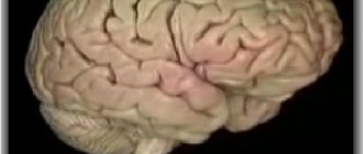
Composition and functions of gray matter
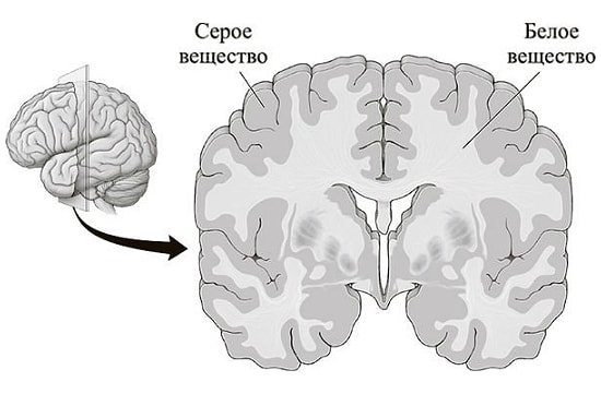
The gray matter of the brain consists of neurons, glial cells, neuropil (a substance that looks like felt, mainly consisting of dendrites), and thin capillaries.
Contrary to popular belief, it is not gray, but brownish in color, surrounded by white matter formed from myelin fibers. It differs significantly in composition: the gray matter contains 84% water, 16% dry matter, half of which, that is, 8%, consists of proteins, 5% lipids and 1% mineral compounds. White contains 70% water, 30% dry matter, only one third of which (9%) consists of proteins, as much as 17% are lipids and 2% are minerals.
Gray matter determines muscle movements, sensory perception (visual, auditory, tactile), cognitive functions (memory, attention, thinking), verbal communications and emotional-sensory response.
to contents ^
Diencephalon
It is located between the corpus callosum and the fornix, and on the sides it fuses with the telencephalon. The dorsal section consists of the visual tuberosities, on the upper part of which there is the epitubercle, and in the ventral part there is the inferior tuberosity region.
The gray matter here consists of nuclei that are associated with centers of sensitivity. White matter is represented by conducting pathways in different directions, guaranteeing the connection of formations with the cerebral cortex and nuclei. The diencephalon also includes the pituitary gland and pineal gland.
Location in the cerebral cortex and basal ganglia
The large hemispheres of the telencephalon are separated by a fissure, and they are connected by the corpus callosum and commissures. Most of them are a cloak, the surface of which is called the neocortex - the new cortex, this is truly the phylogenetically newest structure, where the centers of all HMFs are localized, including those that allowed the formation of the second signaling system, which, according to Darwin's theory, makes humans the highest link in evolution. It is covered with well-known convolutions, which form a complex pattern of grooves and ridges. The thickness of the gray matter here is only 1.3 - 4.5 mm, or six neural layers.
Each hemisphere is divided into large sections - lobes. There are five lobes that form lobules - smaller sections, and convolutions. The pattern that the convolutions form varies in shape for each person, even in two halves of the same brain it looks different.
Useful to know: Human bone marrow and its structure
This substance is also located in the basal ganglia, also called the subcortical formations, or the old brain. It is believed that animal instincts are localized here, since the old brain allows you to automatically make decisions in a difficult situation.
The olfactory brain, the most ancient formation, is also located here. The olfactory organ is the main sense organ in fish and animals. When the distant ancestors of humans came out of the water onto land, those species survived that based their behavior on the basis of smell: it helped to find food, a sexual partner, and run away from the enemy in time. Then, gradually, other structures developed from this part of the brain, in particular, the limbic system, which is responsible for emotions. Now the sense of smell no longer plays a vital role; a person can survive without it, however, his emotions will be significantly impoverished.
to contents ^
Structure
The morphological structure of the cerebral hemispheres within the brain suggests the presence of a cortical layer consisting of gray matter and subcortical sections formed by white matter. Within the mass of white matter there are local areas of gray - the nuclei. White matter serves as the basis for nerve fibers:
- Associative. They unite different functional areas within one hemisphere.
- Commissural. They connect areas of different hemispheres, often symmetrically located.
- Conductive. They connect the parts of the brain - the brain and the spinal cord - thereby forming a single central nervous system network.
The structure of the cortex covering the cerebral hemispheres suggests the presence of grooves (depressions relative to the surface) and convolutions (elevations relative to the surface). In anatomy, the area of the cortex covering the cerebral hemispheres increases due to the presence of grooves. The duration and depth of the furrows are individual characteristics of a person.
The cortical layer, formed from gray matter, contains control centers that are responsible for higher mental functions, which are interconnected with processes such as cognitive and mental activity. The cortex, covering the cerebral hemispheres within the brain, coordinates the functions of the body, forming adaptive responses to external influences. The cortex is formed by 6 types of cells.
The cytoarchitectonics (location of cells in the tissue) of the cortex suggests the presence of molecular, granular (external, internal), pyramidal (external, internal), multiform plates. Within the brain, the surfaces of the hemispheres are distinguished: superolateral (covers the upper lateral areas), medial (located in the middle region), basal (adjacent to the base of the skull).
The superolateral surface has a convex shape and is adjacent to the bone structures that form the cranial vault. The flat medial surface is located opposite the similar surface of the second hemisphere. Each hemisphere consists of 5 lobes. The frontal lobe of the cortex, covering the cerebral hemispheres, is the largest part of the telencephalon. The central sulcus, anatomically transversely crossing the brain, is the border between the frontal and parietal lobes within the cerebral hemispheres.
The Sylvian fissure, also known as the lateral fissure, runs within the cerebral hemispheres perpendicular to the central one, separating the temporal lobe of the telencephalon from two segments - the frontal and parietal. The parieto-occipital sulcus is the border of the parietal segment on the posterior side. The insular lobe within the hemispheres of the brain is located deep in the Sylvian fissure. The insular lobe is visible if you lift the areas of the frontal and temporal lobes that cover it.
The Sylvian fissure, also known as the lateral fissure, which separates the temporal lobe from the brain structures of the frontal and parietal, is one of the largest. It is formed at the 14th week of intrauterine development. Small grooves dividing the lobes of the cerebral cortex into convolutions are formed at 24-38 weeks, forming a peculiar relief of the surface of the hemispheres, individual for each person.
The sulci and gyri of the 3rd order continue to develop after birth, most intensively in the 1st year of life. In the frontal lobe of the cortex within the cerebral hemispheres, a precentral groove runs parallel to the central one, separating and limiting the precentral gyrus - the center where the initiation and regulation of conscious motor activity, directed by volitional effort, occurs.
The precentral gyrus is perpendicularly crossed by the frontal sulci (superior, inferior). They divide the frontal gyri into segments. In the posterior part of the frontal gyrus, located below, there is Broca's center, which is responsible for speech function. In the parietal lobe, the postcentral sulcus runs parallel to the central one.
The postcentral gyrus, located between these grooves, is a center of sensitivity, which is responsible for the body’s reactions to pain, tactile, and temperature influences. The temporal grooves (superior, inferior) lie in the temporal lobe and are located parallel to the Sylvian fissure.
The temporal sulci are divided into segments by the temporal gyri, where in the upper part there is a center responsible for hearing function and Wernicke's center, responsible for speech function. The calcarine groove, which provides visual function, lies in the occipital lobe, where it borders on the parieto-occipital groove.
Location in other parts of the brain
The medulla oblongata regulates all automatic functions, protective and pro-survival reflexes (for example, heartbeat and sneezing). There is also a center that distributes tension between certain muscles responsible for holding the body in any position. The gray matter here forms the nuclei of the olive, several pairs of the lower head and vagus nerves, as well as the reticular formation, the sphenoid and thin bodies.
The hindbrain consists of the pons and the cerebellum. The cores of the bridge are dispersed between fibers directed in different directions. The cerebellar cortex has three sections: external (molecular), ganglionic and granular (granular). The outer layer contains several scattered cell nuclei. The ganglion contains a number of Purkinje cells, their processes extend deep into the cerebellum. The white matter contains the subcortical nuclei: spherical, mushroom, dentate, tent nucleus.
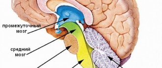
The midbrain has a cavity in its center filled with central gray matter. The so-called aqueduct is located here; at its bottom there are several nuclei, the processes of which mainly innervate the organs of vision, as well as the nuclei of the III and IX cranial nerves. In front of it are the cerebral peduncles, divided by the substantia nigra into a base and a tegmentum, where the red nucleus stands out. Together with the substantia nigra, they belong to the extra pyramidal system and control unconscious movements and muscle tone with the help of the hormone dopamine. The visual and auditory centers are located in the colliculi of the midbrain roof.
Useful to know: How to improve blood circulation in the brain: recommendations, medications, exercises and folk remedies
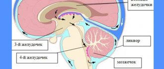
The nervous tissue of the diencephalon forms its four parts. The first of them is the Thalamus, where the nuclei of the thalamus lie. The epithalamus - the second part - includes the leash, its commissure and triangle, as well as the epiphysis, otherwise called the pineal gland. It is interesting for its mystical reputation - esotericists believe that it is this gland that causes clairvoyance and other supernatural abilities. In fact, the pineal gland produces the hormone melatonin, which is responsible for regulating sleep and wake cycles. The third part, the Metathalamus, contains the geniculate bodies. The latter, the Hypothalamus, has two sections. In the anterior one there is the so-called gray tubercle, and in the posterior one there are the mastoid bodies, containing two nuclei.
to contents ^
Nerve fibers
These fibers are multibillion-dollar processes of neurons that conduct nerve impulses in the brain and spinal cord.
The main part of the nerve fiber is the neuron process itself, which subsequently forms the fiber axis. To a large extent it is an axon. The thickness of a human neuron fiber is on average 25 micrometers.
Neuron fibers are divided into:
- myelin
- Unmyelinated
The peripheral and central nervous system is determined by the predominance of myelin fibers. Neuron fibers lacking myelin are usually located in the sympathetic part of the autonomic nervous system.
The main function of neural fibers is the transmission of nerve impulses. To date, scientists have studied only two types of its transmission:
- Pulse (provided by electrolytes and neurotransmitters)
- Pulseless
Notes
- ↑Villee K., Dethier V. Nervous system // Biology (biological processes and laws) = Villee CA, Dethier VG Biological Principles and Processes, 1971. - M.: Mir, 1975. - P. 495-522. — 824 p.
- ↑ 12Bykov V.L. Particular human histology. - St. Petersburg: SOTIS, 2001. - 304 p. — 3,000 copies. — ISBN 5-85503-116-0
- ↑Purves, Dale, George J. Augustine, David Fitzpatrick, William C. Hall, Anthony-Samuel LaMantia, James O. McNamara, and Leonard E. White Neuroscience. 4th ed.. - Sinauer Associates, 2008. - P. 15–16. — ISBN 978-0-87893-697-7
- ↑Kolb {amp}amp; Whishaw: Fundamentals of Human Neuropsychology (2003) page 49
- ↑Voronova N.V., Klimova N.M., Mendzheritsky A.M. Anatomy of the central nervous system. - M.: Aspect Press, 2005. - 128 p. — ISBN 5-7567-0388-8
- ↑Taki Y, Kinomura S, Sato K, Goto R, Wu K, Kawashima R, Fukuda H (2011). “Correlation between gray/white matter volume and cognition in healthy elderly people.” Brain and Cognition (Elsevier Inc.) 75 (2): 170-176. PMID 21131121.
- ↑Li M, Cui L, Deng W, Ma X, Huang C, Jiang L, Wang Y, Collier DA, Gong Q, Li T. (2011). “Voxel-based morphometric analysis on the volume of gray matter in bipolar I disorder.” Psychiatry Research: Neuroimaging191(2):92-97. PMID 21236649.
- ↑Almeida, OsvaldoSmoking causes brain cell loss and cognitive decline. NeuroImage (February 9, 2011). Retrieved November 10, 2012.
- ↑Bill Hathaway.Past abuse leads to loss of gray matter in brains of adolescents (English). Yale University (news.yale.edu) (5 December 2011). Retrieved November 10, 2012.
Brain lesions
Thanks to the achievements of modern science, it has become possible to conduct high-tech brain diagnostics. Thus, if there is a pathological focus in the white matter, it can be detected at an early stage and therapy can be prescribed in a timely manner.
Among the diseases that are caused by damage to this substance are its disorders in the hemispheres, pathologies of the capsule, corpus callosum and syndromes of a mixed nature. For example, if the hind leg is damaged, one half of the human body can be paralyzed. This problem may develop with sensory disturbances or visual field defects. Malfunctions of the corpus callosum lead to mental disorders. In this case, the person ceases to recognize surrounding objects, phenomena, etc., or does not perform purposeful actions. If the lesion is bilateral, swallowing and speech disorders may occur.
The importance of both gray and white matter in the brain cannot be overstated. Therefore, the earlier the presence of pathology is detected, the greater the chance that treatment will be successful.