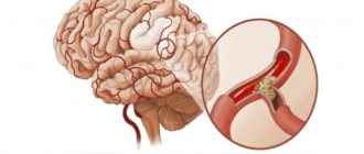
Everyone needs to know what first aid is for a scalp injury, since any delay in case of brain damage can lead to dire consequences. Any head injury is classified as a traumatic brain injury, with the exception of damage to the skin and muscle tissue around the skull.
During a head injury, first aid should be provided immediately, since damage to blood vessels can result in severe blood loss, and in the case of a skull fracture, sharp bone fragments can disrupt the structure of the brain. Concussions are also dangerous; in severe cases, a person can even die from complications of the injury.
Causes
Head injuries, for which first aid should be provided at the scene of an accident, occur at home, at work, during sports or fights, during falls and during car accidents. All segments of the population are susceptible to trauma, regardless of age and gender.
Among athletes, especially those involved in combat sports, TBIs are slightly more common. The skull bones of children under one year of age are mobile and elastic, therefore skull fractures due to falls in infants practically do not occur. Sometimes such injuries occur due to gunshot wounds, for example, during a criminal attack, careless handling of a gun while hunting, or during suicide attempts. The most common cause of head injuries is being under the influence of alcohol.
Classification
There are several types of head injuries and depend on what exactly was damaged and how severely it was damaged:
- Concussion, compression or contusion of the brain;
- Fractures of different parts of the skull;
- Torn, chopped, punctured and incised wounds of the skin, subcutaneous tissue and muscle tissue.
Many traumatic brain injuries have three degrees of severity, which can be determined by the condition of the victim and the clinical picture. Doctors also divide injuries into open, closed and penetrating injuries. Regardless of the type of injury, timely provision of first aid for head injuries can protect against complications and consequences, as well as save a person’s life.
Traumatic brain injury (TBI), intensive care, resuscitation, transportation
Severe (critical)
traumatic brain injury is characterized by a deep and prolonged coma, accompanied by a violation of the vital functions of the body (III-IV degrees).
Terminal conditions can develop with extensive crushing of the brain substance, the formation of intracranial and intracerebral hematomas, dislocation (shear)
and compression of the brain stem. The location of the damage is of great importance. Traumatic brain injury is especially severe when combined with damage to skeletal bones and internal organs.
The death of patients can be caused by both direct traumatic damage to vital structures of the brain and subsequent complications, which primarily include breathing disorders, cerebral edema and intracranial hypertension. Respiratory disorders
develop in all patients with severe brain injury.
They usually occur early after injury
and can be caused by a disorder
of the central regulation of breathing
, increasing tracheobronchial obstruction and pneumonia, often of aspiration nature.
Cerebral edema and increased cerebrospinal fluid pressure are rarely detected in the first hours; they usually develop from 18 to 24 hours after injury.
Although in case of traumatic brain injury the prognosis is determined not so much by the severity of intracranial hypertension as by the localization and volume of injury, the level of cerebrospinal fluid pressure, and especially its dynamics, are of great importance for determining resuscitation tactics.
We should also not forget that if intracranial pressure becomes higher than mean arterial pressure, cerebral circulation is practically blocked and the brain dies.
In the clinical picture of severe traumatic brain injury, along with deep coma (often accompanied by convulsions)
Symptoms of local damage to the central nervous system are often detected. Breathing may be arrhythmic or sharply rapid and deep. Less commonly, bradypnea or primary respiratory arrest occurs.
Blood pressure is increased in the absence of concomitant blood loss. The pulse may be slow in the first hours (especially in the presence of an intracranial hematoma),
then persistent tachycardia develops.
A prolonged comatose state is accompanied by persistent hyperthermia,
both of central origin and caused by purulent-septic complications.
Dysfunction of the gastrointestinal tract (intestinal paresis) is also common.
Disturbances in water and electrolyte balance are characteristic, primarily hypokalemia.
Many patients experience hypercoagulation (increased intravascular coagulation).
The primary task of resuscitation for critical traumatic brain injury is the elimination of hypoxia and correction of acid-base balance.
The method of choice is artificial pulmonary ventilation (ALV) in the mode of moderate hyperventilation, which has a pronounced therapeutic effect, reducing liquor pressure by 30% (V.A. Negovsky, 1971).
Against the background of mechanical ventilation, it is necessary to correct impaired metabolic processes. To prevent cerebral edema, maintaining the oncotic pressure of the plasma is of great importance, for which concentrated protein solutions are administered to patients. Introduction of osmotic agents (urea, manitol).
Administration of saluretics is indicated only for high intracranial pressure.
Traumatic brain injuries are divided into closed - (concussion, bruise, compression by hematoma, fragments of skull bones)
,
open - (gunshot, stab, chopped, penetrating, non-penetrating) and
Fracture of the base of the skull. A). Concussion (commotion):
swelling, pinpoint hemorrhages, hyperemia of the meninges, and venous congestion occur.
Symptoms of injury:
characterized by loss of consciousness for several minutes (sometimes without loss of consciousness). The victim complains of headaches, nausea, vomiting, and dizziness. A characteristic symptom is retrograde amnesia, i.e. loss of memory for previous events.
Treatment
– inpatient 2-3 weeks. The prognosis is favorable.
b). Brain contusion (concussion)
.
There are 3 degrees of injury:
– mild degree
– resembles a concussion, but there are mild focal symptoms;
– average degree
– loss of consciousness up to several hours + focal symptoms + meningeal symptoms;
– severe
– loss of consciousness up to several weeks, pronounced focal symptoms and impaired vital functions. The condition of the patients is severe, rapid breathing, repeated vomiting, deep coma.
The victim must be hospitalized in the intensive care unit.
Treatment:
This category of patients is carried out in the intensive care unit of the neurosurgical department from 3 weeks to 1.5-2 months.
V). Compression of the brain (compression):
compression occurs as a depressed fracture of the skull bones or hematoma, often combined with a brain contusion. Loss of consciousness due to compression can last up to 2 hours or without loss of consciousness.
The general condition of the patient can be characterized as satisfactory
or
moderate severity.
The victim suffers from headaches, nausea, vomiting, and strabismus. The victim is conscious and in contact.
This period is called the light interval.
Then, after a few hours or days (as a result of the accumulation of blood - hematoma), the condition worsens: stupor turns into coma, bradycardia increases and focal symptoms appear: paralysis, anisokaria (different pupil sizes).
Treatment:
urgent hospitalization in the neurosurgical department, surgical intervention (removal of bone fragments, hematomas).
G). Fracture of the base of the skull:
With this injury, rupture of the membranes of the brain, bruise, compression, rupture of cranial nerves, and focal necrosis occurs. The victim experiences the following symptoms: loss of consciousness, nausea, vomiting, headache, bleeding and liquorrhea from the ears, nose, as well as the “glasses” symptom.
The patient's condition is serious. Careful transportation is necessary. Urgent hospitalization in a neurosurgical or trauma department.
Treatment:
surgical or conservative within 5-6 weeks.
Transportation for TBI.
1. Carefully remove the victim.
2. Convenient to lay down, free from restrictive clothing.
3. Transport in a horizontal position, gentle.
4. When vomiting, turn your head to the side.
5. For an open wound, treat with an antiseptic and use an aseptic bandage.
6. In case of bleeding from the nose and ears - toilet the nose, ears, tamponade with a sterile napkin.
7.Raise
lower jaw to avoid tongue retraction.
Intensive therapy and resuscitation for epistatus (epilepsy, “falling sickness”).
It is characterized by repeated paroxysmal convulsions and is a polyetiological, chronic disease.
Provoking factors are: psychological trauma, alcohol intake, infections, flashing bright light. There are several forms of epistatus, the most dangerous being generalized status. It starts suddenly.
Clonic or tonic-clonic convulsions occur, which do not stop for a long time and are accompanied by severe breathing disorders (sometimes apnea, respiratory arrest) or of the Cheyne-Stokes type.
Pulmonary edema is common, aspiration of saliva and mucus into the respiratory tract is possible, blood pressure rises sharply, all this leads to severe brain damage and the longer the status lasts, the more severe and extensive the damage is (electrolyte disorders, aspiration pneumonia, acute arrhythmias are possible).
Intensive therapy.
1. Ensure free passage of the airways (use a mouth dilator, tongue depressor.)
2. Remove dentures, mucus, insert an air duct into the respiratory tract.
3. Put clothes under your head to prevent injury.
4. Anticonvulsant therapy (seduxen, sodium hydroxybutyrate, relanium sodium thiopental).
5. Oxygen therapy.
6. Fighting cerebral edema.
7. Preparation for spinal puncture and artificial ventilation.
In conclusion, I consider it necessary to remind that no resuscitation methods will help the patient if there is an unresolved cause of brain hypoxia.
Source: https://studopedia.su/10_102697_cherepno-mozgovaya-travma-chmt-intensivnaya-terapiya-reanimatsionnie-meropriyatiya-transportirovka.html
Characteristics and symptoms
Signs of TBI depend on the type of injury. Each damage has its own characteristics:
- incised wound - pain occurs, smooth edges are present, bleeding;
- chopped wound - deeper with brain damage;
- puncture wound - has a canal, infectious processes often develop;
- bruised wound - leads to tissue damage and necrosis;
- gunshot wound - can be both superficial and deep with damage to the skull and brain.
First aid
Providing first aid must be carried out in a certain sequence based on the condition of the victim and the type of injury. To do this, you need to use only sterile bandages and cotton wool, and act very carefully. Infection in a wound can lead to encephalitis or meningitis. If a person hits their head and is unconscious, the following steps should be taken:
- Check for pulse and breathing;
- Place the victim in a lying position on his side to prevent vomit from entering the respiratory tract and causing the tongue to stick;
- Call an ambulance;
- In the absence of breathing and pulse, carry out resuscitation measures consisting of artificial respiration and chest compressions, alternating thirty quick but strong pressures on the heart area and two breaths into the mouth, closing the victim’s nose and tilting his head back slightly.
Transportation of victims with traumatic brain injury in a state of shock
Traumatic brain injury (TBI) means mechanical damage to the bones of the skull and intracerebral structures (the substance of the brain, meninges and blood vessels). Depending on the type of injury, a distinction is made between concussion and brain contusion, open and closed TBI.
In the first case, there is a reversible impairment of brain function with general cerebral symptoms (nausea, vomiting, headache). With brain contusions, things are more serious. Here, brain tissue is damaged to one degree or another, and cases of intracranial hematomas (effusion of blood into the cranial cavity) are common.
The insidiousness of traumatic brain injury lies in the fact that doctors are often not always able to objectively assess its severity at the prehospital stage without special studies.
Even with extensive damage to the brain substance with the presence of an intracerebral hematoma, cerebral symptoms are initially mild, and the patient may not complain about anything at all. Doctors call this period of imaginary well-being a bright interval.
The duration of the light interval varies from several hours to several days; cases of its duration of up to six months have been described.
Deterioration occurs quickly, in a matter of minutes, especially in children.
The patient, who previously felt great, begins to complain of general weakness, a unilateral decrease in muscle strength and sensitivity in the extremities.
Subsequently, consciousness disorders of varying degrees appear, up to coma, and different pupil sizes (anisocoria). All this speaks of a serious complication of TBI – cerebral edema due to intracerebral hematoma.
Even the slightest suspicion of a hematoma is an absolute indication for transporting the victim to a neurosurgical or trauma department. Transportation is carried out on a flat, rigid stretcher. The head end should not be raised.
In case of an open traumatic brain injury with head wounds, these wounds are treated and a bandage is applied on top. For timely administration of medications, venous access is required, to ensure which the elbow vein is punctured with a special disposable catheter - a thin polymer tube.
An intravenous infusion system is connected to the catheter.
Patients with severe TBI who are in a comatose state with disorders of vital functions deserve special attention. Here, transportation should be carried out not with 1, but with 4 catheters. In addition to venous access, a catheter is placed in the bladder, a probe is placed in the stomach through the nose, and an endotracheal tube.
The last manipulation is carried out strictly according to indications and requires certain equipment and skills for the doctor. Through the endotracheal tube, air and oxygen are supplied to the patient's lungs using a special device.
Throughout the entire journey to the hospital, blood pressure, cardiac activity and blood gas composition are continuously monitored using a monitor and sensors attached to the patient.
Injuries accompanied by bone fractures and bruises of internal organs require competent and gentle transportation of the victim to a medical facility. Any careless or incorrect movement can cause serious harm to the patient, minimizing all emergency efforts.
From our article you will learn what methods of transporting victims exist depending on the injury, as well as how to carry them out correctly.
Types and methods of transportation
Transportation of victims involves the work of specially trained medical personnel. However, in emergency situations, when it is impossible for an ambulance to reach the scene of the incident, rescuers must independently transport the patient to the hospital.
There are several important factors to consider before transporting a casualty:
- Determine the method of moving the patient;
- Prepare the wounded and specialized devices for movement;
- Choose the most convenient route;
- Ensure the safety of the victim;
- Carry out constant monitoring of the functioning of the patient’s vital systems and organs;
- Determine the method of loading the patient into a vehicle.
The choice of method of transporting the victim is based on the type and location of the injury and the general condition of the patient.
Independent transportation of a patient to a medical facility is permitted only in 2 cases:
- The place where the injury occurred poses a potential threat to the life of the victim and the rescuer;
- The ambulance cannot reach the place where the accident occurred.
Depending on the circumstances that caused the injury, the following types of transportation are distinguished, the features of which are presented in the table.
If the situation does not require emergency transfer of the patient from a place dangerous to his life, you need to use the rules for moving victims, which we will discuss below.
General provisions for transportation
Before moving the patient, you need to carefully examine his condition:
- Check for consciousness;
- Assess pulse and respiration (if possible, blood pressure);
- Examine the patient's head, spine and chest for damage.
If there is a suspicion of injury to the brain, spine or spinal cord, you can only move the victim yourself in emergency situations! If you cannot do without transportation, try to carefully carry the patient into the car in the position in which he was previously.
Source: https://golovnoj-mozg.ru/travmy/transportirovka-postradavshih-s-cherepno-mozgovoj-travmoj-v-sostoyanii-shoka
| Patients with brain injury may require transport within a medical center or to another center for clinical and non-clinical reasons. During transport, patients are isolated from clinic resources and are exposed to additional dangers. Poorly prepared transportation can worsen the patient's condition. Various organizations have established a number of protocols for safe patient transport, most of which emphasize the importance of well-trained staff and careful planning (both at the clinic and patient level), appropriate equipment, and regular monitoring of all phases of transport. Indications for transporting a patient with traumatic brain injury (TBI)Transfer to another clinic : • There are not enough intensive care unit beds in the clinic where the patient is admitted. • The need for special examination or treatment not available in this clinic. • Transfer for treatment at the place of residence. Inside the clinic : • Neuroimaging (CT, MRI). • Neuroradiological procedures such as aneurysm embolization. • Surgical interventions. • Transfer from one hospital department to another for further management (for example, from the intensive care unit to the intensive care unit or from the intensive care unit to the general ward). Principles of transporting a patient with traumatic brain injury (TBI)The principles of transportation are the same for all patients . The systematic approach is based on the ACCEPT principle (Assessment, Control, Communication, Evaluation, Preparation and Packing, Transportation): Assessment of a patient with traumatic brain injury (TBI) : • Neurosurgical patients may have multiple injuries or be critically ill due to secondary injuries or sepsis. • Transfer personnel may be seeing the patient for the first time, so careful reassessment is necessary. It is important to avoid the temptation to simply work as a courier. The standard of care for the patient during transport should not differ from that provided in the clinic. To do this, you need to be well informed about the patient's condition and his medical history. Control : • A person responsible for transportation is appointed in the team. • He must ensure that all necessary requirements for safe transport are met and ensure that therapy is continued. Communication : • All people involved in the process must be informed accordingly. • This includes responsible consultants (surgeon and resuscitator) of the primary and receiving hospitals, relatives and ambulance team members. • Documentation of the transport process is very important for the continuation of treatment in the receiving clinic, for monitoring and improving the quality of care and medico-legal aspects in adverse circumstances. Transport risk assessment (evaluation) of a patient with traumatic brain injury (TBI) : • Transporting the patient is part of the treatment and involves risks. The following is carefully assessed: • the possibility of carrying out in principle; • the urgency of carrying out. • Accompanying personnel and method of transportation will be determined by the urgency of the transportation. |


