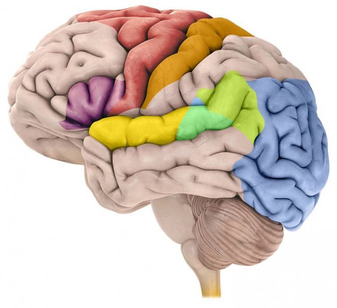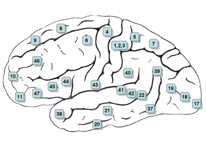
Lateral surface of the brain with numbered Brodmann's areas.
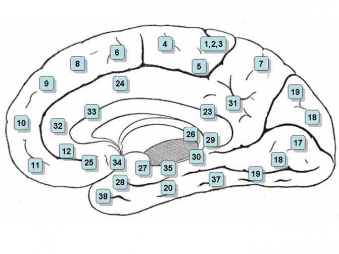
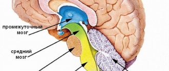
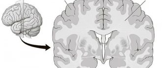
Central part of the brain with numbered Brodmann areas.
Paul Brodmann
- sections of the cerebral cortex, differing in their cytoarchitecture (structure at the cellular level). There are 52 Brodmann cytoarchitectonic areas.
In 1909, the German neurologist Korbinian Brodmann published[1] maps of the cytoarchitectonic fields of the cerebral cortex. Brodmann was the first to create maps of the cortex. Subsequently, O. Vogt and C. Vogt (1919-1920), taking into account the fiber structure, described 150 myeloarchitectonic areas in the cerebral cortex. At the Brain Institute of the USSR Academy of Medical Sciences (now the Scientific Center for Neurology of the Russian Academy of Medical Sciences) I.N. Filimonov and S.A. Sarkisov created maps of the cerebral cortex, including 47 cytoarchitectonic fields [2].
Despite criticism[3], Brodmann's fields are the best known and most often cited when describing the neuronal organization of the cerebral cortex and its functions.
The assignment of a particular area of the cortex to a specific field was based on a histological examination - Nissl staining. Certain fields correspond to areas of the brain responsible for certain functions.
A. W. Campbell proposed a division of fields into primary, secondary and tertiary. The primary and secondary fields (nuclear zone of the analyzer) receive impulses directly from the thalamus, while the tertiary fields receive impulses only from the primary and secondary fields. Primary fields produce a specific analysis of impulses of a certain modality. Secondary fields interact between different analyzer zones. Tertiary fields play a decisive role in complex types of mental activity - symbolic, speech, intellectual.[4]
Paul Brodmann
- Fields 3, 1 and 2 - somatosensory area, primary zone. Located in the postcentral gyrus. Due to the commonality of functions, the term " fields 3, 1 and 2
" (front to back) - Field 4 is the motor area. Located within the precentral gyrus
- Area 5 is the secondary somatosensory area. Located within the superior parietal lobule
- Area 6 - premotor cortex and supplementary motor cortex (secondary motor area). Located in the anterior sections of the precentral and posterior sections of the superior and middle frontal gyri.
- Field 7 is the tertiary zone. Located in the upper parts of the parietal lobe between the postcentral gyrus and the occipital lobe
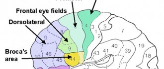
- Field 8 - located in the posterior parts of the superior and middle frontal gyri. Includes the center of voluntary eye movements
- Area 9 - dorsolateral prefrontal cortex
- Area 10 - anterior prefrontal cortex
- Area 11 - olfactory area
- Field 12 -
- Field 13 -
- Field 14 -
- Field 15 —
- Field 16 -
- Field 17 - nuclear zone of the visual analyzer - visual area, primary zone
- Field 18 - nuclear zone of the visual analyzer - center of perception of written speech, secondary zone
- Field 19 - nuclear zone of the visual analyzer, secondary zone (assessment of the meaning of what is seen)
- Area 20 - inferior temporal gyrus (center of the vestibular analyzer, recognition of complex images)
- Area 21 - middle temporal gyrus (center of the vestibular analyzer)
- Field 22 - nuclear zone of the sound analyzer
- Field 23 -
- Field 24—error detector
- Field 25 —
- Field 26 -
- Field 27 -
- Field 28 - projection fields and associative zone of the olfactory system
- Field 29 -
- Field 30 —
- Field 31 -
- Area 32 is the dorsal zone of the anterior cingulate cortex. Receptor area of emotional experiences.
- Field 33 -
- Field 34 -
- Field 35 —
- Field 36 -
- Field 37 - Acoustic-gnostic sensory speech center. This field controls labor processes through speech and is responsible for understanding speech. Facial Recognition Center.
- Field 38 -
- Area 39 - angular gyrus, part of Wernicke's area (center of the visual analyzer of written speech)
- Area 40 - marginal gyrus, part of Wernicke's area (motor analyzer of complex professional, labor and everyday skills)
- Field 41 - nuclear zone of the sound analyzer, primary zone
- Field 42 - nuclear zone of the sound analyzer, secondary zone
- Field 43 - taste area
- Field 44 - Broca Center
- Area 45 - triangular part of Brodmann area (musical motor center)
- Field 46 - motor analyzer of combined rotation of the head and eyes in different directions
- Field 47 - nuclear zone of singing, its speech motor component
- Field 48 -
- Field 49 —
- Field 50 —
- Field 51 -
- Field 52 is the nuclear zone of the auditory analyzer, which is responsible for the spatial perception of sounds and speech
Brain map
The new structural and functional map divides the cerebral cortex into 180 sections.
The cerebral cortex is extremely complex - its different parts differ from each other both in function and in cellular structure. Naturally, those who began to study the brain very soon needed a “terrain map” for the cerebral cortex, and the system of cytoarchitectonic fields, published by the German neurologist Korbinian Brodmann back in 1909, became a kind of gold standard here.
- 1
- 2
These fields differ in cell morphology and in the way the cells in them are stacked relative to each other (that is, in cellular cytoarchitecture). Brodmann fields turned out to be extremely useful, but still had some significant disadvantages.
Firstly, Brodmann himself built his map on the material of just one brain, taken from a deceased person. Subsequently, the structure of the cortical fields was clarified using more diverse material, and functions were added to the pure morphological parameters: what one area is responsible for, what another is responsible for, etc. However, the more neuroscientists learned about the brain, the clearer it became that the cerebral cortex needs to be re-mapped using several features simultaneously.
This work was undertaken by Matthew F. Glasser and his colleagues from Washington University in St. Louis, Oxford, the University of Minnesota and the University of Nijmegen. They took a set of magnetic resonance imaging (MRI) data accumulated as part of the Human Connectome project (recall that the goal of the Human Connectome project is to fully describe the structure of connections in our brain).
The researchers were interested in the results of structural MRI, which allows one to establish, for example, the thickness of certain areas of the cortex and other similar features, and functional MRI, with which one can see the function of one or another area of the brain. At the same time, the brain can rest during scanning, and then we will discern its basic functional topography, or it can perform some task - and then we will see which areas are working on a specific procedure. To construct a new cortical map, we used fMRI data obtained from seven tasks, ranging from audio tests to math problems.
Thus, the algorithm that searched for functional fields in the cortex had to operate with several parameters at once, structural and functional. As a result, it was possible to detect as many as 180 fields in each hemisphere, 83 of which had previously been described in the literature, but 97 turned out to be hitherto unknown.
The algorithm worked with the results of MRI scans of 210 volunteers of the Human Connectome project, and the question immediately arose: would it be possible to determine the same zones in other people? Wouldn't it turn out that a map of 180 fields makes sense only for those two hundred people on whom the above-mentioned algorithm was trained?
But when they tried to analyze a set of MRI data from “strangers”, their cortical zones were determined almost the same way. Moreover, the authors of the work were also able to determine individual differences between certain areas. (Just in case, let’s clarify that individual differences do not mean that one’s brain is structured one way and another’s – another, just that the zones can work with different efficiency and be less developed; similarly, if we see a tall person and a short person next to each other , we are not saying that they have a different structure plan.)
It is clear that the new map (described in the Nature article) will be useful in both basic science and medicine. True, it also has its drawbacks, related, first of all, to the fact that MRI still does not have a high enough spatial resolution, that is, the cerebral cortex can actually be divided into an even larger number of fields.
On the other hand, it remains to be seen how the new 180 zones are structured at the level of cells, synapses and their molecular characteristics. And finally, let's not forget about the recent work that called into question thousands and thousands of MRI scan results - let's hope that the new cortical map will not suffer too much from this revelation.
Notes
- Brodmann Korbinian.
Vergleichende Lokalisationslehre der Grosshirnrinde : in ihren Principien dargestellt auf Grund des Zellenbaues. — Leipzig: Johann Ambrosius Barth Verlag, 1909. - Sapin M. R., Bilich G. L.
Human anatomy. - M.:: "Higher School", 1989. - P. 417. - 544 p. — 100,000 copies. — ISBN 5-06-001145-3. - Gerhardt von Bonin & Percival Bailey.
The Neocortex of Macaca Mulatta. — Urbana, Illinois: The University of Illinois Press, 1925. - E. D. Chomskaya.
Neuropsychology, 4th edition. — Peter, 2008.
| Brodmann's cytoarchitectonic fields at Wikimedia Commons |
2.3. Basic principles of brain structure
The brain as a substrate of mental processes is a single system, a single whole, consisting, however, of various sections and zones that play different roles in the implementation of mental functions.
All data (anatomical, physiological and clinical) indicate the leading role of the cerebral cortex in the cerebral organization of mental processes.
In neuropsychology, based on the analysis of neuropsychological data (i.e., the study of disturbances in mental processes in various local brain lesions), a general structural and functional model of the brain as a substrate of mental activity was developed. This model, proposed by A. Luria, characterizes the most general patterns of the functioning of the brain as a whole and is the basis for explaining its integrative activity. According to this model (Fig. 3), the entire brain can be divided into three main structural and functional blocks: a) an energy block, or a block for regulating brain activity levels; b) a block for receiving, processing and storing exteroceptive (coming from outside) information; c) a block of programming, regulation and control over the course of mental activity. Each higher mental function is carried out with the participation of all three blocks, each of which makes its own contribution to its implementation. The blocks are characterized by certain structural features, the physiological principles underlying their work, and the role they play in the implementation of mental functions.
The first energy block regulates two types of activation processes: general generalized changes in brain activation, which are the basis of various functional states, and local selective activation changes necessary for the implementation of higher mental functions.
The functional significance of the first block in ensuring mental functions primarily consists, as mentioned above, in the regulation of activation processes, in ensuring that
First Signal System: Location
The centers of the first signaling system are located in Brodmann's fields, which are present in both animals and humans. They are responsible for a simple reaction to an external stimulus, the formation of sensations and ideas. These centers are present in both the right and left hemispheres of the cerebral cortex. Brodmann fields of the first signaling system are present in humans from birth and normally do not undergo changes throughout life.
These fields include:
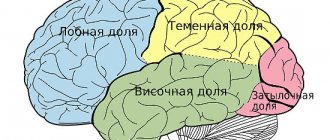
- 1 - 3 - are located in the parietal lobe of the cerebral cortex behind the central gyrus;
- 4, 6 - located in the frontal lobe anterior to the central gyrus, they contain Betz pyramidal cells;
- 8 - this field is located anterior to the 6th, closer to the frontal part of the frontal cortex;
- 46 - located on the outer surface of the frontal lobe;
- 41, 42, 52 - located on the so-called Heschle convolutions, on the basal part of the temporal lobe of the brain;
- 40 - located in the parietal lobe behind 1 - 3 fields, closer to the temporal part;
- 17 and 19 - located in the occipital part of the brain, most dorsal from the other fields;
- 11 is one of the most ancient structures, located in the hippocampus.