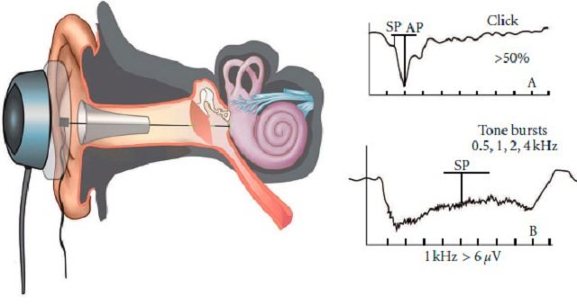
Electrocochleography is one of the methods for objective diagnosis of hearing. The essence of the method is to evaluate the characteristics of electrical signals - auditory potentials coming from hair cells - receptors of the hearing organ and auditory nerve. Auditory potentials are the converted energy of a sound wave, which in the form of electrical impulses enters the auditory centers of the brain and causes auditory sensations in a person. In order to obtain and evaluate these potentials, an external stimulus is required - sound of certain frequencies at different decibel levels. There are two ways to record such auditory evoked potentials:
- Transtympanic - in this case, a special needle electrode is installed through the eardrum onto the wall of the cochlea. This method is invasive and requires preparation and anesthesia. This method is used very rarely in large clinics with special equipment. They resort to it during neurosurgical or hearing-improving operations, for intraoperative study of evoked auditory potentials.
- Extratympanic - in this case, foam inserts wrapped in gold foil are used, which are installed in the patient’s ear canal. Through these inserts a sound stimulus is supplied. This method is much more often used in outpatient practice. In some cases, the results of this method may be less accurate than with an invasive research method.
Pros and cons of the procedure
The advantages of this study are:
- accuracy, simplicity and accessibility of the method for studying hearing threshold levels;
- clarifying the nature of patients’ deafness (sensorineural or conductive);
- the opportunity to obtain information about the functioning of the hairs of the cochlea, as well as all parts of the auditory analyzer;
- no contraindications, except in cases where pain relief is necessary, and patients have a hypersensitivity reaction to drugs for local anesthesia.
When?
A study should be carried out if there are complaints of muscle weakness, loss of limbs, muscle twitching, muscle spasms or cramps. Electromyography is mandatory if myotonia, myopathy, amyotrophic lateral sclerosis, myoclonus, muscular dystonia, tremor, etc. are suspected.
Studying the functional state of the muscle, the degree of its involvement in the process, the preservation of innervation or determining the volume of reinnervation - these are the main issues that are resolved when conducting an electromyographic study. EMG is widely used to differentiate between neurogenic and primary muscular diseases. This helps to diagnose them at an early stage, resolve issues related to the pathogenesis (origin of the disease) of certain forms of neuromuscular diseases, and also allows you to very accurately trace all stages of development and the severity of denervation syndrome in the muscle (a set of changes that occur in postsynaptic neurons, organs and tissues after the loss of nervous influences on these structures).
Disturbances of neuromuscular transmission underlie a relatively small number of different pathological conditions. Diagnosis of various variants of muscle pathology caused by impaired transmission of excitation from nerve to muscle is relevant, as the effectiveness of therapeutic interventions increases and the need arises for differentiated prescription of various forms of therapy.
One of the most frequent and important issues that must be resolved when conducting an electromyographic study is the topical diagnosis. Even the most sophisticated methods of clinical research cannot give such accurate conclusions about the level of damage to the axon (the process of the nerve cell along which nerve impulses travel from the cell body to the innervated organs and other nerve cells) that electromyography provides.
Here is a list of some typical diseases that require the use of electromyography (EMG) and electroneurography (ENG) for diagnosis:
- neuritis;
- neuropathy (alcoholism, diabetic);
- neuritis of the radial, ulnar and peroneal nerves;
- neuritis of the facial nerve;
- herpes, intercostal neuralgia;
- pain in the arm and neck;
- carpal tunnel syndrome.
Scope of application, indications
Electrocochleography data are valuable diagnostic material for confirming a number of diseases of the hearing system:
- Meniere's disease;
- pathologies of the auditory nerve (neuropathy or neuroma);
- sound perception disorders, hearing loss, hearing loss.
Indications for the procedure are:
- frequent headaches and dizziness in combination with hearing loss or distortion (noise, splashing, crackling);
- sensations of pain, discomfort and heaviness in the ears;
- injuries in the parotid space or damage to the structures of the inner ear, as well as general ones - concussions, bruises, contusions;
- patient complaints of progressive hearing loss;
- inflammatory diseases of the hearing aid, including those occurring with the formation of pathological effusion (serous, purulent, hemorrhagic);
- anatomical defects in the structure of the ear;
- suspected Meniere's disease;
- dystrophic changes in the inner ear (adhesions, scars, necrosis).
The study also applies:
- for assessing hearing in easily excitable children and people whose profession requires good hearing abilities (musical instrument tuners, signalmen, singers, etc.);
- to differentiate hearing impairment caused by inflammation of the ear with pathologies of the nerve plexuses and cells (sensorineural hearing loss) or damage to the conduction system of the ear (conductive hearing loss);
- for the purpose of monitoring the results of treatment.
How?
The test is carried out either using electrodes attached to the surface of the skin over the muscle being tested, or with needle electrodes that are inserted superficially into the muscle.
The main goals of EMG as a method of functional diagnostics are:
- identifying the level of damage to the neuromuscular system;
- determination of the topic of the lesion and the extent of the process;
- determination of the nature of the lesions;
- determination of the severity of the pathological process.
Needle EMG - study of motor unit potentials - is used for muscle diseases (progressive muscular dystrophy, polymyositis and other myopathies), diseases of peripheral nerves (mononeuropathy, polyneuropathy), diseases of spinal cord motor neurons (amyotrophic lateral sclerosis, spinal amyotrophy, etc.)
There are a number of other studies that are used to assess sensitivity disorders in diseases of the peripheral nerves, myasthenia gravis, myasthenic syndromes, etc. Various modifications of the method make it possible to assess the condition of the muscles of the arms, legs, torso, face, tongue, and to identify special types of muscle activity characteristic of certain neurological diseases.
Nerve conduction testing using EMG is a non-invasive procedure and therefore does not cause discomfort during its implementation.
It can provide the following information: the presence of nerve damage, the duration of the nerve damage (is it an acute or chronic process), the location of the nerve damage (relative to the periphery and center), the severity of the nerve damage, whether the process of restoring the normal functioning of the nerve is underway.
The duration of the study is from 30 to 60 minutes.
Progress of the study
The procedure is carried out in two ways.
- Transtympanic, in which the electrode is fixed on the inner wall of the labyrinth under a microscope. The procedure requires the use of local anesthesia. Its data is very accurate, but due to its subtlety it is rarely prescribed, usually to people with profound hearing impairment. Examination stages:
- preparatory: examination of the ear using an otoscope, its sanitation;
- anesthetic: administration of anesthetics (injection or instillation of solutions), and general anesthesia is used for preschool children;
- main: insertion of electrodes under visual control using an otoscope or microscope (surgical) and fixation of inserts (special devices for recording impulses), sending signals (tones or clicks), recording data;
- final, deciphering the results.
- Extratympanic, no anesthesia is required here, since the technique is non-invasive. The electrodes are attached at the beginning of the ear canal and on the outer ear (lobe or mastoid), and the ground electrode is located in the area of the 7th cervical vertebra.
The examination is carried out in a lateral lying position, with the patient's ear on top. Typically, the electrocochleography procedure takes no more than 30-60 minutes.
Story
Electrography
invented by Chester Carlson. He and his assistant Otto Korney obtained the first print in their home laboratory in New York on October 22, 1938[1][2]. A patent for this technology was received on October 6, 1942[3]. For a long time, Carlson unsuccessfully tried to introduce his invention, proving that it was absolutely necessary for business, but everywhere he was refused, citing the fact that his invention was too bulky and heavily stained the sheets, and besides, a person could cope much better with the task of copying. Luck smiled at him in 1944 at the Battelle Institute, located in Ohio. There he was offered to improve the technology and even found the exact word for the name of this process - “electrophotography”. After which the license for further development and production of copiers was acquired by the Haloid Company. It was then that it was decided that the word “electrophotography” was too scientific and could scare away a potential buyer. A local philologist professor helped in finding a better name. He coined the term “xerography” from ancient Greek. ξερός “dry” and γράφω “writing”, and then the inventor Carlson himself thought of shortening the word to a simple “copier”. As a result, in 1948, the first copiers appeared on the market, and the first model was simply called Model A. After the release of the first fully automatic model, the Xerox 914, in 1959, Haloid changed its name to Xerox Corporation.
Independently of Chester Carlson, in 1948, in Germany, the inventor Dr. Eisben founded the company Develop Corp to produce a copier of his own design. The company founded by Eisben continues to produce copying equipment today, without recognizing Carlson’s primacy, since it received 16 patents for its founder’s invention.
Decoding the results
The examination results are a curved line with recorded changes (peaks and valleys) of electrical impulses. The doctor analyzes the ratio of the summation potential to the auditory and microphone potential, as well as violations of the norms of electrophysiological indicators.
The interpretation of the results is carried out by an otolaryngologist; on the electrocochleogram he can see disturbances in the conductivity of frequency waves, a decrease or increase in their amplitude, a change in the microphone potential (its norm is 80 dB NHL). Thus, in Meniere's disease, its diagnostic sign is the ratio of SM (summation potential) to AP (action potential of the auditory nerve), which exceeds the norm (more than 0.5 units). With central auditory disorders, AP peaks change, and a decrease in amplitude or loss of electric waves often are a sign of neuropathy.
Electrocochleography is currently actively used in municipal and private medical institutions and is one of the advanced methods for detecting hearing impairment.
Romanovskaya Tatyana Vladimirovna
What is ECoG and why is the study needed?
Electrocochleography (ECoG) - this study is carried out to record the earliest auditory evoked potentials - the cochlea and within the cochlear part of the auditory nerve, occurring within 2-3 milliseconds after the presentation of a short sound stimulus, usually a click.
Normally, ECoG is recorded when stimulated by clicks with a level of more than 60 dB nHL. For successful recording, the active electrode is placed as close as possible to the cochlea of the inner ear.
In other words, ECoG is a high-tech method designed for audiological examination of the human auricle.
It is used to diagnose Meniere's disease, neuropathy and neuroma of the auditory nerve and other auditory disorders.
Meniere's disease is a disease of the ear that leads to increased production of liquid endolymph, the secreted fluid produces excessive pressure on the cells, which leads to hearing impairment and loss of concentration.
Indications for such diagnostics:
- constant feeling of ear fullness;
- dizziness and nausea;
- hearing impairment (one or two-sided);
- otitis and otosclerosis;
- ear injury, which could lead to deterioration in the functioning of the 2nd sense organ;
- eardrum injury.
The main advantage of ECoG is that this diagnostic method has virtually no contraindications. The only thing is that it is not recommended for patients to undergo this examination if they have an individual intolerance to the anesthetic agents used during the diagnosis.