Imagine a map with every star in the galaxy. The map is so detailed that it shows what each star looks like, what it is made of, and which other star it is connected to by the great laws of cosmic physics. While we don't yet have such an astronomical map of the skies, thanks to a monumental study published last week in Neuron, we have a similar map of the brain.
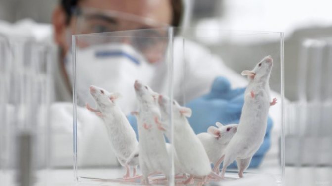
Most experiments are carried out on mice. Universal soldiers of science.
If every neuron were a galaxy, the synapses—the little structures dotted along the serpentine extensions of the neurons—were the stars. A team of scientists from the University of Edinburgh in the UK has built the first detailed map of every synapse in a mouse brain.
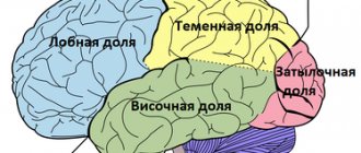
Brain map: the key to the riddle of thinking
Using genetically modified mice, scientists literally caused every synapse to light up under fluorescent light throughout the brain, like stars in the sky. And, just as stars differ from one another, scientists have discovered that synapses are very diverse, but there are patterns that can support memory and thinking.
“The human brain has more synapses than stars in the galaxy. The brain is the most complex object known to us, and understanding its connections at this level will be an important step forward in unlocking its mysteries,” said lead author Dr. Seth Grant from the Center for Clinical Brain Sciences.
Detailed maps revealed a fundamental law of brain activity. Using machine learning, the team categorized the approximately one billion synapses across the brain into 37 categories by type. Here's the point: when sets of neurons receive electrical information, such as choosing between different solutions to a problem, unique subtypes of synapses scattered among different neurons unanimously spark with activity.
In other words, there are different types of synapses. And each type can control a thought, a decision or a memory.
Not surprisingly, neuroscientists reacted very positively to the work.
“Wow,” commented Ben Sanders of the University of Minnesota.
It is "an astonishing paper cataloging the diversity and distribution of synapse subtypes throughout the mouse brain," writes neurogeneticist Kevin Mitchell. This “underscores the fact that synapses are the key computational elements of the nervous system.”
Connectome connection
Scientists' interest in creating a "synaptome" - the first comprehensive catalog of synapses in the mouse brain - grew out of a much larger project: the connectome.
In short, the connectome is all the neural connections within you. As Dr. Sebastian Seung says, the connectome is the biological basis of who you are—your memories, your personality, your thoughts and reasoning. Capture the connectome, and one day scientists will be able to reconstruct you using whole-brain emulation.
And yet, the connectome only describes how neurons functionally talk to each other. Where in the brain is it physically encoded.
This is where synapses come into play. Neuroscientists have long known that synapses transmit information between neurons using chemicals and electricity. There were also hints that synapses differ greatly depending on the proteins they contain, but this difference was generally ignored. Until recently, most scientists believed that the real computation took place in the neural body—the bulbous part of the neuron from which branches emerge.
In order not to miss anything interesting from the world of high technology, subscribe to our news channel on Telegram. There you will learn a lot of new things.
Until now, there was no way to look at the morphology and function of synapses throughout the brain, the authors explain. Typically, we have been focused on mapping these important connection points into small areas.
“The synaptome map can be used to understand whether the spatial distribution of synapses is related to the architecture of the connectome,” the scientists said.
Picking up work.
And if so, future brain emulators will finally find a foothold.
Brain: the work of synapses
Accordingly, neurons do not function individually. They function by gathering in chains and networks. In order for such a network to form, the axon needs to reach the next cell and create contact. These contacts are called synapses, which means 'connection' in Greek. And synapses are the functional and structural unit of the nervous system. That is, in fact, the elementary unit of brain function is not a neuron, but a synapse. It is at the synapse that basic information processes occur and signals are transmitted. Accordingly, while information moves along a nerve cell, it moves in the form of electrical impulses. These impulses are called action potentials, and an action potential is a short burst of electrical current that is directed upward, what is called positivity. And the action potential lasts about one thousandth of a second. And this action potential runs along the membrane of the nerve cell, along the membrane of the axon, reaches the synapse, and then the electrical principle of signal transmission is replaced by the chemical principle. That is, instead of the impulse jumping directly to the next nerve cell, a chemical substance is released from the end of the axon, and this substance already affects the next cell.
Thus, information is transmitted within neurons in electrical form, and between neurons in chemical form. And this substance, which transmits a signal from a neuron to the next cell, plays a huge role in the functioning of the brain, in the functioning of our body. Substances in this category are called mediators. The word "mediator" means 'intermediary'. That is, this substance is an intermediary between a neuron and some next cell. Accordingly, the very idea that there are synapses, contacts between neurons and subsequent cells, appeared at the very end of the 19th century and was very actively discussed at the beginning of the 20th century. There were two greatest histologists - Santiago Ramon y Cajal, a Spaniard, and Camillo Golgi, an Italian. They had a very heated discussion. Golgi believed that a neuron connects directly to a neuron, and then a system is created, as if you have a plug, you insert it into a socket, the signal is continuously transmitted. But Cajal believed that there was still a certain gap between the nerve cells, and in the end he turned out to be right. Although they saw synapses only in the middle of the 20th century, when the electron microscope was invented.
At the same moment, the idea appeared that signals in synapses are transmitted in chemical form - in the form of a transmitter, and again there are two great physiologists, two great names - Otto Lewy and Henry Dale, who received the Nobel Prize for the study of chemical principles work of synapses. Now we present the functioning of the synapse in quite detail, and it all really begins with the fact that the electrical impulse “runs” to the end of the axon. This ending is called the presynaptic ending. At this end there is a mediator ready, which is packed in special membrane vesicles. These bubbles are called vesicles. In order for it to get there, of course, it must first be synthesized. Therefore, the first stage of what is called the life cycle of a mediator is synthesis. That is, you need to make a mediator. As a rule, a mediator arises and is synthesized directly in the presynaptic endings, in these axon endings, and in order for it to arise, you need, firstly, a precursor substance and, secondly, a certain enzyme that will convert this substance into a molecule mediator.
A mediator has appeared, and then it simply cannot get into the vesicle bubble, because the membrane that surrounds the vesicle, although thin (there are two layers of lipids), is quite dense. And in order for the substance to get inside the vesicle, a special transport protein is needed. Proteins of this group are called pump proteins. And so the pump protein takes the transmitter from the cytoplasm of the presynaptic ending and transfers it inside the vesicle. Vesicles can be compared to such packaging for storing and then releasing mediators. Moreover, this package has a fairly standard size, that is, the vesicles in each presynaptic terminal, as a rule, have a more or less fixed diameter, and each contains an average of 7–8, maybe 10 thousand transmitter molecules. These vesicles have accumulated, and they are in the presynaptic terminal, ready to transmit a signal if an action potential arrives. The action potential arrives, and the electrical phenomenon needs to be further transformed, the movement of the electrical impulse into the movement of the transmitter.
Calcium ions serve as mediators in this process. When an action potential arrives, special calcium channels open on the membrane of the presynaptic terminal. This is another category of proteins - channel proteins. Channels look like these cylindrical protein molecules with a passage inside. And this passage, this door into the presynaptic terminal opens at the moment of the arrival of the action potential. There is a special valve there, it opens, and a certain amount of calcium ions enters the presynaptic ending and connects with special motor proteins that ensure the movement of vesicles. The vesicles move towards the end of the axon and burst, and the transmitter ends up in a very narrow intercellular space between the axon and the next cell. This narrow space is called the synaptic cleft.
It is very small, because the transmitter must reach the next cell as quickly as possible in order to transmit information as quickly as possible. Because any trivial thought that arises in our brain is a sequence of firing of dozens of synapses. And imagine if each synapse worked for a long time, transmitting a signal for a long time. Then we would think even slower than we think now, so speed in all these processes is very important. And calcium is important. By the way, one of the ways to activate brain function is to add calcium to the intercellular environment. Therefore, many people probably know that a little more calcium in food is beneficial for a variety of systems in our body: for the immune system, for the heart, for the nervous system, and for brain function. But, for example, magnesium in this system interferes with calcium, so magnesium salts, say magnesium, on the contrary, inhibit brain function and have a clear suppressive effect on the functioning of synapses.
So, the transmitter is released from the end of the axon, from the presynaptic end, then it reaches the next cell, the membrane of the next cell. This membrane is called the postsynaptic membrane. This is the second part of the synapse. Axon, synaptic cleft, postsynaptic membrane. On this postsynaptic membrane, the main actors are receptor proteins. These are special sensitive proteins that are tuned to mediator molecules. That is, this is another category of proteins. In general, proteins are extremely important for the functioning of the brain, for the functioning of the synapse. I have already mentioned pump proteins, channel proteins, and here are also receptor proteins. Accordingly, a receptor protein is a molecular coil that has a hole in its 3D structure. This pit is called the active center, and the mediator fits into this active center like a key into a lock. This is what they call “key-lock-interaction.”
Accordingly, when the mediator enters the active center of the receptor, it turns on the receptor, and then the receptor ensures the transmission of the signal to the next cell. Typically, this occurs due to the synthesis of an additional molecule. This molecule is called a second messenger, another player in this system. Why secondary? Because the primary mediator is the mediator himself. And the second messenger is a molecule that appears already in the postsynaptic cell from the cytoplasm. And inside the cytoplasm of the postsynaptic cell it continues transmitting the signal. Secondary messengers are very important: they can transmit a signal to enzymes, even DNA. But from the point of view of the operation of the synapse, it is important that they transmit a signal to channel proteins, and to channels not for calcium, but, for example, for sodium. There is an axon, it touches the next cell, transmits a signal, the receptors fire, and sodium channels open around the synapse.
Sodium enters through these channels, thanks to the entry of sodium, the postsynaptic cell is excited, and an electrical impulse appears on it, and the signal can be transmitted further. This is the most favorable outcome. If any bit of information has safely passed the synapse and will be transmitted further, as a rule, its occurrence is associated precisely with the input of sodium. Then we say that the next cell is excited, we call this synapse excitatory, and we also call the transmitter that causes the opening of sodium channels an excitatory transmitter. But this is only one of two possible outcomes. The fact is that it is important to carry information into the nervous system, but it is equally important not to carry unnecessary information. Therefore, approximately half of the neurons and half of the synapses use inhibitory rather than excitatory transmitters.
These are mediators that, on the contrary, cause the cell to lose charge and reduce the likelihood of generating an action potential, generating an electrical impulse. In order to do this, the receptor affected by the mediator opens either potassium or chlorine channels in its vicinity. Through potassium channels, potassium leaves the cytoplasm, the cell loses its positive charge, as a result, overall there is less charge in it, the probability of generating impulses decreases, that is, unnecessary signals are not transmitted. Chlorine ions enter through the chlorine channels; they are negatively charged; again, the cell loses its charge, and the probability of an impulse also decreases. As a rule, not one axon, but hundreds and even thousands, approach each nerve cell, and neighboring axons can operate using excitatory and inhibitory transmitters. As a rule, each specific axon, each specific synapse is either excitatory or inhibitory. And accordingly, the transfer of information occurs due to constant competition between excitatory and inhibitory inputs. It turns out that a neuron is already a complex computing machine that compares signals arriving through hundreds and thousands of channels.
And finally, the last stage of the mediator’s work. After the mediator has acted on the receptor protein, it must be removed from there. Otherwise, the signal will be transmitted too long and too strong. In order for this to happen, there are special systems, they are called transmitter inactivation systems, and they remove the transmitter from the receptor. There are two main options: either this is done by a special enzyme protein that literally “bites out” the transmitter from the receptor, or there are pump proteins that return the transmitter to the presynaptic ending, then the transmitter can be reloaded into vesicles and reused. And it turns out that the inactivation system is a system that turns off the transmission of signals in the synapse, and if we break it with the help of, for example, some drugs, then our synapse will work more actively, and this is also one of the very important pharmacological approaches. In general, the study of synapses makes it possible to select drugs that force specific parts of the brain to transmit information more strongly and transmit information more weakly, which is why the study of synapses is the basis of modern psychopharmacology.
https://postnauka.ru/video/66832
Synaptome map
To build the mouse synaptome, the authors developed a design called SYNMAP. They started with genetically modified mice whose synapses glowed different colors. Each synapse is densely packed with different proteins, among which PSD-95 and SAP102 are the most famous gentlemen. Don't be put off by the names. The authors added glowing proteins to them, which acted as flashlights, highlighting every synapse in the brain.
Basically, the scientists first altered the mouse's biology by making its synapses glow under fluorescent light.
They then painstakingly cut the brain into pieces, used a microscope to photograph synapses in different areas of the brain, and pieced the photographs together.
Mouse brain.
The image of the synapses resembles to the untrained eye a densely packed star map, like the one Hubble recently captured. Categorizing every synapse is beyond the ability (and time) of any person, so the scientists used new classification methods through machine learning and developed an algorithm that parses this data—more than 10 terabytes—automatically, without the need for supervision.
Physical connectome
Scientists were first struck by the “expressive patterns” of glowing synapses. One tagged protein, PSD-95, seemed to hang out in the more distal parts of the brain where higher cognitive functions occur. Although the regions overlap, the other glowing protein preferred more interior regions of the brain.
Upon closer inspection, the two glowing proteins appeared to represent different sets of synapses, the authors explained. Each brain region has a characteristic “synaptome signature.” Like fingerprints that vary in shape and size, different brain regions appear to contain synapses that differ in protein composition, size and number.
Using a specially developed machine learning algorithm, the scientists classified the synapses into 37 subtypes. Remarkably, brain regions associated with higher reasoning and thinking abilities also contained the most diverse population of synapses, while "reptilian brain regions" were more homogeneous in synapse content.
Special area.
To see if synapse diversity helps with information processing, the scientists used computer simulations showing how synapses respond to common electrical patterns in the hippocampus, an area of the brain important for learning and memory. The hippocampus is one region that exhibits striking diversity in synaptic subtypes.
Importantly, each type of electrical information processing is transferred to a unique synaptome map - change the input, the synaptome changes.
The work of synapses.
This suggests that the brain can process multiple electrical information using one brain region because different synaptomes are recruited.
Subscribe to our channel in Yandex Zen. There you can find a lot of interesting things that are not even on our website.
Scientists found similar results when they recorded the brain circuits of mice trying to choose between three reward options. Different synaptomes lit up when a choice was correct or incorrect. Like a map of internal thinking, the synaptome painted a vivid picture of what the mouse was thinking when making its choice.
Each behavior activates a different synaptome. Each synaptome is a unique imprint of the thought process.
Structure of a chemical synapse
The vast majority of synapses in the nervous system of the animal kingdom are chemical.
They are characterized by the presence of several common features, although, nevertheless, the size and shape of the pre- and postsynaptic components vary very widely. Synapses in the mammalian cortex have preterminal axons about 100 nanometers thick and presynaptic boutons with an average diameter of about 1 micrometer. The chemical synapse consists of two parts: the presynaptic, formed by the club-shaped extension of the axon terminal of the transmitting cell, and the postsynaptic, represented by the contacting portion of the plasma membrane of the receiving cell. Between both parts there is a synaptic cleft - a gap 10-50 nm wide between the postsynaptic and presynaptic membranes, the edges of which are strengthened by intercellular contacts.
https://www.youtube.com/watch?v=ytcopyrightde
The part of the clavate extension axolemma adjacent to the synaptic cleft is called the presynaptic membrane. The section of the cytolemma of the receiving cell that borders the synaptic cleft on the opposite side is called the postsynaptic membrane; in chemical synapses it is prominent and contains numerous receptors.
In synaptic expansion there are small vesicles, so-called presynaptic or synaptic vesicles, containing either a mediator (a substance that mediates the transmission of excitation) or an enzyme that destroys this mediator. On the postsynaptic, and often on the presynaptic membranes, there are receptors for one or another mediator.
The similar size of presynaptic vesicles at all synapses examined (40–50 nanometers) was initially considered evidence that each vesicle is a minimal cluster whose release is required to produce a synaptic signal. Vesicles are located opposite the presynaptic membrane, which is due to their functional purpose for releasing the transmitter into the synaptic cleft.
The synaptic cleft is the space between the presynaptic membrane and the postsynaptic membrane, 20 to 30 nanometers wide, which contains pre- and postsynaptic connecting structures built from proteoglycan. The width of the synaptic gap in each individual case is determined by the fact that the transmitter extracted from the presynapse must pass to the postsynapse in a time that is significantly less than the frequency of nerve signals characteristic of the neurons forming the synapse (the time of passage of the transmitter from the presynaptic membrane to the postsynaptic membrane is on the order of several microseconds) .
The postsynaptic membrane belongs to the cell that receives nerve impulses. The mechanism for translating the chemical signal of the transmitter into the electrical potential of action on this cell is receptors - protein macromolecules built into the postsynaptic membrane.
Using special ultramicroscopic techniques, a fairly large amount of information on the detailed structure of synapses has been obtained in recent years.
Thus, an ordered structure of crater-like depressions with a diameter of 10 nanometers pressed inward was discovered on the presynaptic membrane. At first they were called synaptopores, but now these structures are called vesicle insertion sites (VAS). MPVs are collected in ordered groups of six separate depressions around the so-called compacted protrusions.
Reprogramming the synaptome
Like computer code, a synaptome appears to underlie a computational outcome—a decision or a thought. What if we change this code?
Reprogramming synapses.
Psychiatric illnesses often have genetic causes that affect proteins in the synapse. Using mice that exhibited symptoms similar to schizophrenia or autism, scientists mapped their synaptomes—and discovered dramatic changes in how different subtypes of synapses in the brain are structured and connected.
For example, in response to normal brain electrical patterns, some synaptic maps were weak while others became abnormally strong in the mutant mice. Mutations can alter the synaptome and potentially lead to psychiatric disorders. That is, some psychiatric diseases “reprogram” the synaptome. Stronger or simply new synaptome maps may be the reason why patients with schizophrenia experience delusions and hallucinations.
The “passive mode of brain functioning” is activated even before birth
Studying the activity of the fetal brain is very interesting: after all, a child is born with a fully functioning brain, and it is very important to find out how certain neural networks are formed during intrauterine development. But how to do that? You can’t insert electrodes into the placenta, and fMRI is usually not suitable: you can’t force the fetus to lie still.
Almost two possible methods that scientists use are EEG of very premature children and model animals, newborn mice and rat pups, which are born less developed than humans. However, of course, these are all half measures. Therefore, scientists from the University of Washington in 2020 decided to try to learn how to do functional magnetic resonance imaging of the fetus in utero, that is, right in the womb. What they came up with was published in the journal Human Brain Mapping.
The fMRI signal processing method, developed by a group led by Colin Studholme, takes into account the movement of the subject of study in the scanner. Moreover, it was previously tested on volunteers who deliberately moved in the scanner.
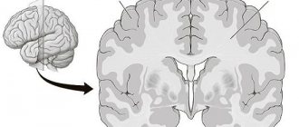
Examples of MR image correction
After the tests, the scientists began research directly. First of all, they turned their attention to the so-called passive mode network of the brain (operational resting neural network, Default Mode Network, DMN). This neural network, discovered and named in the early 2000s by Markus Reichl, is active in a state when a person is not busy performing any task related to the outside world, but, on the contrary, is inactive, resting, daydreaming, or absorbed in himself. The authors of the study were interested in whether this network is already formed before birth, or appears after the baby is born. This is also important because, according to modern concepts, it is the DMN that plays a key role in the functioning of consciousness.
DMN in the fetal brain. Illustration from the article discussed
A study of eight fetuses at 32-37 weeks, as well as premature babies of this period, showed that the DMN is already quite active at this age.
According to Colin Studholme, the new technique can be used in many studies. Especially in studying the harmful effects of alcohol and other psychoactive substances on fetal brain development.
Text: Alexey Paevsky
Detecting default mode networks in utero by integrated 4D fMRI reconstruction and analysis” by Sharmishtaa Seshamani, Anna I. Blazejewska, Susan McCown, Jason Caucutt, Manjiri Dighe, Christopher Gatenby, and Colin Studholme in Human Brain Mapping. Published online August 11, 2020 doi:10.1002/hbm.23303
So you are your synaptome?
Maybe. The essence of you—memories, thoughts—seems to be imprinted in the way different synapses fire in response to input. Like a fingerprint, the synaptome could be read to decipher what you're thinking. However, this research is just the beginning. Neuroscientists have yet to dissect the complex connections between synapses and you.
"This map opens up many new avenues of research that should transform our understanding of behavior and brain disease," Grant says.